Below is a list of over 200 cases arranged by condition or type. Click on the card to display cases in that category.
.jpg)
Cataract
Minimize List
Anterior Chamber Cilium: 81-year-old male with intraocular cilium and dislocated intraocular lens
Capsular Block Syndrome: An Unusual Presentation: 51-year-old pseudophakic female presents with poor vision in the right eye (OD). Fluctuations in vision were first noticed 6 months ago with documented vision as poor as 20/150. She had uneventful cataract surgery 10 years prior with insertion of a Polymethyl methacrylate (PMMA) intraocular lens (IOL) in the capsular bag.
Cataract Formation after Pars Plana Vitrectomy. Patient referred from the retina service for dense cataract formation three days following pars plana vitrectomy. Due to the rapid onset and severity of the cataract, there was concern for an iatrogenic break in the posterior capsule during vitrectomy. (includes video)
Communication Issues Following a Post Operative Surprise: Blurry Vision Following Cataract Surgery
Endophthalmitis: 82-year-old male status post phacoemulsification in the left eye with acute decrease in vision
Intraoperative Floppy Iris Syndrome: IFIS presents problems during cataract surgeries by decreasing the size of the work field and increasing the rate of complications such as iris trauma, posterior capsule rupture, and vitreous loss.
Intralenticular Foreign Body: 23-year-old male with staple in the eye
Intraocular Foreign Body: A Classic Case of Metal on Metal Eye Injury
Intraocular Lens "Time Out": A Systems Based Case
Marfan Syndrome: 28-year-old female with Marfan syndrome and decreased vision/glare in both eyes
Morgagnian Cataract: 59-year-old female with decreased vision at distance and near, more so in the right eye than the left and iincreasing difficulty with glare from bright lights at night, seeing road signs, watching television, and reading the newspaper.
Ocular Syphilis Presenting with Posterior Subcapsular Cataract and Optic Disc Edema: bilateral painless progressive vision loss
Persistent Fetal Vasculature: 19-year-old female with a history of amblyopia in the right eye (OD) due to persistent fetal vasculature (PFV) presented to the University of Iowa Hospitals and Clinics (UIHC), Department of Ophthalmology to re-establish care.
Pembrolizumab (Keytruda®) Induced Uveitis in a Patient with Metastatic Melanoma: 65-year-old male with history of metastatic cutaneous melanoma previously on pembrolizuamb presented to the comprehensive ophthalmology service with blurred vision worse in the left eye for the past 6 months.
Phacoemulsification: Considerations for Astigmatism Management, a tutorial and case
Posterior capsular folds creating a Maddox rod effect following cataract surgery: 73-year-old female presents with glare in the right eye 5 months after cataract surgery
Posterior Polar Cataract. 38-year-old male with blurry distance vision and problems seeing at night and significant glare from oncoming headlights when driving at night.
Toxic Anterior Segment Syndrome (TASS): A System's Based View of a Day in the Life of a Canula
Traumatic Cataract: 11-year-old boy with glass injury to the right eye
True Exfoliation Syndrome:84-year-old male presented with gradually worsening blurry vision in both eyes (OU), left worse than right, over a period of several years.
Minimize List
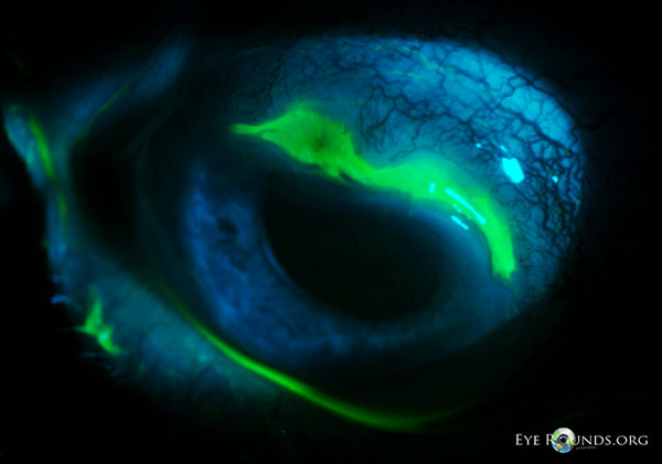
Cornea / External Eye Disease
Minimize List
Acanthamoeba Keratitis: 39-year-old contact lens wearer with persisting keratitis & pain
Acute Corneal Hydrops: 51-year-old male with keratoconus and history of rigid gas permeable (RGP) contact lens wear in the left eye presented with left eye redness, pain, light sensitivity, and tearing for 1 week.
Adenoviral Conjunctivitis: 38-year-old white female with watery, red, irritated eyes; left more than right.
Adult Chlamydial Conjunctivitis: 23-year-old male with 6-week duration of red eye
Atopic Keratoconjunctivitis: 52-year-old male with poor vision, severe ocular itchin, and mucoid discharge. He reports a history of waxing and waning eczema
Bilateral Corneal Edema Associated with Amantadine Use: 42-year-old female with 2-month history of blurry vision, photophobia, and mild, aching pain in both eyes.History of Huntington disease-related movement disorder
Boston Keratoprosthesis: An option for patients with multiple failed corneal grafts
Calcific Band Keratopathy. 71-year-old female referred for evaluation of a corneal scar in the right eye (OD). The scar was present 10 years previously when she was evaluated for painless and slowly progressive vision loss. She attributed the scar to a "fan injury" that she experienced at 20 years of age. Over the last ten years, she has experienced further painless vision loss OD. The vision in the left eye (OS) has been excellent.
Chandler Syndrome: A subtle presentation. (Iridocorneal endothelial syndrome: Chandler Syndrome variant) Patient initially sought care for migraines with mild headache on the right side of her head and halos around lights through her right eye. An ophthalmologist confirmed the narrow angles and placed her on medications to successfully lower her intraocular pressure. Halos around lights and a mild decreased acuity persisted.
Chemical Eye Injury: A Case Report and Tutorial: A guide to emergent management and medical therapy to promote ocular surface rehabilitation
Cogan's Syndrome: 42-year-old Female with Interstitial Keratitis and Vertigo
Congenital Aniridia: 18-year-old male with suspected aniridia with corneal pannus involving both eyes
Conjunctival Melanoma arising from Primary Acquired Melanosis (PAM): 61-year-old Caucasian female presenting for evaluation of corneal mass OS found on routine exam about 2 months previous.
Conjunctival rhinosporidiosis in a temperate climate. 27-year-old male was referred for evaluation of a left medial canthal lesion. The patient first noticed the lesion about one month prior to presentation.
Corneal Endothelial Transplantation: Descemet's Stripping Endothelial Keratoplasty (DSEK)
Corneal Marginal Ulcer: Marginal keratitis with ulceration in a 45 year-old male
Cystinosis: A 12-year-old boy with light sensitivity. Patient had progressive photophobia during outdoor activities. Recently, he became symptomatic indoors. He had intermittent redness and mild foreign body sensation in both eyes. Examination showed the presence of corneal crystals, laboratory testing showed increased levels of leukocyte cystine.
Cystinosis: 4-year-old girl with failure to thrive, severe photophobia, and newly diagnosed renal insufficiency.
Cytarabine-induced Keratoconjunctivitis: 17-year-old boy presented with one day of acute-onset bilateral foreign body sensation, tearing, photophobia, blurriness, and redness.
Deep Lamellar Endothelial Keratoplasty (DLEK) for Fuchs: 72-year-old female with Fuchs.
Descemet’s membrane endothelial keratoplasty (DMEK) for Fuchs’ endothelial dystrophy
Epithelial-Stromal and Stromal Corneal Dystrophies: A Clinicopathologic Review
Erythema Multiforme: 22-year-old male with a persistent red eye for two weeks
Exposure Keratopathy in the Critically Ill: A Case Report, Discussion, and Systems-Based Intervention
Fabry Disease: A key differential in the diagnosis of corneal verticillata
Fuchs Endothelial Dystrophy: 35-year-old female with blurry vision OU lasting hours in the morning
Fungal Keratitis - Fusarium: 41-year-old female contact lens wearer with persisting keratitis
Granular Corneal Dystrophy Discovered Following LASIK: A Clinicopathologic Correlation of Granular Corneal Dystrophy Type II and LASIK. 44-year-old male with progressive decrease in vision in both eyes with "tremendous" glare symptoms. Eight years prior to presentation he underwent Laser-assisted in situ keratomileusis (LASIK) in both eyes.
Herpes Simplex Keratitis: 36-year-old female presented to the Emergency Treatment Center (ETC) with one day of right eye pain, photophobia and decreased vision.
Infectious Crystalline Keratopathy (ICK): 72-year-old female with decreased vision
Interstitial keratitis secondary to Herpes Simplex Virus (HSV): 22- year-old white man with a history of asthma, vitamin D deficiency, and blepharitis presented to the cornea clinic with gradual onset blurred vision in the right eye and photophobia over 6 weeks.
Lattice Corneal Dystrophy Type II – Meretoja's Syndrome: patient with anterior stromal corneal dystrophy, progressive decline in vision with glare
Laser Assisted Keratoplasty: 63-year-old female with history of Fuchs endothelial dystrophy and bilateral pseudophakic bullous keratopathy presents with decreasing vision in the right eye
Megalocornea. 60-year-old male with a history of simple megalocornea reported visual disturbance while changing head position, his vision worsened with his head bent down.He previously had cataract surgery with an iris-sutured IOL.
Ocular Cicatricial Pemphigoid: atypical presentation as pseudopterygium and limbal stem cell deficiency. 78-year-old male with chronic irritation and dry eye unresponsive to aggressive lubrication, punctal occlusion, and prednisolone/sulfacetamide drops
Ocular Manifestations of Stevens-Johnson Syndrome: 13-year-old female treated with trimethoprim-sulfamethoxazole developed mucosal and dermatologic eruptions with ocular involvement.
Ocular Rosacea: 62-year-old female with blurry vision and red, itchy eyes
Perioperative Corneal Abrasions: Systems-based review and analysis. 64-year-old male patient noted severe pain in his right eye while in the post-anesthesia care unit shortly after laparoscopic surgery. A bedside ocular examination was remarkable for a linear, horizontal corneal epithelial defect.
Peripheral Ulcerative Keratitis (PUK): A 79-year-old woman was referred for the evaluation of a corneal ulcer in the right eye (OD). She began experiencing foreign body sensation in the right eye about 2 months prior to presentation after getting poked in the eye while gardening
Peters Anomaly: 2-day-old infant evaluated for bilateral corneal opacity noted at the time of delivery. There were no known maternal infections during the pregnancy and no problems during the perinatal period. The infant did not show evidence of tearing, blepharospasm, photophobia, drainage, or ocular redness.
Phakic intraocular lenses for high myopia: A 24-year-old male with unilateral high myopia presented with anisometropia (unequal refractive error between the eyes) with aniseikonia (binocular differences in image size) and a large right sensory exotropia. His primary concerns were the cosmetic effect of his exotropia and the intolerable double vision and aniseikonia he experienced when his "bad eye decided to work." He wore spectacles with the right eye under-corrected to minimize his symptoms. He did not tolerate contact lenses and was interested in surgical options.
Phlyctenular Keratoconjunctivitis: 12-year-old Female with Staphylococcal Blepharitis
Posterior Polymorphous Corneal Dystrophy (PPMD). 3-year-old male who was referred to the cornea clinic after failing a KidSight screening and was found to have bilateral corneal opacities when evaluated by his optometrist. He had bilateral, diffuse, geographic-shaped, discrete, gray lesions at the level of Descemet's membrane.
Phacoemulsification: Considerations for Astigmatism Management, a tutorial and case
Pseudophakic Bullous Keratopathy: Deep Lamellar Endothelial Keratoplasty (DLEK) and Intraocular Lens (IOL) exchange with Anterior Vitrectomy.
Post LASIK Ectasia: Female patient presented status post bilateral LASIK for myopia. Vision in the right eye was progressively more blurred.
Salzmann's Nodular Corneal Degeneration: 62-year-old woman presents with 3 years of progressive decrease in vision. She has had 3 updates of her spectacle prescription that have not provided satisfactory vision.
Surgical Management of Keratoconus: A 22-year-old male with a history of progressive keratoconus
Superior Limbic Keratitis: A 63-year-old male presents for evaluation of irritation and injection of both eyes, that is refractory to management with lubrication.
Terrien Marginal Degeneration: A 35-year-old man with spontaneous open globe injury.
Thygeson's Superficial Punctate Keratitis: A 23-year-Caucasian male presents with bilateral photophobia, foreign body sensation, and tearing that is worse in the left eye.
Use of Ophthalmic Medications in Pregnancy: A 19-year-old pregnant woman at 19 weeks gestation was referred for evaluation and treatment of suspected Acanthamoeba keratitis.
Vernal Keratoconjunctivitis: 8-year-old asthmatic male with reduced vision
Verticillata: 54-year-old white male with a known history of atrial fibrillation and hypertension on amiodarone.
Vision Loss After Contact Lens-Related Pseudomonas Keratitis. Since the majority of cases of microbial keratitis (mk) in contact lens wearers are due to Pseudomonas, and this specific infection heralds a worse prognosis, we investigated outcomes at our facility to determine the frequency and extent of visual loss in cases of Pseudomonas keratitis occurring in eyes with previously normal visual acuity and no risk factors for MK except contact lens wear.
Minimize List
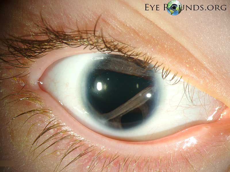
Genetic Eye Diseases
Minimize List
Autosomal Dominant Optic Atrophy: 47-year-old female with chronic, mildly subnormal vision
Best Vitelliform Macular Dystrophy: 30-year-old female referred for bilateral macular lesions.
Homocystinuria: 18-year-old male presented to clinic for regular monitoring of ocular manifestations of methionine synthase deficiency type homocystinuria, specifically cobalamin deficiency of the Cb1G subtype.
Juvenile open-angle glaucoma: 22-year-old Caucasian female referred in 1990 for evaluation of elevated intraocular pressure (IOP)
Lattice Corneal Dystrophy Type II – Meretoja's Syndrome: patient with anterior stromal corneal dystrophy, progressive decline in vision with glare
Leber Hereditary Optic Neuropathy: A 17-year-old male presents with progressive, painless, bilateral vision loss
Malattia Leventinese (Familial Dominant Drusen): 30-year-old female with drusen
Pattern Dystrophy Associated with Myotonic Dystrophy: 56-year-old female presented with a one-year history of increasingly distorted vision OS described as "crinkled"
RPE65-associated Leber Congenital Amaurosis
Unilateral Retinitis Pigmentosa: Visual field changes in a 31-year-old female
Minimize List
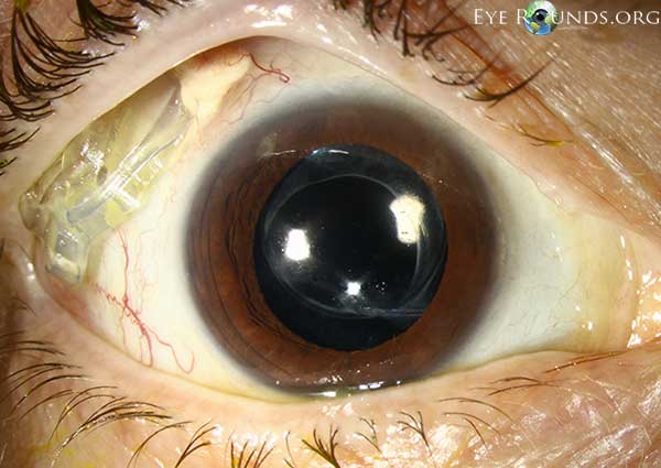
Glaucoma / Iris
Minimize List
Aphakic Glaucoma: 5-year-old female was referred to the University of Iowa Hospitals and Clinics (UIHC) Glaucoma Clinic for evaluation of suspected aphakic glaucoma of the left eye (OS).
Aqueous Misdirection: 88-year-old hyperopic female presented with left eye pain rated 10/10 and associated blurred vision, headache, nausea, and vomiting
Blebitis: 74-year-old with red eye after trabeculectomy
Competition Leads to Healthcare Savings: A systems based case from the VA
Giant reservoir formation from Ahmed seton valve: A 79-year-old man presenting for treatment of a chalazion on the left eye and follow-up care of his right anophthalmic socket. His wife mentioned that he exhibits a right head turn while watching television.
Grant syndrome (Syndrome of precipitates on the trabecular meshwork): 60-year-old male referred for evaluation of elevated intraocular pressure (IOP), right eye with corneal edema. Treated with a laser peripheral iridotomy and started on bimatoprost and brimonidine/timolol. The only pertinent exam findings were pigmented precipitates on the anterior trabecular meshwork OD on gonioscopy.
Hypotony: Late hypotony from trabeculectomy and Ahmed seton with resulting hypotony maculopathy: A 36-year old female presented with poor vision secondary to a low intraocular pressure (IOP) in her right eye.
Iris Cyst: 38-year-old female with iris lesion and gradual decrease in vision
Iris Melanoma: 47-year-old male referred in 1997 for evaluation of iris lesion
Iridocorneal Endothelial Syndrome (ICE) - essential iris atrophy: 63-year-old female with PAS, "iris mass", corectopia, and increased IOP OS.
Juvenile open-angle glaucoma: 22-year-old Caucasian female referred in 1990 for evaluation of elevated intraocular pressure (IOP)
Melanocytoma of the Iris and Ciliary Body: 14-year-old male with a pigmented iris lesion of the left eye noticed on routine eye exam approximately two years earlier. Higher power magnification showed elevation of lesion with mildly irregular surface without surface vessels and the iris central to the lesion pushed into folds.
Nanophthalmos with Ocular Hypertension and Angle-Closure: 37-year-old woman presented as a referral from her local eye doctor who found that her intraocular pressure (IOP) was high on routine eye examination.
Neovascular Glaucoma: 80-year-old man with recurrent vitreous hemorrhages, hyphema, and elevated intraocular pressure after a central retinal vein occlusion in the right eye
Normal Tension Glaucoma: 50-year-old female with progressive visual field loss, glaucomatous disc change and normal IOP
Pigmentary Glaucoma: 24-year-old male with episodic haloes around lights and blurry vision
Phacolytic Glaucoma: 65-year-old male presented with acute onset of pain and redness in his right eye (OD), which had long-standing light perception vision after an explosive injury resulted in a penetrating shrapnel wound and large macular scar
Plateau Iris: A healthy 47-year-old female referred for evaluation for glaucoma exhibitsan uncommon form of primary angle closure glaucoma
Posner-Schlossman Syndrome: 63-year-old male was evaluated on an urgent basis for pain, redness, and "elevated eye pressure" in the left eye (OS).
Primary Congenital Glaucoma (Infantile Glaucoma): 3-year-old female referred for evaluation of increased eye size, left eye
Pseudoexfoliation Glaucoma: 65-year-old male with complaints of painless, gradual loss of vision OS.
Refractory Primary Open-Angle Glaucoma Treated with Ab Interno Canaloplasty (ABiC): A 59-year-old male presented with a history of Granulomatosis with Polyangiitis (GPA) - formerly known as Wegener's Granulomatosis, medically-uncontrolled primary open angle glaucoma (POAG), and pathologic myopia.
Secondary Open-Angle Glaucoma Treated with Gonioscopy-Assisted Transluminal Trabeculotomy (GATT). A 20-year-old female was referred for management of secondary open-angle glaucoma. 3 years prior she had been diagnosed with an unusual vasoproliferative tumor in her left eye. Shortly after treatment with intravitreal steroids and photodynamic therapy, her IOP spiked to 59 mm Hg.
Seton Infection: Serratia Marcescens: 57-year-old white female with 1 day history of redness, swelling, and pain in her right eye.
Sturge-Weber Syndrome: 4-year-old child with a history of seizures and glaucoma.
Minimize List
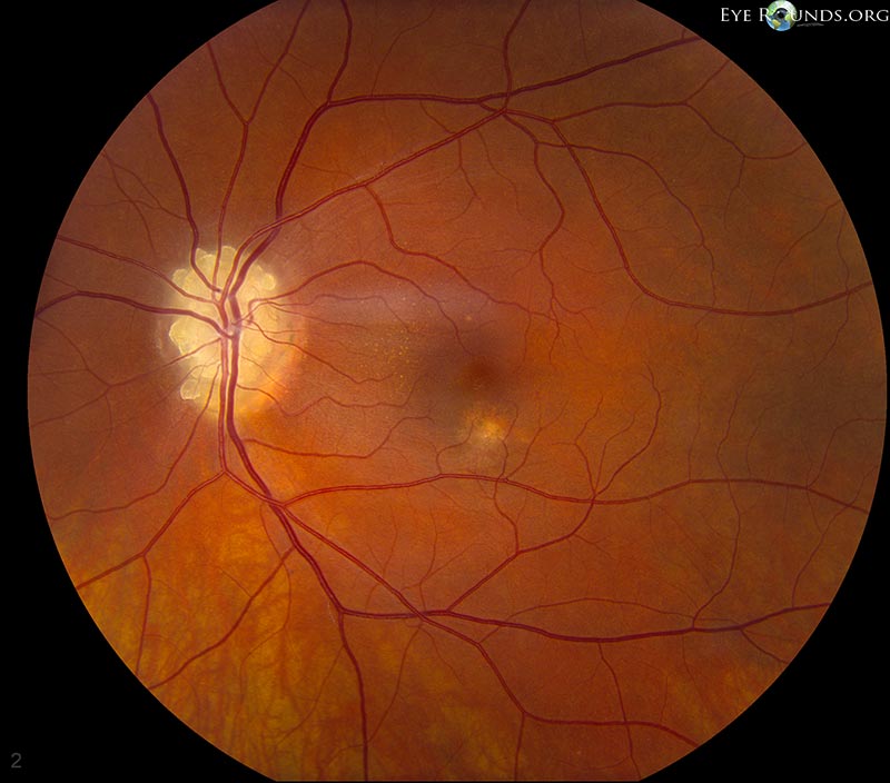
Neuro-Ophthalmology
Minimize List
Acute Demyelinating Encephalomyelitis (ADEM) with associated optic neuritis: 9-year-old girl presents to an outside hospital with fatigue, poor appetite, and decreased activity for 3 weeks.
All-trans retinoic acid (ATRA) induced intracranial hypertension in a patient with acute promyelocytic leukemia: 19-year-old male ungergoing ATRA and Idarubicin induction therapy for APL with one-week complaint of intermittent headaches and transient visual obscurations.
ANCA-associated granulomatous vasculitis (Wegener's granulomatosis): 54-year-old male with binocular vertical diplopia 4 months prior to presentation and progressively worsening. Vision in the right eye was starting to "darken." Prior to presentation, patient was followed by an otolaryngologist for his sinus disease and nosebleeds.
Bilateral optic nerve hypoplasia, 2-month-old with: Highlighting the importance of a multi-disciplinary approach
Cranial Nerve IV (Trochlear Nerve) Palsy: 57-year-old male complaining of vertical diplopia after head trauma.
Dorsal Midbrain Syndrome (Parinaud's Syndrome) with Bilateral Superior Oblique Palsy: 43-year-old male referred for evaluation of binocular diplopia.
Dramatic Visual Recovery in Untreated Indirect Traumatic Optic Neuropathy: Severe vision loss in one eye immediately after motorcycle accident.
Ethambutol Toxicity and Optic Neuropathy: 60-year-old female with bilateral painless central vision loss
Facial Nerve Palsy: Ocular Complications and Management. 72-year-old male with left lower eyelid retraction in left facial nerve palsy secondary to metastatic lymphoma and radiation therapy. This case features 11 oculoplastic surgery videos.
Functional Visual Loss: 37-year-old male with decreased vision progressively worsening over the past year. The patient was referred because he could not read any of the letters on the chart and showed no improvement with refraction.
Horner's Syndrome (due to Cluster Headache): 46-year-old male presenting with HA and ptosis.
Idiopathic Intracranial Hypertension (Pseudotumor Cerebri): 31-year-old obese female presenting with a history of headaches and transient visual obscurations for six months.
Idiopathic Orbital Myositis: A Treatment Algorithm: 19-year-old female who presents with a one-week history of headache, one day of left "eye swelling," and pain that is worse with eye movement.
Internuclear Ophthalmoplegia: 40-year-old man presented to the emergency room complaining of acute onset blurry vision through his left eye (OS).
Leber Hereditary Optic Neuropathy: A 17-year-old male presents with progressive, painless, bilateral vision loss
Myasthenia Gravis: 32-year-old female with progressive LUL ptosis for several years.
Neurofibromatosis Type 1 - Optic Nerve Glioma: 8-year-old white female with acute awareness of complete vision loss, left eye.
Ocular Tilt Reaction: 53-year-old female complaining of vertical diplopia following a stroke and found to have a skew deviation, fundus torsion, and torticollis
Ophthalmoplegic Migraine: 30-year-old male with migraine headaches and occasional diplopia
Optic Nerve Drusen: 19-year-old female with blurred vision
Optic Nerve Hypoplasia: 51-year-old female referred for consideration of cataract surgery. {Related Case: Communicating with patients about alternative therapies: A case of optic nerve hypoplasia in a 14-year-old male with poor vision since birth}
Optic Neuritis: A 40-year-old female noticed visual loss in her right eye accompanied by pain with eye movements and a dull retro-orbital ache, two weeks prior to her clinic visit. She also noted decreased perception of color and contrast.
Orbital inflammation secondary to bisphosphonate therapy: 80-year-old female with right eyelid swelling, pain, decreased vision, diplopia
Parkinson's disease: a review of the ophthalmological features: 67-year-old man presented to the strabismus clinic with increased difficulty reading for the past year. He experienced intermittent, horizontal, binocular diplopia and blurring of his vision while reading, accompanied by fatigue and headaches.
Pituitary Adenoma Causing Compression of the Optic Chiasm: 49-year-old white female with painless progressive vision loss.
Posterior Ischemic Optic Neuropathy: 64-year-old male admitted to the intensive care unit following a spinal surgery reporting profoundly decreased vision in both eyes upon waking the following morning. His corrective lenses did not improve his vision.
Progressive Supranuclear Palsy in a Patient with Progressive Nonfluent Aphasia: A case of an 81-year-old woman with limited eye movements
Pseudoabducens palsy: When a VI nerve palsy is not a VI nerve palsy
Recurrent Neuroretinitis: 29-year-old white female with progressively worsened vision, right eye, diminished central vision, and pain with eye movements.
Sarcoidosis: 53-year-old A female with gradual, painless loss of vision in both eyes. (also see Ocular Sarcoidosis)
See-Saw Nystagmus: 64-year-old male who first noted the onset of oscillopsia five months prior to presentation.
Spinocerebellar Ataxia with Ophthalmoplegia: 46-year-old male presenting with progressive esotropia
Thyroid Eye Disease (Graves' ophthalmopathy): 51-year-old male seen in consultation from the internal medicine service for evaluation and management of "dry and red eyes.
Tumor Necrosis Factor Alpha Antagonist Precipitating Demyelinating Disease: A 34-year-old woman presents with optic neuritis and develops demyelinating brain lesions
Unilateral Optic Nerve Hypoplasia in a patient desiring surgical treatment for exotropia. 17-year-old male with a longstanding sensory left exotropiainquiring about strabismus surgery.
Vertical Oscillopsia: A Case of Superior Oblique Myokymia. A 39-year-old healthy male reported intermittent episodes of "objects bouncing up and down" in his vision in his left eye lasting for seconds to minutes for the previous two months. [video included]
Visual Snow Syndrome A 59-year-old pilot with a history of visual aura since his teenage years and anxiety presented to the neuro-ophthalmology clinic with a primary complaint of seeing a "snow storm" for one year.
Minimize List
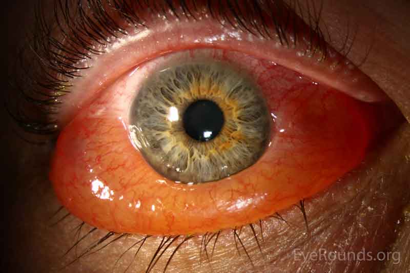
Oculoplastic Surgery, Orbit, Eyelids, Lacrimal
Minimize List
28-year-old woman presented with a 1-month history of progressive left upper eyelid pain
Adenoid cystic carcinoma lacrimal gland: 99-year-old patient presented to the oculoplastics clinic with double vision, left proptosis, and decreased vision on the left, which had progressed slowly over the last year.
Allergic Contact Dermatoblepharitis: 55-year-old female with a Medical History significant for multiple sclerosis presented with a one-week history of itchy eyes and blurry vision
ANCA-associated granulomatous vasculitis (Wegener's granulomatosis): 54-year-old male with binocular vertical diplopia 4 months prior to presentation and progressively worsening. Vision in the right eye was starting to "darken." Prior to presentation, patient was followed by an otolaryngologist for his sinus disease and nosebleeds.
Basal Cell Carcinoma, Morpheaform subtype: 78-year-old male with complaints of enlarging Left Lower Lid lesion.
Bell's Palsy Treated with Facial Nerve Decompression: 68-year-old man presented to the eye clinic with five days of right-sided facial droop, which was associated with the inability to fully close the right eye and slurred speech.
Bilateral Nasolacrimal Duct Obstruction after Adjuvant I-131 for Thyroid Cancer: 53-year-old female with bilateral tearing worsening over the preceding year. She describes tears that run down the front of her face and denies any ocular irritation.
Blepharospasm and Apraxia of Eyelid opening: "I Can't Open My Eyes": 72-year-old man with increasingly frequent difficulty opening his eyes, hw was also bothered by frequent contractions of the muscles around the eyes on both sides of his face which caused forcible eyelid closure.
Canalicular Laceration - Dog Bite: 5-year-old white female presenting with dog bite to left side of face.
Capillary Hemangioma: 4 month-old female with occlusive vascular lesion of right upper eyelid
Carotid Cavernous Fistula: 46-year-old female patient presented twith double vision and visual distortion.
Cavernous Hemangioma: Painless Proptosis in a 62-year-old female: Patient states that over the last year she has noticed that her right pupil is higher than her left
Chalazion: Acute presentation and recurrence in a 4-year-old female
Chronic Progressive External Ophthalmoplegia (CPEO) - Kearns-Sayre Syndrome: 12-year-old boy with painless, progressive ptosis OU over 3 years
Churg-Strauss Syndrome with Orbital Inflammation. 56-year-old male with two-month history of progressive proptosis & drooping of the left upper eyelid and binocular diplopia for several weeks
Ciliary Body Leiomyoma: Clinicopathologic Correlation
Congenital dacryocystocele with spontaneous resolution: Full-term 10 day-old female referred to the pediatric ophthalmology clinic by dermatologist for a bluish mass inferior to the right medial canthus
Congenital Ptosis. Seven month-old male patient with drooping of the left upper lid/left upper eyelid ptosis
Dermoid Cyst: 6 month-old male was referred by his pediatrician for evaluation of a right upper eyelid mass.
Distichiasis: 64-year-old white male with bilateral eye irritation.
Enophthalmos Secondary to Metastatic Breast Cancer. 63-year-old female presenting with progressive double vision and a droopy right eyelid.
Facial Nerve Palsy: Ocular Complications and Management. 72-year-old male with left lower eyelid retraction in left facial nerve palsy secondary to metastatic lymphoma and radiation therapy. This case features 11 oculoplastic surgery videos.
Fibrous Dysplasia: 43-year-old female was referred to the University of Iowa Oculoplastics Clinic by her comprehensive ophthalmologist for the evaluation of worsening right-sided proptosis.
Floppy Eyelid Syndrome. Two cases illustrating the presenting symptoms and comorbidities associated with floppy eyelid syndrome.
Giant reservoir formation from Ahmed seton valve. A 79-year-old man presenting for treatment of a chalazion on the left eye and follow-up care of his right anophthalmic socket. His wife mentioned that he exhibits a right head turn while watching television.
Idiopathic Orbital Myositis: A Treatment Algorithm: 19-year-old female who presents with a one-week history of headache, one day of left "eye swelling," and pain that is worse with eye movement.
Invasive Fungal Orbitorhinocerebral Mucormycosis
Involutional ectropion: Options for surgical management of ectropion. A 72-year-old male presents with epiphora of the left eye, present for one year, worsening over the last month. Also experiencing a foreign body sensation in both eyes and redness along the left lower eyelid.
Involutional entropion: options for surgical management of entropion. A 67 year-old male presents with irritation and "scratchiness" of the right eye. His symptoms have been present for the past 8 months and have been only partially relieved with artificial tears and lubricating ointment at night.
Lymphoma, Orbital. 60-year-old male who was evaluated 1 wk ago for "conjunctivitis" left eye by internal medicine.
Meningiomas. a case and review. A 36-year-old female presented with a "bulging" and apparently swollen left eye noticed about one month prior to her visit.
Merkel Cell Carcinoma of the Eyelid. A 94-year-old female is referred for an evaluation for possible skin cancer on the nasal aspect of her left lower lid. The lesion has been present on the left lower lid for about one month. It has grown rapidly in size over the previous two weeks and is now obstructing her vision
Multiple endocrine neoplasia type 2B: A 27-year-old male presents with multiple eyelid lesions. This patient, with a history of multiple endocrine neoplasia type 2B (MEN2B) was referred by his oncologist for a large lesion on the outer corner of his right eye.
Myeloid Sarcoma: 20-year-old female with a history of intermittent binocular diplopia that worsened with right head tilt and a six week history of progressive fullness of the right upper eyelid. Medical History is notable for Acute Myelogenous Leukemia reported to be in remission.
Nasolacrimal duct obstruction secondary to sarcoidosis: 72-year-old female with sarcoidosis, corneal perforation, and nasolacrimal duct obstruction
Nodular Basal Cell Carcinoma: 49-year-old female with left lower lid lesion
Oculopharyngeal muscular dystrophy: 58-year-old female with gradual worsening of bilateral blepharoptosis.She also has mild dysphagia and proximal muscle weakness
Spontaneous Retrobulbar Hemorrhage with Subsequent Orbital Compartment Syndrome. 80-year-old male presented with diplopia and proptosis of right eye. Complained of sudden vision loss in the right eye. Symptoms began that morning with diplopia when he left home to walk his dog. By the time he returned home, he had profound vision loss and right eye protruding.
Orbital Cellulitis in a Child: 9-year-old female with left nasal pain, swollen, red left eye and diplopia in all gazes
Orbital Eosinophilic Granuloma: 16-month-old male patient with left-sided periorbital swelling beginning three weeks prior to presentation in the setting of an upper respiratory infection
Orbital inflammation secondary to bisphosphonate therapy: 80-year-old female with right eyelid swelling, pain, decreased vision, diplopia
Orbital Lymphatic Malformation (Lymphangioma): 5 year-old female complained of an intermittent frontal headache. Over the next 2 days, she began having daily episodes of emesis and her parents noticed "bulging of her left eye." She was taken to the emergency room where an MRI was performed and demonstrated a left orbital lesion extending into the superior orbital fissure.
Orbital Osteoma: 20-year-old female with a 9-month history of left upper lid droopiness
Periorbital Necrotizing Fasciitis. 87-year-old female with orbital cellulitis resistant to broad-spectrum antibiotics was referred to UIHC. CT imaging which showed fat stranding in the retrobulbar space as well as proptosis of the right eye. Initial cultures from her blood and periocular drainage grew group A beta-hemolytic streptococci.
Pan-endophthalmitis and Orbital Cellulitis: 89-year-old female with a history of gradual redness and proptosis, right eye over 3 weeks.
Punctal and Canalicular stenosis (dacryostenosis) from Docetaxel: 38-year-old female with persisting tearing for 6 months.
Sarcoidosis Affecting the Lacrimal Gland: 39-year-old female with left upper eyelid swelling of 4 months duration
Sebaceous Cell Carcinoma: A Masquerade Syndrome
Secondary Tumors After Hereditary Retinoblastoma: A Case of Orbital Leiomyosarcoma 50 Years After Initial Enucleation and Radiation Therapy..
Silent Brain Syndrome: 32-year-old male with history of excess tearing in both eyes with mucous-like yellowish discharge. Excess tearing henders vision.
Silent Sinus Syndrome: 36-year-old male with sunken left orbit (Enophthalmos)
White-eyed blowout fracture: 17-year-old male with diplopia, pain with eye movement, nausea and vomiting after blunt trauma to the left eye
Minimize List
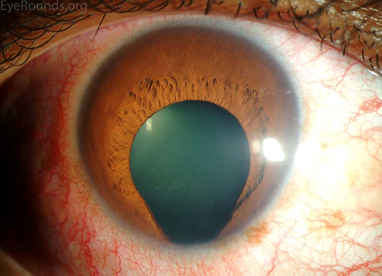
Pediatric Ophthalmology & Strabismus
Minimize List
Acute Acquired Brown Syndrome: 38-year-old Male with Ethmoidal Sinus Mucocele
Anisometropic Amblyopia. 5-year-old male with unequal vision.
2-month-old with bilateral optic nerve hypoplasia: Highlighting the importance of a multi-disciplinary approach
Capillary Hemangioma: 4 month-old female with occlusive vascular lesion of right upper eyelid
Chalazion: Acute presentation and recurrence in a 4-year-old female
Coats Disease: 11-year-old male with poor vision in left eye
Congenital dacryocystocele with spontaneous resolution: Full-term 10 day-old female referred to the pediatric ophthalmology clinic by dermatologist for a bluish mass inferior to the right medial canthus
Congenital Lymphocytic Choriomeningitis Virus (LCMV) infection: 1-week-old male with hydrocephalus and bilateral chorioretinitis
Congenital Ptosis. 7 month-old male patient with drooping of the left upper lid/left upper eyelid ptosis.
Convergence Insufficiency: 15-year-old male with worsening headaches and eye strain while reading, present since he started high school the previous year. He complained of blurry vision while reading and letters running together on the page. He often had to cover one eye to read comfortably
Dermoid Cyst: 6 month-old male was referred by his pediatrician for evaluation of a right upper eyelid mass.
Down Syndrome: 14-month-old male with intermittently crossed eyes
Duane Retraction Syndrome: 31-year-old Male With Globe Retraction
Goldenhar Syndrome (Oculo-Auriculo-Vertebral Spectrum): 6 day-old male with limbal dermoids
Holoprosencephaly and Strabismus. Female child presenting to the eye clinic at age 15 months for eye crossing. She had a history of severe hydrocephalus with seizures and alobar holoprosencephaly at birth.
Infantile (Congenital) Esotropia: 2 month-old female began crossing her eyes shortly after birth. Parents feel that it is worsening. Crossing is worse when patient is tired and alternates between the right eye and the left eye
Juvenile Idiopathic Arthritis with Associated Bilateral Anterior Uveitis in a Four-year-old Girl. Routine preschool screening found central posterior synechiae in both eyes. Patient was found to have macular edema and was referred to rheumatology for systemic evaluation with suspicion of inflammatory etiology.
Management of Amblyopia: Discussing Options for Treatment in the Age of the Internet
Monofixation Syndrome: 11-year-old female referred after inability to improve vision with refraction in the left eye
Non-accidental Trauma: An unresponsive infant with bilateral retinal hemorrhages
Non-accommodative Esotropia and Botulinum Toxin Therapy: 7-year-old boy presented with a history of intermittent, alternating eye crossing for one year that had become constant in the past 2 months.
Orbital Cellulitis in a Child: 9-year-old female with left nasal pain, swollen, red left eye and diplopia in all gazes
Orbital Eosinophilic Granuloma. 16 month-old male patient with left-sided periorbital swelling beginning three weeks prior to presentation in the setting of an upper respiratory infection
Orbital Lymphatic Malformation (Lymphangioma): 5 year-old female complained of an intermittent frontal headache. Over the next 2 days, she began having daily episodes of emesis and her parents noticed "bulging of her left eye." She was taken to the emergency room where an MRI was performed and demonstrated a left orbital lesion extending into the superior orbital fissure.
Parkinson's disease: a review of the ophthalmological features: 67-year-old man presented to the strabismus clinic with increased difficulty reading for the past year. He experienced intermittent, horizontal, binocular diplopia and blurring of his vision while reading, accompanied by fatigue and headaches.
Prostaglandin-Associated Periorbitopathy: Progressive glaucoma and periorbital changes. 52-year-old woman presented with elevated intraocular pressure (IOP) and visual fields demonstrating progressive field loss despite maximum tolerated medical therapy. She had gradual periorbital changes consisting of a “sunken appearance” with a larger eyelid opening.
Refractive Accommodative Esotropia. 4-year-old boy originally presented to ophthalmologist at 2 years of age with crossed eyes. Discussion of the management of Refractive Accommodative Esotropia.
Retinoblastoma: 4 month old boy with a "blur" in one eye since birth
Rhabdomyosarcoma: Eleven-year-old male with two weeks of blurry vision, headaches, and difficulty moving the right eye for the past two weeks
Sagging Eye Syndrome. 77-year-old female with a complaint of intermittent, binocular, horizontal diplopia present for the last six months. Diplopia was only present at distance and no double vision with near tasks. The double vision was worse with lateral gazes in both directions.
Unilateral Optic Nerve Hypoplasia in a patient desiring surgical treatment for exotropia. 17-year-old male with a longstanding sensory left exotropiainquiring about strabismus surgery.
Vernal Keratoconjunctivitis: 8-year-old asthmatic male with reduced vision
Minimize List
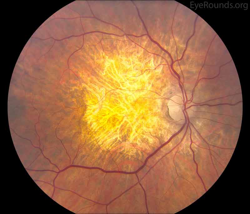
Retina & Vitreous
Minimize List
Acute Macular Neuroretinopathy: 30-year-old woman presented for evaluation of vision loss in both eyes (OU) in the setting of a newly diagnosed transverse venous sinus thrombosis.
Acquired Ocular Toxoplasmosis: 42-year-old female with "fuzzy" vision for two weeks
Acquired Peripheral Retinoschisis: 83-year-old male was referred to our retina clinic for a retinal detachment which was described as close to the macula in his right eye. He was asymptomatic on presentation.
Acute Retinal Necrosis: 84-year-old man with Alzheimer’s dementia presented with floaters and decreased vision in both eyes (OU).
Acute Zonal Occult Outer Retinopathy (AZOOR) and Acute Idiopathic Blind Spot Enlargement Syndrome (AIBSE): 34-year-old female awoke with painless loss of vision left eye 3 weeks prior to presentation.
Age-related Macular Degeneration: Progression from Atrophic to Proliferative: 76-year-old female with 17-year history of atrophic AMD and increased size of existing central scotoma discovered with home Amsler grid testing
Birdshot Choroiditis: 55-year-old female with persistent vitreous floaters
Behcet's Disease: A 32-year-old female with recurrent ocular inflammation
Cat-Scratch neuroretinitis (Ocular bartonellosis): 44-year-old female with non-specific "blurriness" of vision, left eye.
Cataract Formation after Pars Plana Vitrectomy. Patient referred from the retina service for dense cataract formation three days following pars plana vitrectomy. Due to the rapid onset and severity of the cataract, there was concern for an iatrogenic break in the posterior capsule during vitrectomy. (includes video)
Central Retinal Artery Occlusion (CRAO): 81-year-old white male with sudden, painless vision loss left eye.
Changing Trends in Sympathetic Ophthalmia: Sympathetic ophthalmia following repeated vitreoretinal surgery with no history of trauma
Choroidal Hemangioma: Central scotoma in the right eye. Patient with reduced vision and oval-shaped central scotoma in the central visual field of the right eye for two months.
Choroidal Malignant Melanoma: 58-year-old female with pigmented retinal lesion and exudative retinal detachment
Choroidal Osteoma: 10-year-old girl presents for annual follow-up. She reports that she recently failed a school screening
Chorioretinitis Sclopetaria: A Systems Based Approach to Eye Injury Prevention. Patient with BB gun injury to right orbit
Coats Disease: 19-year-old male presented to his local optometrist with a 4-week history of floaters and a shadow in the superotemporal visual field of his left eye
Commotio Retinae: 29-year-old man was referred after sustaining a head injury during an altercation.
Congenital Lymphocytic Choriomeningitis Virus (LCMV) infection: 1-week-old male with hydrocephalus and bilateral chorioretinitis
Cytomegalovirus (CMV) Retinitis: 36-year-old Indian male with HIV and decreasing visual acuity
Endogenous Endophthalmitis: 33-year-old male presented to an outside hospital for suicidal ideation. He had felt unwell for several days, had decreased vision and pain in his left eye, and attributed his suicidality to pain.
Full-thickness Macular Hole (FTMH):68-year-old female with a history of non-exudative macular degeneration referred for vision changes.
Hydroxychloroquine (Plaquenil) Toxicity: 57-year-old female with bilateral central photopsias for two years. Currently treated with prednisone and methotrexate for Sjogren syndrome and inflammatory arthritis and was previously treated with hydroxychloroquine for 10 years, stopped one year ago.
Interferon Retinopathy: 60-year-old male with hepatitis C virus infection presents with new floaters in both eyes. Treated with peginterferon and ribavirin for 6 weeks prior to visit.
Idiopathic Juxtafoveal Telangiectasia Type II (Macular Telangiectasia type 2): 43-year-old male with decreased vision and a central scotoma OU for the past 10 years, which has been getting progressively worse
Malattia Leventinese (Familial Dominant Drusen): 30-year-old female with drusen
Multilayered hemorrhage in Acute Myeloid Leukemia (AML): 35-year-old female presented with severe, acute bilateral vision loss one day after admission to the hospital for weakness.
Multiple Evanescent White Dot Syndrome (MEWDS): 24-year-old female with one week duration of central scotoma, right eye
Ocular Ischemic Syndrome: 7-year-old female presented to clinic with decreased visual acuity in her left eye.
Optic Nerve Drusen: 19-year-old female with blurred vision
Pathologic Myopia: 69-year-old woman presented for evaluation of decreased vision and "wavy lines" in the right eye for approximately one week.
Pattern Dystrophy Associated with Myotonic Dystrophy: 56-year-old female presented with a one-year history of increasingly distorted vision OS described as "crinkled"
Polypoidal Choroidal Vasculopathy (PCV): 57-year-old Caucasian male referred for retinal thickening and subretinal fluid in right eye greater than left eye. The patient noted a stable gray spot and visual distortion OD for the previous several weeks. He had stable chronic floaters but no flashes of light, eye pain, or other ocular symptoms.
Progression of Proliferative Diabetic Retinopathy During Pregnancy. A woman with type 1 diabetes was evaluated, one week after an otherwise uncomplicated cesarian section, for decreased vision and multiple new floaters in both eyes. Onset of dramatically decreased vision in both eyes began at the time of child delivery.
Purtscher's Retinopathy: 22-year-old male with vision loss after trauma
Presumed Ocular Histoplasmosis (POHS): 30-year-old male with blurred central vision
Punctate Inner Choroidopathy with Choroidal Neovascular Membrane
Recurrent Neuroretinitis: 29-year-old white female with progressively worsened vision, right eye, diminished central vision, and pain with eye movements.
Retinal Artery Macroaneurysm (RAMA): Sudden painless decreased vision in the right eye.
Retinochoroidal coloboma: 54-year-old female with blurred vision
Serpiginous Choroiditis: 60-year-old male with 15 year history of serpiginous choroiditis reports a 2 day history of a new scotoma noticed on Amsler Grid testing of his right eye.
Stickler Syndrome: 10-year-old male with family history of retinal detachment and optically empty vitreous.
Sickle Cell Retinopathy: 18-year-old African-American male with known sickle cell disease is referred to the vitreoretinal service for evaluation of two days of blurry vision and new-onset floaters in the left eye that developed while playing basketball.
Unilateral Retinitis Pigmentosa: Visual field changes in a 31-year-old female
Valsalva Retinopathy: Vision loss after asthma attack: 32-year-old female patient presented with a "black spot" in the vision of her left eye.
Vascular Tufts of the Pupillary Margin: 68-year-old female presented to the retina clinic with a history of recurrent hyphema in both eyes (OU). She had multiple instances of spontaneous, non-traumatic hyphema for which she had eventually sought medical attention.
Vitamin A Deficiency and Nyctalopia: 55-year-old male with gradual onset of night blindness
Vitreous Syneresis: An Impending Posterior Vitreous Detachment (PVD): 60 year-old female with newly reported flashing lights and "large and stringy" floaters in the left eye.
Waldenstrom Macroglobulinema-Associated Retinopathy: A 61 year-old male presenting with worsening vision in both eyes over the past 2 years, poorly controlled diabetes, persistent DME, and proliferative diabetic retinopathy.
Minimize List
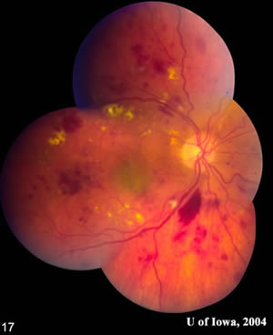
Systemic Disorders
Minimize List
Accelerated Hypertension : 67-year-old female with hypertension and diabetes mellitus with painless vision loss
Congenital Lymphocytic Choriomeningitis Virus (LCMV) infection: 1-week-old male with hydrocephalus and bilateral chorioretinitis
Cystinosis: A 12-year-old boy with light sensitivity. Patient had progressive photophobia during outdoor activities. Recently, he became symptomatic indoors. He had intermittent redness and mild foreign body sensation in both eyes. Examination showed the presence of corneal crystals, laboratory testing showed increased levels of leukocyte cystine.
Herpes Zoster Post-herpetic Neuralgia: 68-year-old male with decreased vision
Interferon Retinopathy: 60-year-old male with hepatitis C virus infection presents with new floaters in both eyes. Treated with peginterferon and ribavirin for 6 weeks prior to visit.
Invasive Fungal Orbitorhinocerebral Mucormycosis
Juvenile Idiopathic Arthritis with Associated Bilateral Anterior Uveitis in a Four-year-old Girl. Routine preschool screening found central posterior synechiae in both eyes. Patient was found to have macular edema and was referred to rheumatology for systemic evaluation with suspicion of inflammatory etiology.
Kayser-Fleischer Ring: A Systems Based Review of the Ophthalmologist's Role in the Diagnosis of Wilson's Disease
Kyphotic Patient Presents for Cataract Surgery: A Systems Based Case. Patient unable to assume typical supine position used for cataract surgery
Metastatic Choroidal Lesion: 61-year-old male presents with pain and blurry vision, right eye
Minocycline-Induced Hyperpigmentation. 69-year-old male complaining of chronic, progressive blue-black, painless hyperpigmentation of bilateral lower extremities in the setting of chronic lower extremity venous stasis and long-term, minocycline use for meibomian gland dysfunction and blepharitis.
Multiple endocrine neoplasia type 2B: A 27-year-old male presents with multiple eyelid lesions. This patient, with a history of multiple endocrine neoplasia type 2B (MEN2B) was referred by his oncologist for a large lesion on the outer corner of his right eye.
Myasthenia Gravis: 32-year-old female with progressive LUL ptosis for several years
Ocular Rosacea: 62-year-old female with blurry vision and red, itchy eyes
Ocular Sarcoidosis: A systems-based approach to diagnosis and treatment (also see Sarcoidosis)
Ocular Syphilis Presenting with Posterior Subcapsular Cataract and Optic Disc Edema:30-year-old female with progressive bilateral vision loss
Orbital inflammation secondary to bisphosphonate therapy: 80-year-old female with right eyelid swelling, pain, decreased vision, diplopia
Sarcoidosis Affecting the Lacrimal Gland: 39-year-old female with left upper eyelid swelling of 4 months duration
TTP-HUS: 29-year-old male with HA, hematuria, and visual obscurations.
Unilateral Optic Nerve Granuloma: Patient with bilateral panuveitis and unilateral optic nerve sarcoid granuloma
Vitamin A Deficiency and Nyctalopia: 55-year-old male with gradual onset of night blindness
Minimize List
Trauma
Minimize List
Canalicular Laceration - Dog Bite: 5-year-old white female presenting with dog bite to left side of face.
Carotid Cavernous Fistula: 46-year-old female patient reporting double vision and visual distortion
Chorioretinitis Sclopetaria: A Systems Based Approach to Eye Injury Prevention. Patient presenting with decreased vision after BB gun injury to right orbit
Firework-Related Ocular Injuries: 48-year-old male presented July 4th with a firework injury to his left eye which when the patient leaned over a firework that had malfunctioned. The firework launched and directly contacted the left orbit. The patient experienced severe eye pain and had immediate loss of vision in the left eye (OS).
Intralenticular Foreign Body: 23-year-old male with staple in the eye
Intraocular Foreign Body: A Classic Case of Metal on Metal Eye Injury
Patient Non-Compliance: Physician responsibility. 50-year-old male with penetrating globe injury
Purtscher's Retinopathy: 22-year-old male with vision loss after trauma
Traumatic Cataract: 11-year-old boy with glass injury to the right eye
White-eyed blowout fracture: 17-year-old male with diplopia, pain with eye movement, nausea and vomiting after blunt trauma to the left eye
Minimize List
Tumor / Oncology
Minimize List
Adenoid cystic carcinoma lacrimal gland: 99-year-old patient presented to the oculoplastics clinic with double vision, left proptosis, and decreased vision on the left, which had progressed slowly over the last year.
Basal Cell Carcinoma, Morpheaform subtype: 78-year-old male with complaints of enlarging left lower lid lesion.
Bilateral Diffuse Uveal Melanocytic Proliferation (BDUMP): 89-year-old male presents with multiple choroidal nevi, both eyes.
Capillary Hemangioma: 4 month-old female with occlusive vascular lesion of right upper eyelid
Cavernous Hemangioma: 31-year-old caucasian female with a painless, "dark spot" in her left visual field. (also see: Painless Proptosis in a 62-year-old female)
Ciliary Body Leiomyoma: Clinicopathologic Correlation
Cholesterol Granuloma: 38-year-old male was referred to the University of Iowa oculoplastics service for evaluation of a right orbital mass.
Conjunctival Lymphoma: 51-year-old male noticed a raised bump on the nasal portion of his eye a month prior to presentation
Dermoid Cyst: 6 month-old male was referred by his pediatrician for evaluation of a right upper eyelid mass.
Enophthalmos Secondary to Metastatic Breast Cancer. 63-year-old female presenting with progressive double vision and a droopy right eyelid.
Fungal Endophthalmitis: Hematogenous seeding in an immune suppressed patient with positive fungal and bacterial blood cultures.
Iris-Ciliary Body Melanoma: 57-year-old female with iris lesion
Iris Cyst: 38-year-old female with iris lesion and gradual decrease in vision
Iris Melanoma: 47-year-old male referred in 1997 for evaluation of iris lesion OS.
Leiomyosarcoma: 53-year-old male with a bump along his left upper eyelid accompanied by mild discharge, but no pain, duration of one month. Patient's ocular history is significant for bilateral retinoblastoma diagnosed at the age of 2 years, at which time he underwent bilateral enucleation followed by 6 weeks of radiation.
Lymphoma, Orbital: 60-year-old male who was evaluated 1 wk ago for "conjunctivitis" OS by internal medicine.
Melanocytoma of the Iris and Ciliary Body: 14-year-old male with a pigmented iris lesion of the left eye noticed on routine eye exam approximately two years earlier. Higher power magnification showed elevation of lesion with mildly irregular surface without surface vessels and the iris central to the lesion pushed into folds.
Meningiomas: a case and review. A 36-year-old female presented with a "bulging" and apparently swollen left eye noticed about one month prior to her visit.
Merkel Cell Carcinoma of the Eyelid. A 94-year-old female is referred for an evaluation for possible skin cancer on the nasal aspect of her left lower lid. The lesion has been present on the left lower lid for about one month. It has grown rapidly in size over the previous two weeks and is now obstructing her vision
Myeloid Sarcoma: 20-year-old female with a history of intermittent binocular diplopia that worsened with right head tilt and a six week history of progressive fullness of the right upper eyelid. Medical History is notable for Acute Myelogenous Leukemia reported to be in remission.
Nodular Basal Cell Carcinoma: 49-year-old female with left lower lid lesion
Ocular Surface Squamous Neoplasia (OSSN): 22-year-old male with concerns of a new, enlarging conjunctival/corneal mass on his left eye
Orbital Eosinophilic Granuloma. 16-month-old male patient with left-sided periorbital swelling beginning three weeks prior to presentation in the setting of an upper respiratory infection
Orbital Osteoma: 20-year-old female with a 9-month history of left upper lid droopiness.
Painless Proptosis in a 62-year-old female: Patient states that over the last year she has noticed that her right pupil is higher than her left (case presentation: Cavernous hemangioma
Pituitary Adenoma Causing Compression of the Optic Chiasm: 49-year-old white female with painless progressive vision loss.
Retinoblastoma: 4 month old boy with a "blur" in one eye since birth
Rhabdomyosarcoma: Eleven-year-old male with two weeks of blurry vision, headaches, and difficulty moving the right eye for the past two weeks
Sarcoidosis Affecting the Lacrimal Gland: 39-year-old female with left upper eyelid swelling of 4 months duration
Sebaceous Cell Carcinoma: A Masquerade Syndrome
Minimize List

Uveitis
Minimize List
Behcet's Disease: A 32-year-old female with recurrent ocular inflammation
Birdshot Choroiditis: 55-year-old female with persistent vitreous floaters
Churg-Strauss Syndrome with Orbital Inflammation. 56-year-old male with two-month history of progressive proptosis & drooping of the left upper eyelid and binocular diplopia for several weeks
HLA-B27-associated Acute Anterior Uveitis. Recurrent anterior uveitis in HLA-B27 positive Ulcerative Colitis (IBD) with multiple positive auto-antibodies
Intermediate uveitis associated with hidradenitis suppurativa. 40-year-old male was referred to the University of Iowa for worsening floaters in his left eye starting two months prior to presentation.
Juvenile Idiopathic Arthritis with Associated Bilateral Anterior Uveitis in a Four-year-old Girl. Routine preschool screening found central posterior synechiae in both eyes. Patient was found to have macular edema and was referred to rheumatology for systemic evaluation with suspicion of inflammatory etiology.
TB Uveitis: 48-year-old A female with complaint of photophobia, tearing, and eye pain in both eyes.
Tubulointerstitial Nephritis and Uveitis Syndrome (TINU): This 18-year-old female presented with a four month history of foggy vision, floaters, and eye redness/irritation in both eyes (OU). She developed mild light sensitivity in both eyes as well as mild, circumferential headaches during the four months leading up to ophthalmologic evaluation.
Minimize List

Vascular Disorders
Minimize List
ANCA-associated granulomatous vasculitis (Wegener's granulomatosis): 54-year-old male with binocular vertical diplopia 4 months prior to presentation and progressively worsening. Vision in the right eye was starting to "darken." Prior to presentation, patient was followed by an otolaryngologist for his sinus disease and nosebleeds.
Carotid Cavernous Fistula: 46-year-old female patient presented to the Oculoplastics Clinic reporting double vision and visual distortion.
Cavernous Hemangioma: Painless Proptosis in a 62-year-old female: Patient states that over the last year she has noticed that her right pupil is higher than her left
Retinal Artery Macroaneurysm (RAMA): Sudden painless decreased vision in the right eye.
Minimize List

We thank the Iowa Eye Association: Friends and Alumni of the Univeristy of Iowa Department of Ophthalmology and Visual Sciences
Author. Title. EyeRounds.org. date posted; Available from: http://www.eyerounds.org/cases/filename.htm.
example: Mullaney S, Vislisel J, Maltry A, Boldt HC. Choroidal Malignant Melanoma: 58-year-old female with pigmented retinal lesion and exudative retinal detachment. EyeRounds.org. July 8, 2014; available from https://eyerounds.org/cases/190-choroidal-malignant-Melanoma.htm
Featured Case:
MOG-IgG Associated Optic Neuritis
Miller Fisher Syndrome: Acute-onset diplopia and bilateral ophthalmoplegia
(MOG)-IgG Associated Optic Neuritis: Progressive vision loss left > right
Charles Bonnet Syndrome: “I see people outside my home.”
Diabetic White Cataract: Bilateral white cataracts in a patient with untreated diabetes
Primary Vitreoretinal Lymphoma (PVRL): Six months of persistently blurry vision in the left eye