This site uses tracking information. Visit our privacy policy. Click to agree to this policy and not see this again.

Ophthalmology and Visual Sciences

Optic Nerve Hypoplasia:

51-year-old female referred for consideration of cataract surgery

Optic Nerve Hypoplasia:

51-year-old female referred for consideration of cataract surgery
Tim Johnson, MD, PhD
February 21, 2005

Chief Complaint: 51-year-old female referred for consideration of cataract surgery.

History of Present Illness: Poor vision OS since childhood, and patient having more difficulty with distance vision.

PMH: hypertension, "borderline" diabetes, bipolar disorder, schizophrenia, "glaucoma".

MEDS: Seroquel, Sonata, trazodone, Lexapro, Valium, furosemide, Zaroxylyn, Travoprost.

ALLERGIES: none. FH: cataracts, arthritis, diabetes, thyroid disease.

EXAM

  • Best corrected visual acuities: 20/40 OD (20/60 before mRx of -6.00+3.50X115), HM OS.
  • Pupils: 2.7 log unit RAPD OS; VF: full to CF OD, LP OS all quadrants
  • EOM: large left exotropia
  • IOP: normal OU
  • DFE: see below.
  • SLE: anterior segments normal with 1+ nuclear sclerosis cataracts OU (mild).

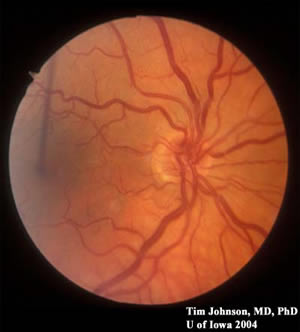
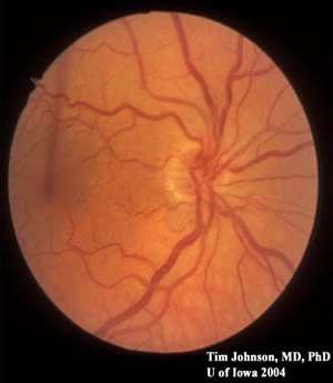
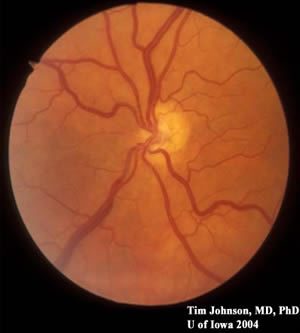
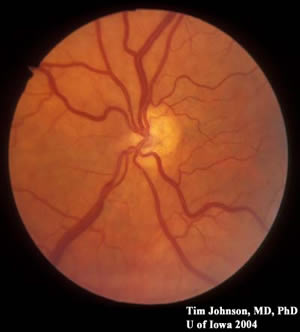
Figure 1: Stereo photos of the right optic nerve. Note the small optic nerve with crowded vessels and a pale ring around the nerve. The veins are 120-150 microns in diameter; thus, this optic nerve is less than 1 mm in diameter (average optic nerves are ~1.5 mm in diameter with ~1.2 million nerve fibers).

Figure 2: Stereo photos of the left optic nerve. The nerve looks pale, but there is only a small area (stump) of nerve rim where the vessels enter the posterior pole. The vessels are extremely crowded, and the nerve is surrounded by a large pale ring

OCT findings (OCT image not available)

OD

  • Average ON NFL thickness: 66 microns
  • Disc Area: 1.484 mm2

OS

  • Average ON NFL thickness: no value
  • Disc Area: 0.459 mm2

Discussion

Optic Nerve Hypoplasia

This patient presented with long-standing vision loss OS from optic nerve hypoplasia (OS>>OD), and her cataracts were mild. She was diagnosed with "glaucoma" and started on travoprost by her previous doctor. The appearance of the optic nerves supports optic nerve hypoplasia, most likely since birth. Both optic nerves are small (less than 1.484 mm2 OD) with normal mean disc area being 2.89 mm2. Additionally, the left optic nerve is severly hypoplastic with only a stump of nerve tissue with crowded retinal vessels exiting the nerve. In glaucoma, we see cupping with bayoneting of the vessels. If there is no evidence of glaucoma (increased intraocular pressure, changes in the optic nerve head, or visual field changes), then her glaucoma medications should be stopped. The patient was informed that: she was born with optic nerve hypoplasia, her vision was affected because of this, and her cataracts were not visually significant.

Diagnosis: Optic Nerve Hypoplasia

EPIDEMIOLOGY

  • Affects all races and socio-economic groups.
  • boys = girls
  • Prevalence 127 : 100,000 (Sweden)
  • Congenital problem.
  • Etiology is uncertain: defective chiasm/midline development leading to retrograde degeneration versus defective differentiation of ganglion cells.
  • Risk factors: young maternal age, first parity, maternal smoking, preterm birth, may be associated with fertility drugs and use of antidepressant medications, associated with other developmental defects, e.g. CNS maldevelopment and/or aniridia.

SIGNS

  • Fundus exam notable for small optic nerves with "double ring sign" (small nerve surrounded by a pale peripapillary halo).
  • MRI scans of the head and orbits are indicated to rule-out mid-line abnormalities (de Morsier’s syndrome): absent septum pellucidum and agenesis or thinning of corpus callosum.
  • May be associated with hypopituitary symptoms if mid-line defects present: diabetes insipidus, hypothyroidism, hypercorticoidism, and growth hormone deficiencies.

SYMPTOMS

  • Variability in vision loss: mild to severe no light perception.
  • May be associated with nystagmus.
  • Behavioral problems due to visual impairment.
  • Not aware of people or objects.
  • Visual field defects with perimetry.
  • Failure to thrive with feeding issues.
  • Pituitary hormone deficiency.
  • Associated with other congenital defects.

TREATMENT

  • If there are pituitary abnormalities, then consultation with pediatric endocrinologist for hormonal supplementation is necessary.
  • There is no treatment for the optic nerve hypoplasia. If patient is monocular, then education about safety glasses and prescription of polycarbonate lenses are indicated.

Differential Diagnoses

  • Optic nerve hypoplasia
  • Coloboma
  • Optic atrophy, genetic vs tumor
  • Other congenital anomaly of optic nerve

References
  1. Bennett JL. Developmental neurogenetics and neuro-ophthalmology. J Neuroophthalmol. 2002 Dec;22(4):286-96.
  2. Blohme J, Bengtsson-Stigmar E, Tornqvist K. Visually impaired Swedish children. Longitudinal comparisons 1980-1999. Acta Ophthalmol Scand. 2000 Aug;78(4):416-20.
  3. Hellstrom A, Wiklund LM, Svensson E. The clinical and morphologic spectrum of optic nerve hypoplasia. J AAPOS. 1999 Aug;3(4):212-20.
  4. Tornqvist K, Ericsson A, Kallen B. Optic nerve hypoplasia: Risk factors and epidemiology. Acta Ophthalmol Scand. 2002 Jun;80(3):300-4.
Suggested citation format:

Johnson AT: Optic Nerve Hypoplasia: 51-year-old female referred for consideration of cataract surgery. February 21, 2005; Available from: http://www.EyeRounds.org/cases/case10.htm.


last updated: reviewed 11-12-2008

Image Permissions:
Creative Commons

Ophthalmic Atlas Images by EyeRounds.org, The University of Iowa are licensed under a Creative Commons Attribution-NonCommercial-NoDerivs 3.0 Unported License.