This site uses tracking information. Visit our privacy policy. Click to agree to this policy and not see this again.

Ophthalmology and Visual Sciences

Multiple Evanescent White Dot Syndrome (MEWDS):

24-year-old female with one week duration of central scotoma, OD

Multiple Evanescent White Dot Syndrome (MEWDS):

24-year-old female with one week duration of central scotoma, OD
Sudeep Pramanik, MD, MBA, Hilary A. Beaver, MD
April 8, 2005, updated April 9, 2007

Chief Complaint: One week duration of central scotoma, OD.

History of Presenting Illness: The patient is an adult white female in her mid-twenties who experienced sudden awareness of a painless, central scotoma in her right eye while reading one week ago. There has been no pain or progression in the last week. There was no known trigger. She reports normal color vision. She denies headache. She denies preceeding viral illness.

Medical History: 1) An MRI six years previously showed enlargement of the pituitary gland with no focal adenoma. Endocrine evaluation showed normal pituitary function. 2) Motor vehicle accident, 1 year previously. No loss of consciousness. Computerized tomography (CT) without contrast was normal at that time. Other history was non-contributory.

Review of systems was otherwise negative except as noted.

Medications: Benzidine for acne. No steroid use.

EXAM OCULAR

  • Visual Acuity: OD—20/50; OS—20/20
  • Manifest Refraction: Improved vision to 20/40, OD.
  • Critical Flicker Fusion: 21.4 +/- 3.8 Hz OD, and 32.6 +/-0.6 Hz OS
  • Extraocular motility: Full
  • Pupils: equally reactive, 0.3 log unit afferent pupillary defect (APD), OD.
  • Intra-ocular pressure: 11 mmHg, OD; 10 mmHg, OS.
  • External and anterior segment examination: Normal, OU. No cell nor flare.
  • Dilated fundus exam (DFE): OD—Orange, granular retinal pigment epithelium (RPE) changes in the central macula and multifocal white dots in the outer retina (see Figure 1); OS—Normal fundus (see Figure 2).
Figure 1. Stereo fundus photos, OD. Note the orange, granular changes in the central RPE and multifocal white dot changes.

Figure 2. Stereo fundus photos, OS. Essentially normal fundus.

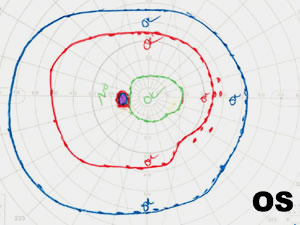
Goldman visual field (GVF) testing was performed and revealed a slightly enlarged blind spot and mildly reduced paracentral visual field overall, OD (see Figure 3).
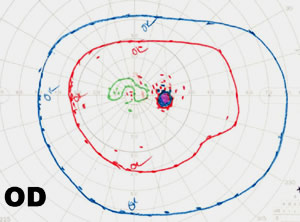
Figure 3.
3A: GVF, OS, is essentially normal. 3B: GVF, OD. Enlarged blind spot with reduction of the I1e isopter centrally.

Discussion

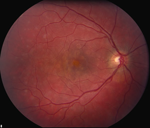
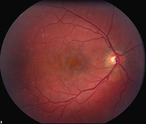
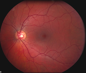
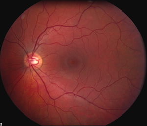
MEWDS (Multiple Evanescent White Dot Syndrome) is a rare, unilateral, self-limiting inflammatory disease that afflicts young women more than men in a 4:1 ratio. Despite the lack of treatment, the prognosis is excellent. Our patient is the typical age and gender. She had no preceeding viral illness, which can be present in approximately 1/3 of MEWDS patients. Visual field testing reveals enlargement of the blind spot, central, and/or paracentral scotomas. Fluorescein angiography of active lesions may be normal early on and demonstrate a "wreath-like" late hyperfluorescence of the white dots. ERG during the active phase may show a reduced a-wave, consistent with inflammation at the level of the photoreceptors. Typically, the visual acuity recovers, but the granularity of the foveal RPE remains.

Diagnosis: MEWDS (Multiple Evanescent White Dot Syndrome)

EPIDEMIOLOGY

  • Affects young women 20-45 years of age
  • Typically unilateral (bilateral cases documented)
  • No known racial or hereditary associations

OCULAR SIGNS

  • Visual acuity 20/40-20/400
  • Orange-yellow granularity to the RPE underlying the fovea
  • Small, transient white dots at the level of the RPE
  • Small relative afferent pupillary defect possible
  • Mild vitreous cells
  • Mild optic disc edema/hyperemia possible

SYMPTOMS

  • Sudden, painless, monocular decline in central acuity
  • Photopsias
  • Dyschromatopsia
  • Central/paracentral scotomas
  • Preceeding viral illness in about 1/3 of cases

TREATMENT

  • None—resolves spontaneously
  • Most patients achieve normal acuity and fields in weeks to months
  • Photopsia, scotomas, dyschromatopsia may rarely persist
  • Recurrence is unusual
  • No systemic workup is necessary

Differential Diagnoses

  • Acute Idiopathic Blind Spot Enlargement Syndrome (some feel this may be the same disease)
  • Acute Posterior Multifocal Placoid Pigment Epigheliopathy
  • Acute Retinal Pigment Epithelitis
  • Birdshot Retinochoroidopathy
  • Multifocal Choroiditis
  • Sarcoid
  • Diffuse Unilateral Subacute Neuroretinitis
References
  1. Aaberg TM. Multiple evanescent white dot syndrom., Arch Ophthalmol 1998;106:1162-1163.
  2. Jampol LM, Wiredu A. MEWDS, MFC, PIC, AMN, AIBSE, and AZOOR: one disease or many? Retina 1995;15: 373-378.
  3. Quillen DA, Blodi BA. Ch 9. Inflammatory Diseases. In: Clinical Retina. Chicago: AMA Press. 2002.
Suggested citation format:

Pramanik S, Beaver HA. Multiple Evanescent White Dot Syndrome (MEWDS): 24-year-old female with one week duration of central scotoma, OD. EyeRounds.org. April 8, 2005; Available from: http://www.EyeRounds.org/cases/37-MultipleEvanescentWhiteDotSyndromeMEWDS.htm.


last updated: 04-09-2007

Image Permissions:
Creative Commons

Ophthalmic Atlas Images by EyeRounds.org, The University of Iowa are licensed under a Creative Commons Attribution-NonCommercial-NoDerivs 3.0 Unported License.