
Chief Complaint: Sudden painless decreased vision in the right eye
History of Present Illness: The patient is an 85-year-old female who noted sudden-onset darkening of her central vision surrounded by a gray ring-like pattern in her right eye. She had no associated photopsias, metamorphopsia, or new floaters. She denied any eye pain, diplopia, redness, or headache. She had no history of ocular trauma, transient ischemic attacks, or neurologic symptoms.
Medical History: Hypertension, hypothyroidism, osteopenia, hypercholesteremia
Medications: Timolol drops OU. Systemic medications include aspirin, levothyroxine, losartan, simvastatin, risedronate, tolterodine tartrate.
Family History: Noncontributory
Social History: Patient smokes half a pack of cigarettes per day and does not consume alcohol.
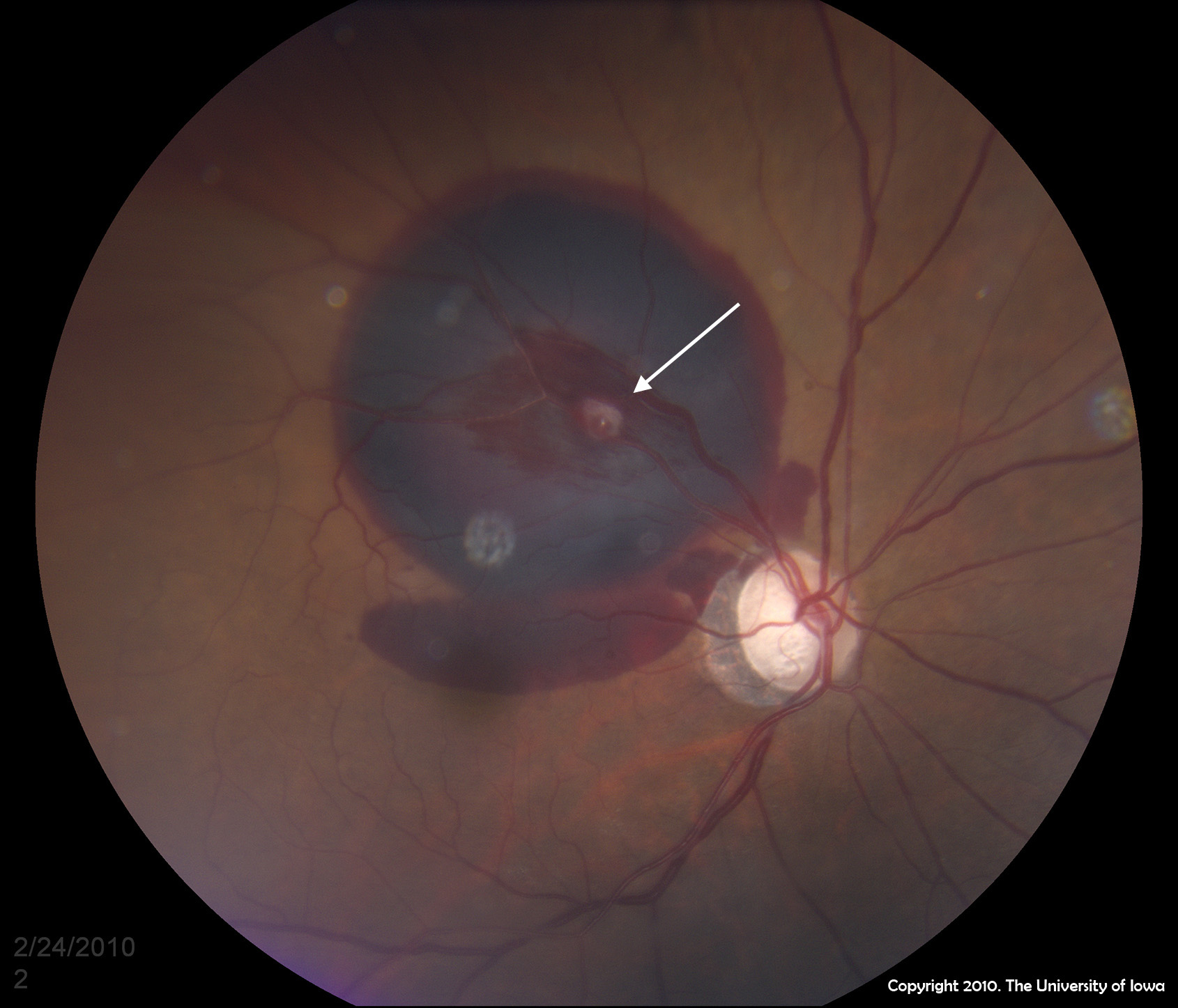
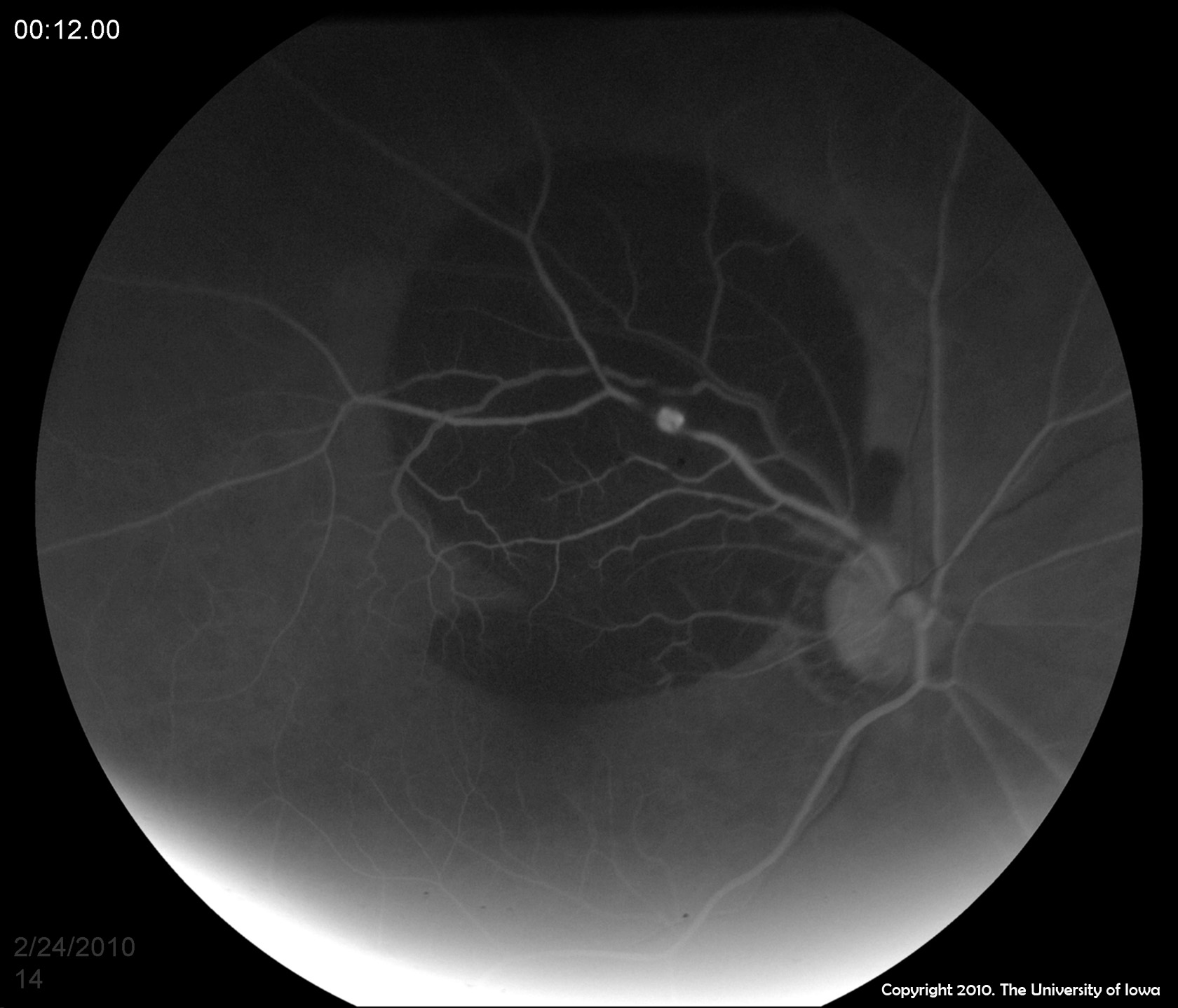
| 1A:Fundus photo of the right eye showing large area of subretinal hemorrhage with hemorrhage extending into the macula. Note the white spot along the superotemporal artery which corresponds to the hyperfluorescent lesion in the angiogram.(Click image for higher resolution.) | 1B: FFA of the right eye in the early arteriolar phase showing early hyperfluorescence along the superotemporal arcade over the subjacent blockage from subretinal hemorrhage.(Click image for higher resolution.) |
 |
 |
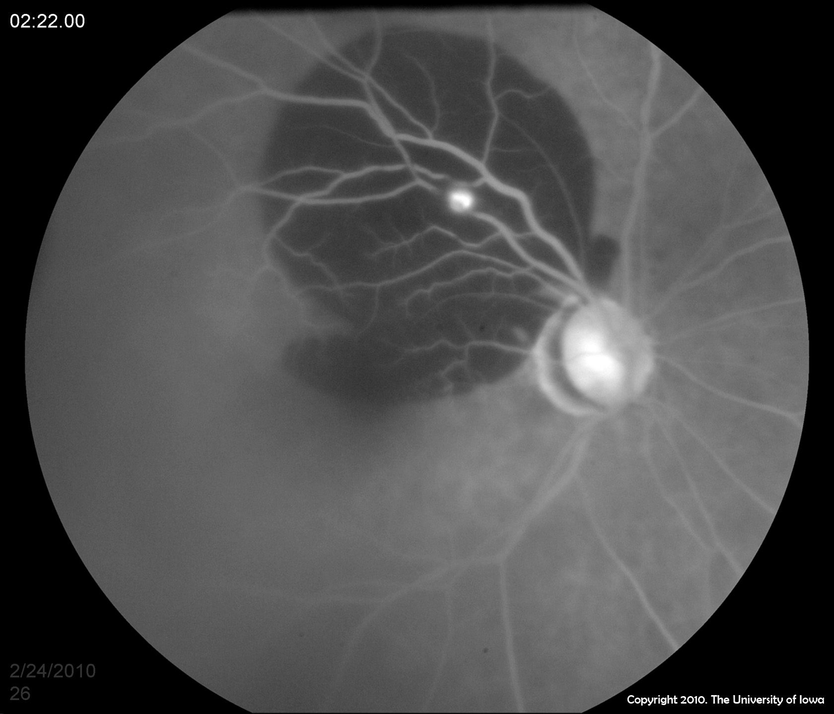
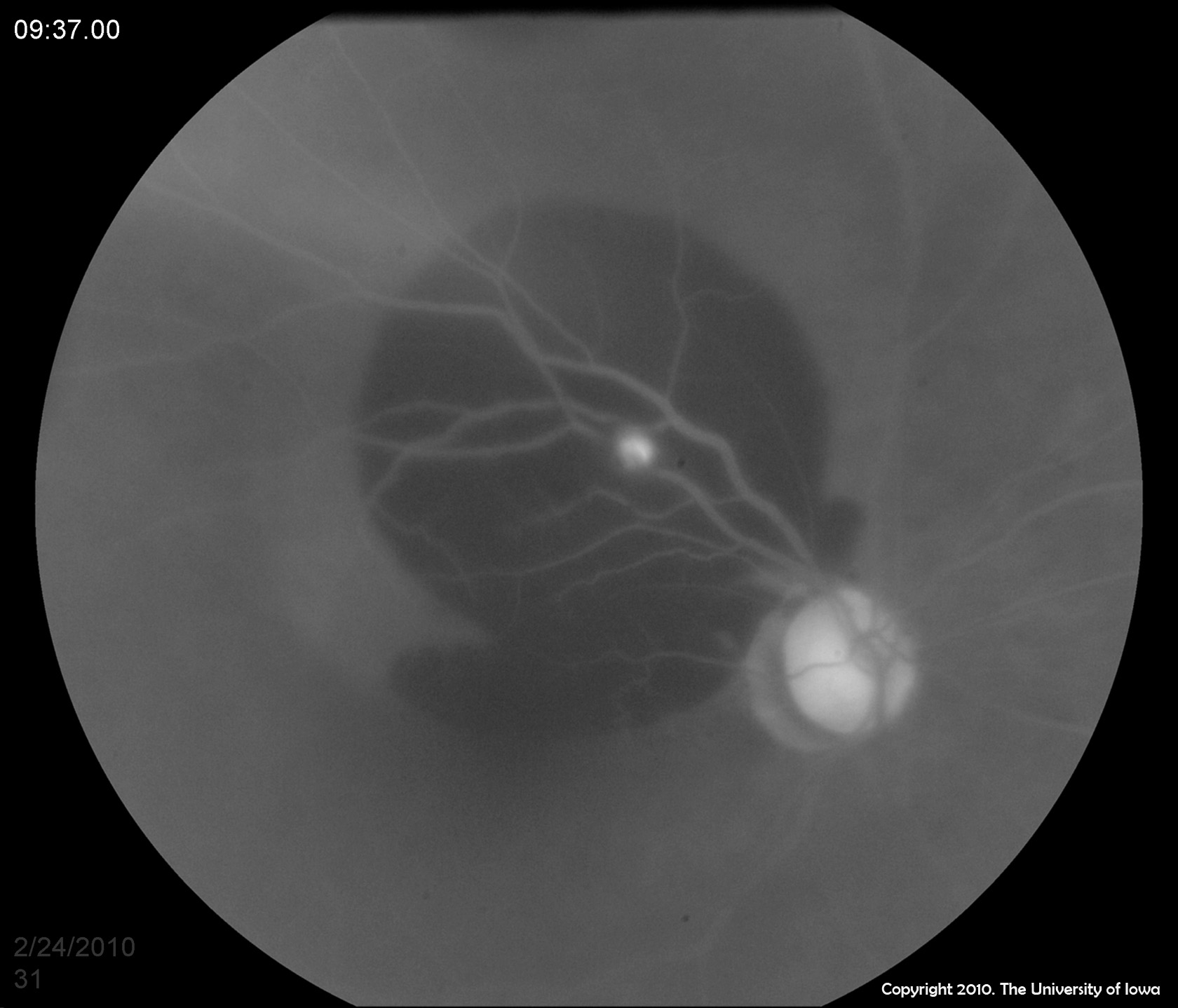
| 1C: Mid-venous phase of FFA shows increased hyperfluorescence.(Click image for higher resolution.) | 1D: Late or washout phase of FFA shows persistent lesion hyperfluorescence.(Click image for higher resolution.) |
 |
 |
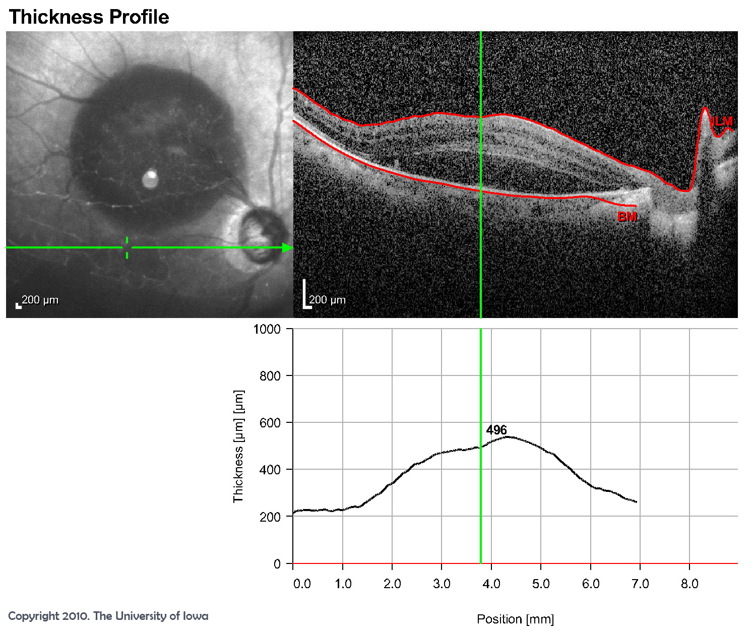
| 2A: OCT of the right eye showing subretinal fluid beneath the fovea.(Click image for higher resolution.) | 2B: OCT of the right eye showing extensive area of intraretinal edema and subretinal fluid (blood) with no choroidal abnormality.(Click image for higher resolution.) |
 |
 |
A retinal artery macroaneurysm (RAMA) is usually described as an idiopathic, acquired dilation of a major retinal arteriole. Typically these develop within its first three bifurcations, at branch points, or areas of arteriovenous crossing. The most commonly reported site for RAMAs is the superotemporal arteriole. Less commonly they may develop from the cilioretinal arteries or on the optic nerve head. The underlying pathophysiology of macroaneurysms is not fully understood. One hypothesis is that arteriosclerosis leads to vessel wall fibrosis. The resulting decrease in wall elasticity, combined with elevated luminal pressure results in aneurysmal dilation. An alternative or accessory hypothesis is that emboli (which have been associated with vessels harboring macroaneurysms) or intra-arterial thrombosis leads to mechanical damage of the endothelium or adventitial vessel wall, which predisposes the vessel to aneurysm formation. Chronic venous stasis from hypertension and arteriosclerosis may also play a role (Rabb et al. 1988).
RAMAs usually develop in patients after the 6th decade of life (mean age of 57-71 years). However, there are published reports of patients with RAMAs in patients as young as 16 (Fernandez 1920, Rabb et al. 1988). Females have a strong predilection toward the development of RAMAs. In larger studies 71-80% of patients were female (Rabb et al. 1988). RAMAs are usually singular and unilateral. Ten percent of cases are reportedly bilateral and 20% present with multiple aneurysms along the same vessel or on multiple vessels. Systemic hypertension is the most commonly associated risk factor. This factor is present in 31%-81% of affected patients depending on the study cited (Fernandez 1920, Rabb et al. 1988). Other risk factors reported in the literature have not been consistently observed in all studies and include elevated lipid levels, systemic vasculitis such as polyarteritis nodosa, sarcoidosis, diabetes, rheumatoid arthritis, and Raynaud's phenomenon.
Classically, patients with RAMAs present with sudden painless loss of vision. Many, however, are asymptomatic, and their RAMAs are discovered on routine exam, especially if the aneurysm is not associated with exudates, edema, or subretinal hemorrhage involving the macula. Vision loss from a RAMA is usually attributable to macular involvement including macular edema or hemorrhage in any of the retinal layers or vitreous. RAMAs may remain unchanged for many years, but most will eventually undergo thrombosis, fibrosis, and/or involution. Most patients have preservation or return of their vision unless they have extensive subfoveal hemorrhage or chronic macular edema. Ultimately the site and severity of the hemorrhage and macular edema determine visual outcomes (Rabb et al. 1988).
The key fundoscopic finding is the presence of blood at multiple layers including the preretinal, intraretinal, subretinal, and sub-ILM spaces and the vitreous or the presence of an hourglass hemorrhage (described by Schatz et al.), which represents simultaneous preretinal and subretinal hemorrhage. Other common but less pathognomonic findings include exudative retinopathy consisting of yellow/white hard lipid exudates in a circinate pattern surrounding the aneurysm. Pulsations of the aneurysm can be appreciated in 10% of cases (Rabb et al. 1988, Schatz et al. 1980). Macular edema without exudates is less commonly seen, as are neurosensory detachments. Fluorescein angiography typically demonstrates an immediate filling of the aneurysm, which may leak throughout the study. If retinal hemorrhage is present, an area of blocked fluorescence around the aneurysm may be observed (see Figure 2). The involved artery may be narrowed and irregular, and the surrounding capillaries may demonstrate leakage (Rabb et al. 1988).
Because many patients spontaneously recover a significant amount of vision and the majority of aneurysms involute without intervention, most patients can safely be observed. Hypertension and other systemic risk factors should be treated adequately. Indications for laser treatment include vision loss due to chronic macular exudates or edema (Rabb et al. 1988). Laser photocoagulation directly to the macroaneurysm using xenon arc, argon, or dye yellow has been documented in the literature to improve vision in some patients (Rabb et al. 1988, Abdel-Khalek and Richardson 1986, Hudomel and lmre 1973). However, some studies note that direct photocoagulation to the macroaneurysm does not improve visual outcomes and may lead to branch retinal artery occlusion (Brown et al. 1994). Indirect laser treatment to the area surrounding the macroaneurysm may also improve visual outcomes in some patients with macular edema (Robertson 1973, Palestine et al. 1982, Francois 1979). Laser hyaloidotomy using a neodymium-doped yttrium aluminium garnet (Nd:YAG) laser for subhyaloid hemorrhage has been reported but is controversial due to the risk of vitreous hemorrhage or damage to the macula (Tassignon et al. 1989). In the setting of vitreous hemorrhage of unclear etiology, pars plana vitrectomy has been performed. Surgical evacuation of subretinal hemorrhage has also been utilized (Ibanez HE et al. 1995, Humayun M et al. 1998).
EPIDEMIOLOGY
|
SIGNS
|
SYMPTOMS
|
TREATMENT
|
Birkholz ES, Johnson AT, Russell SR. Retinal Artery Macroaneurysm. EyeRounds.org. posted July 7, 2010; Available from: http://www.EyeRounds.org/cases/113-RAMA.htm.

Ophthalmic Atlas Images by EyeRounds.org, The University of Iowa are licensed under a Creative Commons Attribution-NonCommercial-NoDerivs 3.0 Unported License.

Address
University of IowaLegal
Related Links
