
Chief complaint: Progressive glaucoma and periorbital changes
A 52-year-old woman presented to the University of Iowa Glaucoma Service with elevated intraocular pressure (IOP) and visual fields demonstrating progressive field loss in the left eye (OS) despite maximum tolerated medical therapy. She had been involved in an automobile accident in 1995 and suffered a traumatic cataract and iridodialysis secondary to blunt eye injury in the left eye. Elevated IOP was noted shortly thereafter in the left eye only. She had been treated since that time with brimonidine 0.2% and dorzolamide 2%/timolol 0.5% for angle recession glaucoma. Two years prior to referral, bimatoprost 0.03% was added to the regimen. Two weeks prior to presentation she was found to have an IOP of 28 mmHg OS with progressive field changes and so was placed on oral acetazolamide 500 mg by mouth twice daily and referred for possible glaucoma surgery. She had significant side effects from the acetazolamide and also complained of gradual left-sided periorbital changes over the preceding 2 years consisting of a “sunken appearance” with a larger eyelid opening.

The patient underwent trabeculectomy with mitomycin-C in the left eye for progressive glaucoma and all medications were discontinued.
Prostaglandin analogues (PGAs) are the most effective outflow drugs approved for clinical use.(1) Once daily dosing of either latanoprost, bimatoprost or travoprost produces 28-33% IOP reduction in a meta-analysis of 28 randomized clinical trials.(2,3) Known adverse effects of PGAs include conjunctival hyperemia, eyelash lengthening, iris darkening and periocular skin pigmentation. In addition, they may be associated with anterior uveitis, cystoid macula edema and reactivation of herpetic eye disease, although these lack a direct causal relationship with the medication.(4) More recently, a constellation of changes referred to as prostaglandin-associated periorbitopathy (PAP) has been associated with the use of PGAs.(5, 6) (see Figures 4 and 5) PAP includes deepening of the upper eyelid sulcus, upper lid ptosis, loss of the inferior orbital fat pads and enophthalmos. A cross-sectional survey done recently concluded that PAP also includes loss of lower lid steatoblepharon and levator dysfunction.(7) While these changes were often dismissed as age-related adnexal changes in bilateral users, they became clearly evident due to monocular PGA use.
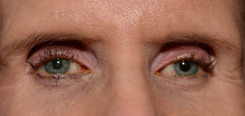
Figure 4. Severe orbital fat atrophy in a patient with prolonged bilateral PGA use. Note the very deep superior sulcus and "sunken" eye appearance.
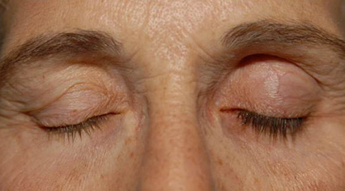
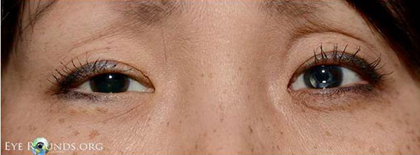
Figure 5. Left-sided PAP and hypertrichosis in a patient using PGA monocularly. Note the superior sulcus and loss of the lid crease.
The exact mechanism of PAP is not known, though many have been proposed and studied. Peplinski and Albiani first reported upper eyelid sulcus deepening and dermatochalasis involution in three patients treated with bimatoprost unilaterally in 2004. They attributed the altered periorbital appearance to the direct effect of PGA on Müller’s muscle mediated by the prostaglandin F (PF) receptor.(5) Filippopoulos et al postulated that loss of the preaponeurotic fat explains the loss of the upper eyelid fullness. This proposed mechanism is also mediated by PF receptor activation, which has been associated with inhibition of preadipocyte differentiation. The antiadipogenic effects of PF receptor activation were further supported by in vitro studies.(6, 8, 9)
Park et al. first described the histologic changes in PAP.(10) Biopsies were taken of the preaponeurotic orbital fat in patients with superior sulcus deepening using monocular or binocular bimatoprost, travoprost or latanoprost. They found that the periocular fat atrophy was characterized by shrinking of the individual adipocytes with loss of lipid content and not a loss of number of adipocytes. Although the number of samples was small, a significant difference was noted between the PGA-treated eyes and the untreated eyes and among the three PGAs; the fat atrophy was highest in the bimatoprost group followed in order by the travoprost and latanoprost groups.
Fortunately, these changes seem to occur late and are reversible. Yang et al. detected a significant deepening of the superior sulcus only after 1 or 2 years of use and this change resolved around 15 months after discontinuation. Another study found recovery from PAP as soon as 2 months after stopping PGA.(11) It has been noted that PAP is often more significant or noticeable in the Asian population, where a deep superior eyelid sulcus is not common as the levator complex is inserted low onto the tarsus.(12)
Latisse® is a topical cosmetic preparation of bimatoprost that is approved for the treatment of hypotrichosis. It carries the same risks of periocular changes. Certainly, these patients should be warned of this potentially disfiguring side effect.(13)
Because early clinical signs of PAP are subtle, the diagnosis may often be delayed until periorbital changes have progressed quite significantly. Given the great variety of glaucoma medications available, it may be reasonable to consider a different first line therapy in young patients or those being treated monocularly to avoid PAP. When PAP does occur, we would recommend discontinuing the PGA, if possible, to eliminate both the cosmetic and functional sequelae.
Tabaza L, Welder JD, Alward WLM. Prostaglandin-Associated Periorbitopathy. EyeRounds.org. October 14, 2013; available from https://eyerounds.org/cases/181-prostaglandin-periorbitopathy.htm

Ophthalmic Atlas Images by EyeRounds.org, The University of Iowa are licensed under a Creative Commons Attribution-NonCommercial-NoDerivs 3.0 Unported License.

Address
University of IowaLegal
Related Links
