This site uses tracking information. Visit our privacy policy. Click to agree to this policy and not see this again.

Ophthalmology and Visual Sciences

Pediatric Spectacle Prescription and Retinoscopy Made Simple: A Tutorial

Pediatric Spectacle Prescription and Retinoscopy Made Simple: A Tutorial

Contributors: Jacob Evans BS, Tyler Risma, MD, Alina Dumitrescu, MD

The University of Iowa
Department of Ophthalmology and Visual Sciences

July 13, 2017


Introduction

This review covers simple optical principles of retinoscopy, describes an easily learned technique for retinoscopy with reliable results, and guides decisions based on those results through basic prescribing guidelines as they apply to children.

The estimated prevalence of refractive amblyopia varies widely by ethnic and socioeconomic groups but is estimated to be somewhere between 0.75% and 2.5% of children worldwide.[1-4] The economic impact of pediatric vision loss is difficult to estimate and is most evident only after a child reaches adulthood. However, a study from 2007 estimated 158.1 million global cases of visual impairment resulted from uncorrected or under-corrected refractive error; of these, 8.7 million were blind. This created an estimated economic productivity loss of $268.8 billion. [5]

The pediatric population with refractive error needs regular assessment and prescription adjustment due to eye growth. The cornea reaches adult size by the age of 2 years, but the eye continues to grow in anteroposterior length until age 7-8 years old. The infant eye averages 16.6 mm in anteroposterior diameter (length), and grows to an adult size averaging 22.0-24.8 mm. [6,7] Since these two variables do not have parallel growth, refractive error changes as children age.

Determining and then correcting a child's refractive error can prevent or treat amblyopia which ultimately helps to avoid irreversible vision loss. Giving children the best possible vision, including correcting refractive error when needed, allows them to succeed scholastically. Visual deficits can also affect a child's daily function and normal play which may decrease his or her confidence. Children may be unaware of less severe vision problems, particularly if anisometropia is present.

In these situations where traditional subjective refraction methods are impossible, including in young children or patients of any age with limited ability to communicate or cooperate, a method for objective refraction is required. This is also useful in cases of atypical refractive error or implausible subjective refraction results. Since retinoscopy is the gold standard for objective refraction, mastering this skill is essential.

Retinoscopy allows the observer to accurately measure a patient's refractive error by determining the spherical power, cylindrical power, and cylindrical axis which focus the patient's eye at optical infinity.  This is done by shining diverging light into the patient's eye from the retinoscope and finding the refraction which focuses that light on the patient's retina.

Retinoscopy results also provide information about risk factors for additional ocular morbidities. Patients with myopia have higher rates of other significant ocular disease, such as retinal detachment and glaucoma.[8,9] Patients with severe hyperopia are more likely to develop accommodative esotropia (see "Special Considerations for Spectacles in Children" section) and amblyopia.[10] A subgroup of children with crowded anterior segment and high hyperopia have an increased risk of angle closure glaucoma.   

There are a few valuable trends to keep in mind when determining refractive error and prescribing spectacles for children. First, refractive error typically moves from hyperopia toward myopia as children age. Roughly 80% of children between 2 and 6 years old are hyperopic and about 10% of children need refractive correction before 8 years old.[11] Myopia then develops between 6 – 9 years old and frequently increases throughout adolescence.[11] Second, astigmatism is relatively common in babies but decreases in prevalence during the first few years of life.[11] The newborn eye averages a K value (diopters) of 51.2, while the adult eye averages a K value of 43.5 diopters.[12] Thus, although the focusing power of the eye decreases with age, there is a simultaneous trend away from hyperopia -- meaning the eye needs less additional corrective power -- because of simultaneous axial eye length increase as the patient ages.

Multiple methods of retinoscopy exist and have been extensively described.  Here we aim to simply describe the basic concept and specifically one common method: plus cylinder neutralization retinoscopy. Please refer to other resources for a more exhaustive description of additional techniques and their applications.


Plus cylinder neutralization retinoscopy

  1. Tools
    • Retinoscope types
Figure 1: Welch Allyn retinoscope. Leaving the sleeve down on this retinoscope (and other non-Copeland retinoscopes) provides the plane mirror effect.
Figure 2: Copeland retinoscope. Putting the sleeve up provides the plane mirror effect.
Figure 3: Black convex lens ("plus power") and red concave lens ("minus power") retinoscopy paddles.
    • Lenses
      • Loose lenses: better tolerated by infants/toddlers
      • Retinoscopy paddles: quickest to use (Figure 3)
      • Phoropter: only for cooperative patients
  • Setup
    • Cycloplegic retinoscopy ("wet") vs non-cycloplegic ("dry")
      • Advantages of cycloplegia
        1. Paralyzed accommodation, which prevents underestimation (i.e. overly myopic/less hyperopic) of the patient's refractive error
        2. Dilated pupil, making it easier to see the retinoscopic reflex
      • Un-dilated retinoscopy can be performed in cooperative, presbyopic patients and is not recommended in children other than for dynamic purposes (e.g. to assess accommodative ability)
    • Dark room
    • Working distance
      • Dependent on the examiner's arm length and preference
      • A common working distance is 67 cm which gives a working distance lens of +1.50 D.
  • The Basic Technique

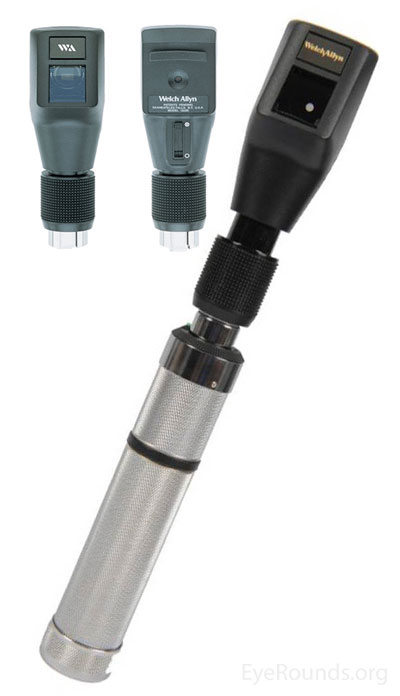
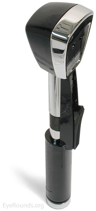
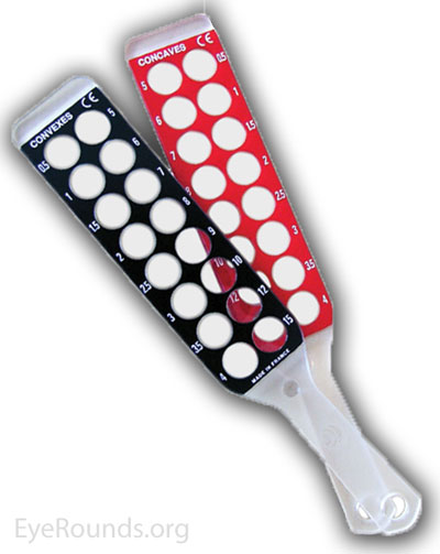
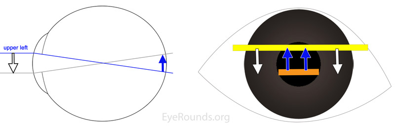
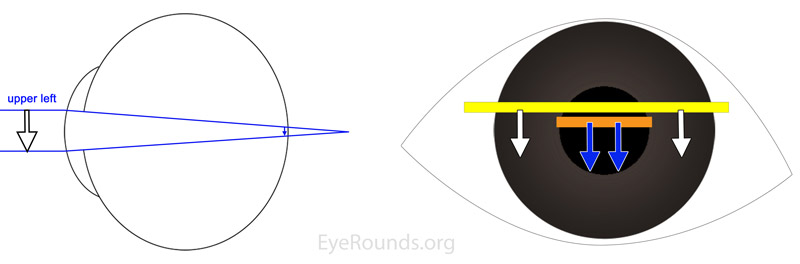
    The examiner should place himself aligned with the pupil while the patient is looking at a distant object. The examiner then obtains a red reflex with a "streak" of light and passes that streak of light perpendicular to the axis of the streak.  By observing the pattern of "with" or "against" motion, the examiner can interchange a lens with more plus power or less plus power to the point where the retinoscopic reflex is "neutralized."  At this point, instead of moving against or with the movement of the streak, the reflex will appear as a diffuse, even light that changes very little as the examiner moves the retinoscope.  For plus-cylinder refractions, the examiner starts by neutralizing the meridian with the lower hyperopic power, thereby leaving the meridian 90 degrees away with "with motion." The examiner then neutralizes this second meridian, noting the axis, and calculates the power difference between the two lenses needed for neutralization (plus cylinder power). The examiner then "takes out the working distance" from the lowest hyperopic power which gives the final spherical refraction.

  • Against motion vs. With motion
    •  "Against Motion": There is too much plus power in the combined optical system of the eye and the corrective lens, thus the light from the retinoscope is focused in front of the retina as seen by the intersection of the rays in Figure 4 on the left. Therefore, the retinoscopic light reflex seen by the examiner moves in the opposite direction of the retinoscope streak, as illustrated in Figure 4 on the right.
Figure 4: Left: Diagrammatic cross-section of a myopic eye, with rays of light entering the eye from the left and focusing anterior to the retina. The light that strikes the superior cornea is directed toward the inferior retina and the light that strikes the inferior cornea is directed toward the superior retina. Right: The light reflex visible in the pupil (illustrated by the orange bar) will move "against" the direction of the streak of light (illustrated by the yellow bar) from the retinoscope. Thus, as the streak moves downward, the pupillary reflex moves upward.
Figure 5: Left: Diagrammatic cross-section of a hyperopic eye, with rays of light entering the eye from the left and focusing posterior to the retina. The light that strikes the superior cornea is directed toward the superior retina and the light that strikes the inferior cornea is directed toward the inferior retina. Right: The light reflex visible in the pupil (illustrated by the orange bar) will move "with" the direction of the streak of light from the retinoscope (illustrated by the yellow bar). Thus, as the streak moves downward, the pupillary reflex also moves downward.
  • "With Motion": There is not enough plus power (or, alternately, too much minus power), causing the retinoscopic light reflex to focus behind the retina as seen by the intersection of the rays in Figure 5 on the left. Therefore, the light reflex seen by the examiner will move in the same direction that the retinoscope streak is moved.  This is called "with" motion and is demonstrated in Figure 5 on the right.
  • Examples: (all of the following examples will assume a working distance of 67 cm, which correlates with a working distance lens of +1.50 D; in practice, this must be individualized for each examiner's arm length)
    • Emmetropia: The simplest patient for conceptualizing retinoscopy is an emmetrope
      • With no lens in front of an emmetropic eye, an examiner would see "with" motion in all meridians
      • With a +1.50 D lens the light streak would be neutralized in all meridians. 
      • With a +2.00 D lens the examiner sees against motion and by putting "less plus" and switching back to a +1.50 D lens the streak is again neutralized
      • After taking out the working distance (by subtracting +1.50), the examiner is left with a refraction of +0.00 D, or plano.
    • Hyperopia (+2.00)
      • With no lens in front of a hyperopic eye, the examiner would similarly see "with motion" in all meridians
      • The examiner adds more plus power until the streak is neutralized with a +3.50 D lens in all meridians
      • After taking out the working distance the final Rx is +2.00 D sphere (no astigmatism correction)
    • Mild Myopia (-1.00)
      • With no lens in front of a patient with mild myopia, the typical examiner would also see "with motion" in all meridians
      • The examiner adds plus power until the streak is neutralized in all meridians with a +0.50 D lens
      • After taking out the working distance the examiner determines a final Rx of -1.00 D sphere
    • Moderate Myopia (-4.00)
      • With no lens in front of a patient with moderate myopia, the examiner sees "against" motion in all meridians
      • The examiner adds minus-powered lenses until the streak is neutralized in all meridians with a -2.50 D spherical lens 
      • After taking out the working distance the examiner determines a final Rx of -4.00 D sphere
    • "With the rule" astigmatism ( +1.00 +1.50 x 090) 
      • In most cases of with the rule (WTR) astigmatism, the cornea is steeper in the vertical meridian and flatter in the horizontal meridian, which means more plus lens power will be required to neutralize the horizontal meridian than to neutralize the vertical
      • With no lens in front of this patient with WTR astigmatism, the examiner should first streak the vertical then the horizontal meridian
        1.  The examiner "streaks the vertical meridian" by passing a horizontal streak of light up and down along the vertical meridian and would see "with motion," which would neutralize with a +2.50 D lens
        2. The examiner then moves on to "streak the horizontal meridian" by passing a vertical streak of light side to side along the horizontal meridian, which in this patient would neutralize with a +4.00 D lens
        3. This indicates that an additional +1.50 D is required to neutralize the horizontal meridian as compared to the vertical, which represents the cylindrical power
        4. In this case the streak was placed at exactly 90 degrees which indicates the axis; a cylindrical lens gives power in the axis 90 degrees away, ergo, a cylindrical lens of power +1.50 and axis 90, gives +1.50 D of power in the horizontal (180 degree) meridian
        5. Taking out the working distance (from +2.50) gives +1.00 D and the final Rx is +1.00 +1.50 x 090
    • Against the rule astigmatism ( +2.00 +1.00 x 180)
      • In most cases of against the rule (ATR) astigmatism, the cornea is steeper in the horizontal meridian and flatter in the vertical meridian, which means more plus lens power will be required to neutralize the vertical meridian than to neutralize the horizontal. 
      • With no lens in front of this patient with ATR astigmatism, the examiner should first streak the horizontal and then the vertical meridian
        1. The examiner "streaks the horizontal meridian" by passing a vertical streak of light side to side along the horizontal meridian and would see "against motion," which would neutralize with a +3.50 D lens
        2. The examiner then moves on to "streak the vertical meridian" by passing a horizontal streak of light up and down along the vertical meridian, which in this patient would neutralize with a +4.50 D lens
        3. This indicates that an additional +1.00 D is required to neutralize the vertical meridian as compared to the horizontal, which represents the cylindrical power
        4. In this case the horizontal streak (for the vertical meridian) was placed at exactly 180 degrees which indicates the axis; a cylindrical lens gives power in the axis 90 degrees away, ergo, a cylindrical lens of power +1.00 and axis 180 gives +1.00 D of power in the vertical (90 degree) meridian
        5. Taking out the working distance (from +3.50) gives +2.00 D and the final Rx is +2.00 +1.00 x 180
  • Tips and Hints
      • Because most patients with astigmatism have the WTR type, most examiners begin by "streaking the vertical meridian" (with a horizontal streak of light). After noting the lens power required to neutralize the vertical meridian, the examiner then moves on to "streak the horizontal meridian" (with a vertical streak of light). If the examiner notes that the patient has ATR astigmatism, it is typically easier to neutralize the horizontal meridian first, and then the vertical meridian second. This keeps the refraction in plus-cylinder format and allows the examiner to simply note the orientation of the streak on the second neutralization and use that as the axis on the prescription.
      • If against or with motion is not obvious, a high refractive error should be suspected and a high plus (+10) or minus (-10) lens can be used to attempt to clarify the reflex's movement, followed by neutralization retinoscopy as described in this guide.

    Deciding when to prescribe corrective lenses

    Once a child's refractive error has been determined, the next decision is whether or not to prescribe corrective lenses. When a child is less than 9 years old, considerations include whether the refractive error is normal for a child's age, and whether the uncorrected error will cause amblyopia or interfere with the child's visual function and alignment. Whether wearing corrective lenses will interfere with emmetropization is controversial. Some providers will prescribe a little less power than needed to encourage emmetropization of the eye, perhaps because the rate of emmetropization is related to the total initial refractive error in infants.[13] A recent study found that prescribing the smallest amount of hyperopic correction needed to allow near-focusing does not impede emmetropization.[14] In daily practice, for hyperopic patients, lower than full plus prescription seems to be better tolerated and accepted by children except in cases of accommodative esotropia, where the full cycloplegic correction is necessary to minimize or eliminate strabismus.


    Suggested indications for prescribing spectacles in pediatric populations [15,16]

    The following guidelines come from the American Academy of Ophthalmology's preferred practice patterns. These guidelines represent the minimum values at which spectacle prescription is recommended for isolated refractive error, specifically in the absence of amblyopia or strabismus which should lower the threshold for spectacle prescription.   

    Isoametropia

    Myopia

    • <1 year: > -5 D
    • 1-2 years: > -4 D
    • 2-3 years: > -3 D
    • >4 years: > -1.5 D or if symptomatic [17]

    Hyperopia

    • <1 year: > +6 D
    • 1-2 years: > +5 D
    • 2-3 years: > +4.5 D
    • >4 years: > +4 D or if symptomatic [17]

    Guidelines For Spectacle Correction of Isolated Isoametropia in Young Children

    grpah of guidleines for spectles in young children

    Hyperopia with esotropia

    • <1 year: > +2.5 D
    • 1-2 years: > +2 D
    • 2-3 years: > +1.5 D

    Astigmatism

    • <1 year: >3 D
    • 1-2 years: >2.5 D
    • 2-3 years: >2 D
    • >4 years: >1.5 D or if symptomatic[17]

    Anisometropia

    Myopia

    • <1 year: > -4 D
    • 1-2 years: > -3 D
    • 2-3 years: > -3 D

    Hyperopia

    • <1 year: >2.5 D
    • 1-2 years: >2 D
    • 2-3 years: >1.5 D

    Astigmatism

    • <1 year: >2.5 D
    • 1-2 years: >2 D
    • 2-3 years: >2 D

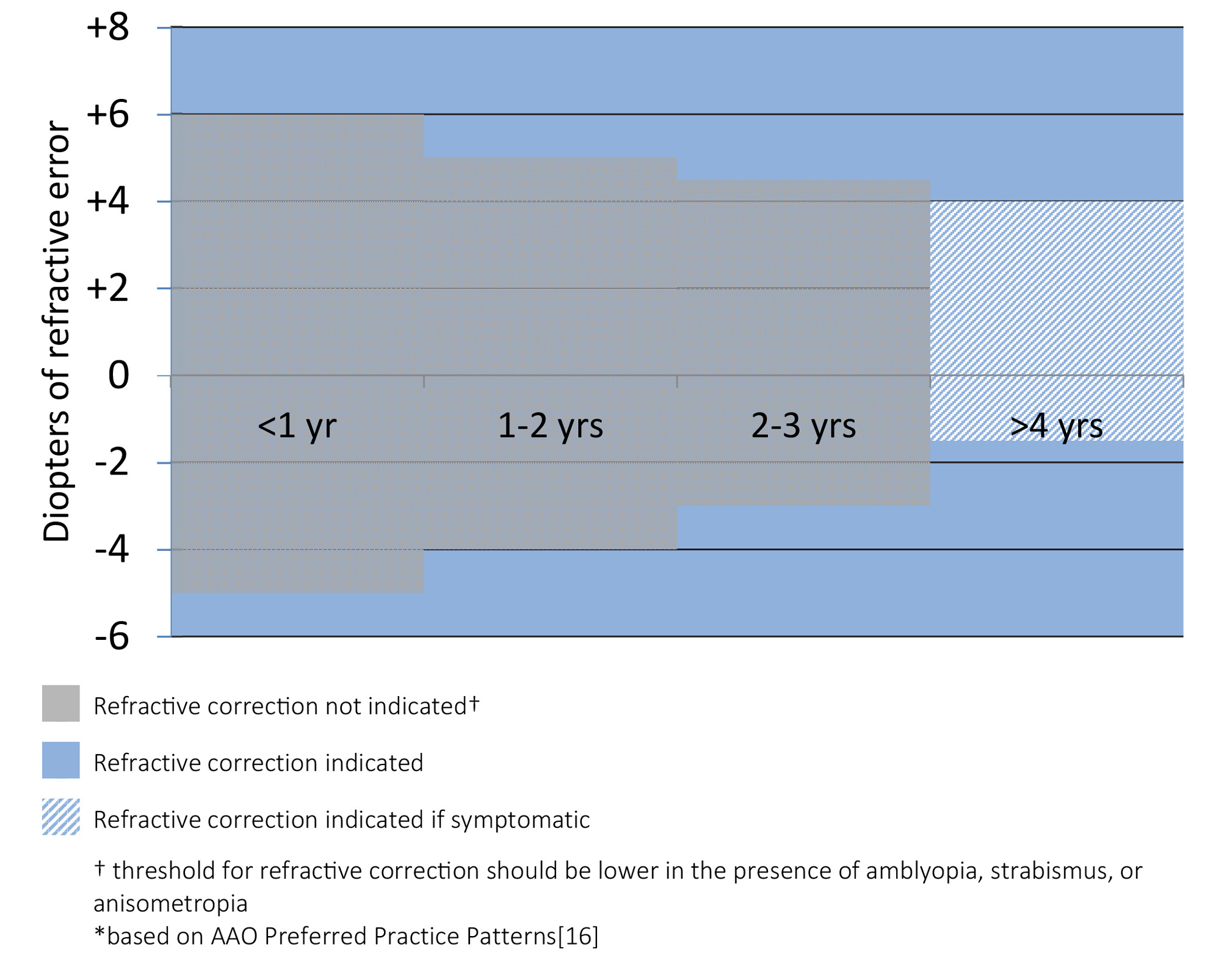
    Prescribe anisometric difference at any age if amblyopia is present


    Special considerations for spectacles in children [18-20]

    Intermittent exotropia

    Lens power may be reduced ("minus lens therapy") from the cycloplegic refraction, even for minor prescriptions, to induce accommodative convergence and reduce exotropia.[18]

    Accommodative esotropia

    1. Determine the accommodative convergence/accommodation ratio, or AC/A ratio.
    2. In children with a normal AC/A ratio (<5:1), the full cycloplegic refraction is prescribed [19]
    3. In children with a high (>5:1) AC/A ratio, prescribing the full cycloplegic distance correction can correct the distance deviation completely, but the near deviation may persist. These children may be prescribed bifocals to help correct the near deviation as well. It is important to ensure that the bifocals split the pupil in children. [19]

    Aphakia or pseudophakia

    Prescribe the full amount of correction with +2 to +3 D (retinoscopy will show -2 to -3D) to allow near activities, since infants are primarily interested in objects near them. At around 1.5-2 years of age, bifocals can be considered.

    Anisometropic or astigmatic amblyopia

    Correct the full anisometropia, astigmatism, and myopia to cycloplegic refraction. Correct hyperopia that is >3 D to either full cycloplegic refraction, or to a level that is under corrected by as much as 1.5 D. Using this prescribing guideline (without the need for occlusion therapy or other therapies) has been shown to resolve anisometropic amblyopia in roughly one third of cases..10-21]


    References

    1. Xiao O, Morgan IG, Ellwein LB, He M. Prevalence of amblyopia in school-aged children and variations by age, gender, and ethnicity in a multi-country refractive error study. Ophthalmology 2015;122:1924-31.
    2. Wang Y, Liang YB, Sun LP, et al. Prevalence and causes of amblyopia in a rural adult population of chinese the handan eye study. Ophthalmology 2011;118:279-83.
    3. Attebo K, Mitchell P, Cumming R, et al. Prevalence and causes of amblyopia in an adult population. Ophthalmology 1998;105:154-9.
    4. Friedman DS, Repka MX, Katz J, et al. Prevalence of amblyopia and strabismus in white and african american children aged 6 through 71 months the baltimore pediatric eye disease study. Ophthalmology 2009;116:2128-34.e1-2.
    5. Smith T, Frick K, Holden B, et al. Potential lost productivity resulting from the global burden of uncorrected refractive error. Bull World Health Organ 2009;87:431-37.
    6. Riordan-Eva P. Chapter 1. Anatomy & embryology of the eye. In: Riordan-Eva P, Cunningham ET (eds). Vaughan & asbury's general ophthalmology, 18e. New York, NY: The McGraw-Hill Companies, 2011.
    7. Bekerman I, Gottlieb P, Vaiman M. Variations in eyeball diameters of the healthy adults. Journal of Ophthalmology 2014;2014:5.
    8. Lai TY. Retinal complications of high myopia. Med Bull 2007;12:18-20.
    9. Mitchell P, Hourihan F, Sandbach J, Wang JJ. The relationship between glaucoma and myopia: The blue mountains eye study. Ophthalmology 1999;106:2010-5.
    10. Blouza AJ, Loukil I, Mhenni A, et al. Prise en charge de l'hypermétropie de l'enfant. J Fr Ophtalmol 2007;30:255-59.
    11. Fredrick DR. Chapter 17. Special subjects of pediatric interest. In: Riordan-Eva P, Cunningham ET (eds). Vaughan &amp; asbury's general ophthalmology, 18e. New York, NY: The McGraw-Hill Companies, 2011.
    12. Gordon RA, Donzis PB. Refractive development of the human eye. Arch Ophthalmol 1985;103:785-89.
    13. Saunders KJ, Woodhouse JM, Westall CA. Emmetropisation in human infancy: Rate of change is related to initial refractive error. Vision Res 1995;35:1325-8.
    14. Somer D, Karabulut E, Cinar FG, et al. Emmetropization, visual acuity, and strabismus outcomes among hyperopic infants followed with partial hyperopic corrections given in accordance with dynamic retinoscopy. Eye 2014;28:1165-73.
    15. Summers CG, et al. Preferred practice pattern guidelines. Amblyopia. San Francisco, CA: American Academy of Ophthalmology, 2012.
    16. Summers CG, et al. Preferred practice pattern guidelines. Pediatric Eye Evaluations. San Francisco, CA: American Academy of Ophthalmology, 2012.
    17. Miller JM, Harvey EM. Spectacle prescribing recommendations of aapos members. Journal of pediatric ophthalmology and strabismus 1998;35:51-2.
    18. Caltrider N, Jampolsky A. Overcorrecting minus lens therapy for treatment of intermittent exotropia. Ophthalmology 1983;90:1160-5.
    19. Rogers GM, Longmuir SQ. Refractive Accommodative Esotropia. EyeRounds.org. January 26, 2011; Available from: https://eyerounds.org/cases/129-accommodative-esotropia.htm
    20. Cotter SA, Edwards AR, Wallace DK, et al. Treatment of anisometropic amblyopia in children with refractive correction. Ophthalmology 2006;113:895-903.
    21. Steele AL, Bradfield YS, Kushner BJ, et al. Successful treatment of anisometropic amblyopia with spectacles alone. J AAPOS 2006;10:37-43.

    Suggested Citation Style

    Evans J, Risma T, Dumitrescu A. Pediatric Spectacle Prescription and Retinoscopy Made Simple. EyeRounds.org. posted July 13, 2017; Available from: https://eyerounds.org/tutorials/pediatric-spectacle-prescription-and-retinoscopy/


    last updated: 07/13/2017