

How to Grade Papilledema
Grading and recognition of the features of papilledema have clinical importance for diagnosis, differentiating papilledema from pseudopapilledema and other causes of optic disc edema, and for monitoring a patient over time, especially with respect to their treatment. Optical coherence tomography (OCT) has advantages of being quantitative and provides insight into features that may or may not be obvious on clinical examination and has become an important adjunct to the clinical exam. Appearance of the optic nerve with fundoscopy is still the mainstay of clinical diagnosis and monitoring of papilledema. Many points of care do not have OCT capabilities or fundus photography and clinicians must rely on their clinical exam of the fundus.
Therefore, it is very important to be able to recognize features of papilledema as they appear on fundus exam and to have a standardized grading scale of its severity. The Frisen grading scale has become the standard for evaluating the severity of papilledema. Standardized grading also helps assess the risk of visual loss, which is higher as the severity of papilledema increases. With the advent of semi-automated fundus photography, grading of papilledema from photographs is needed. There is intense clinical research and interest into automated approaches to evaluate papilledema from fundus photographs and OCT and these goals have become bolstered by recent investigations into the use of different forms of artificial intelligence to evaluate image features.
This module will help clinicians and researchers to accurately and consistently grade papilledema. Future modules are being developed for differentiating papilledema, pseudopapilledema and optic disc drusen and for the use of OCT to diagnose and monitor these entities.
Grading Papilledema - Examples
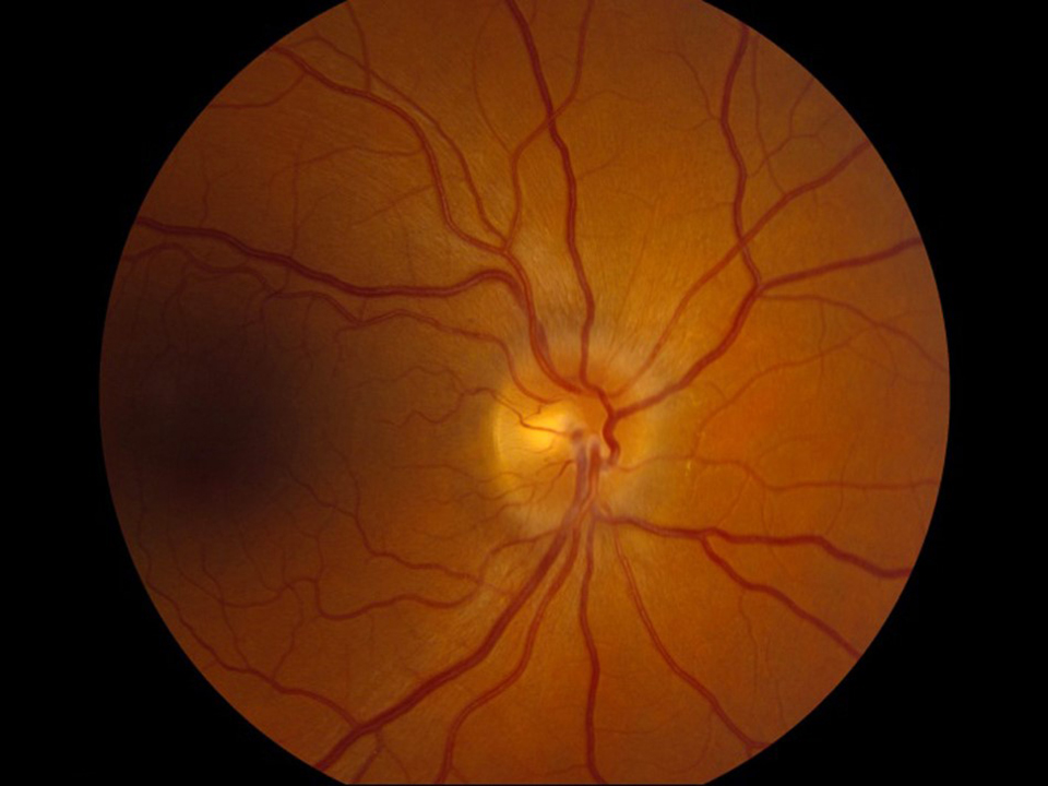
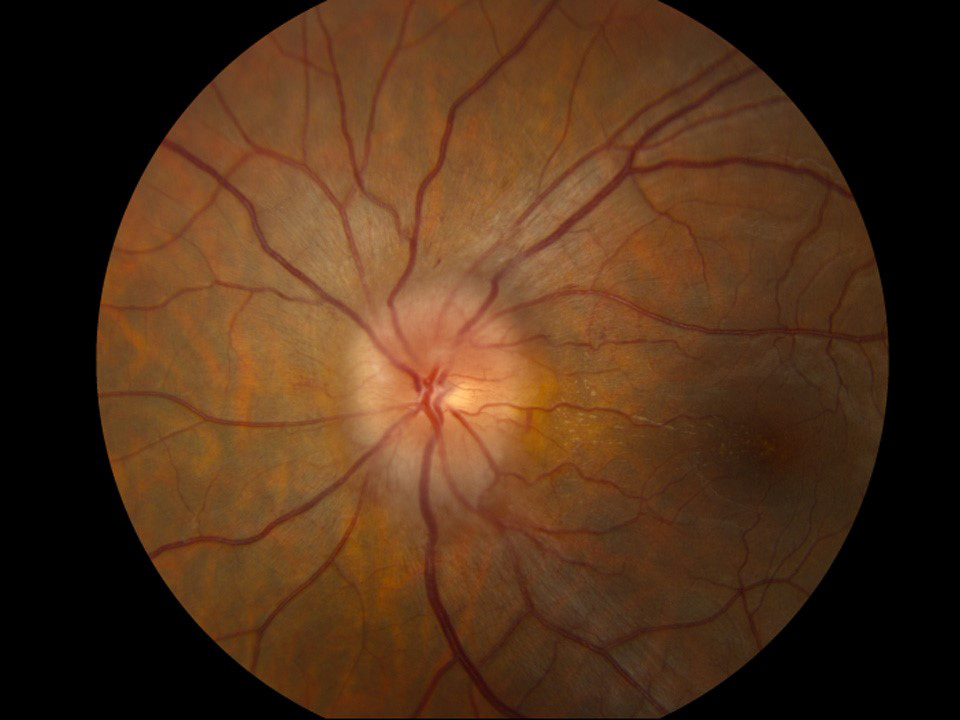
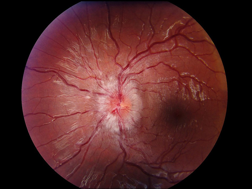
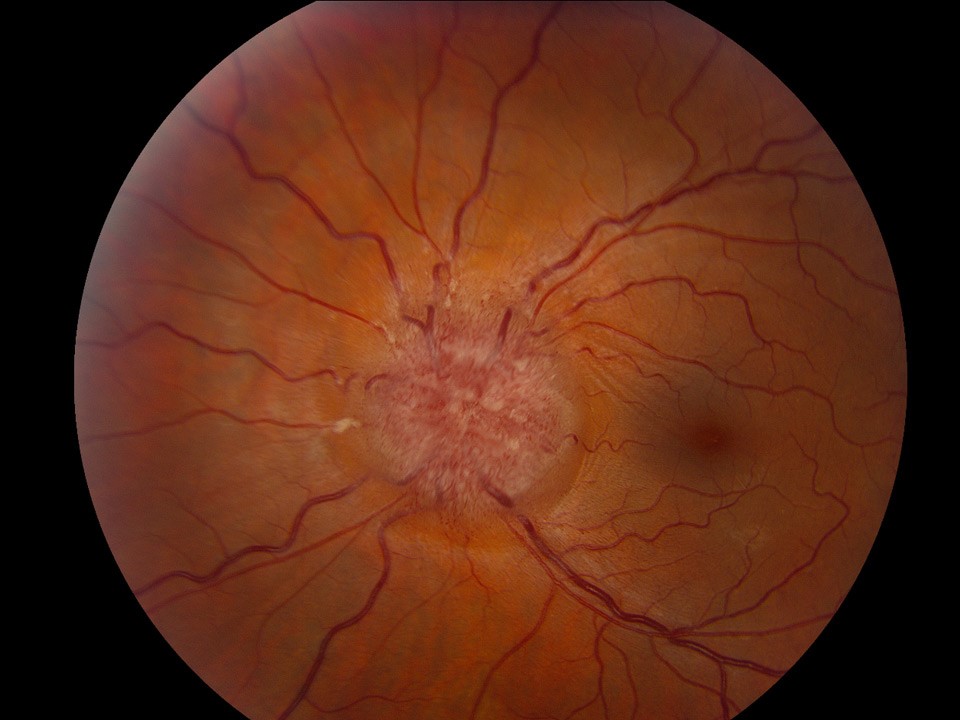
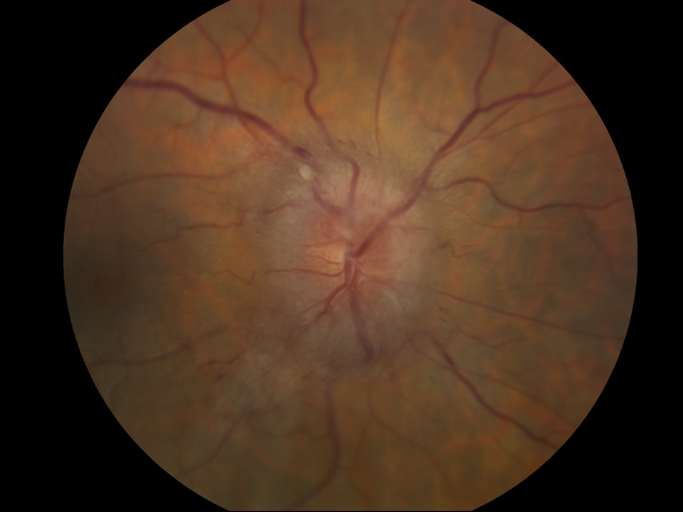
Grade 1
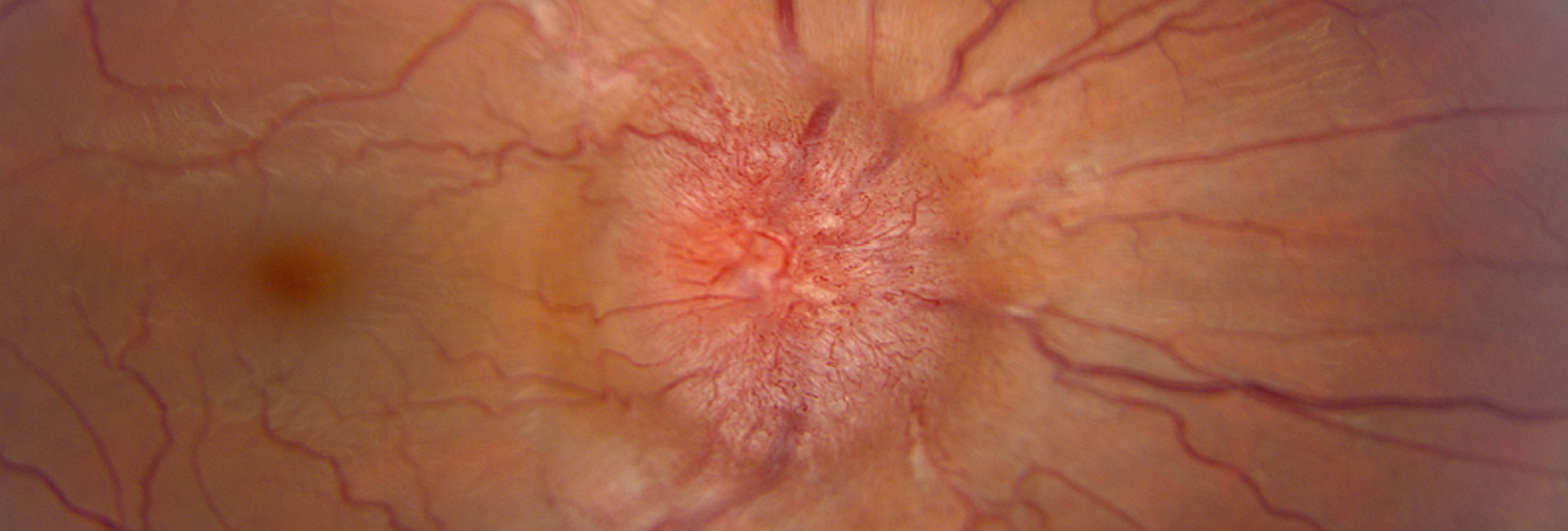
C-shaped halo with scalloped or feathered borders with a temporal gap that obscures underlying retinal details.
Disruption or thickening of normal retinal nerve fiber layer anatomy (striations seen with higher magnification).
Click the large image to highlight areas of importance. Change large image by clicking on a smaller examples below the main image.
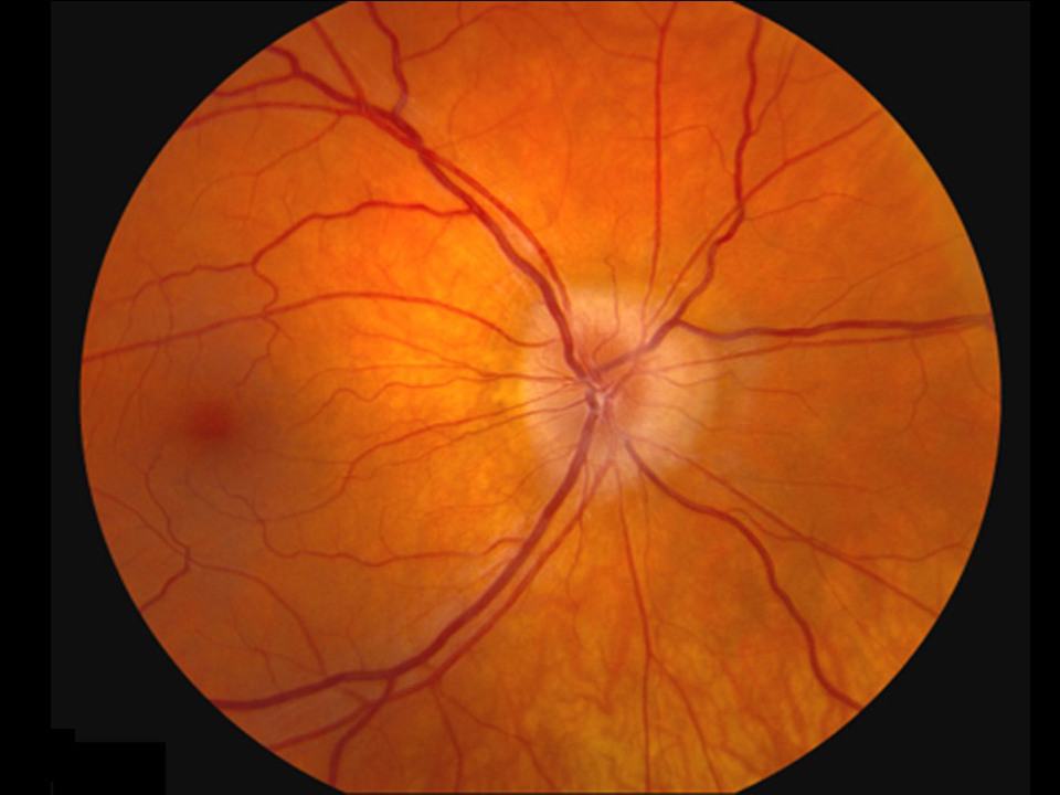
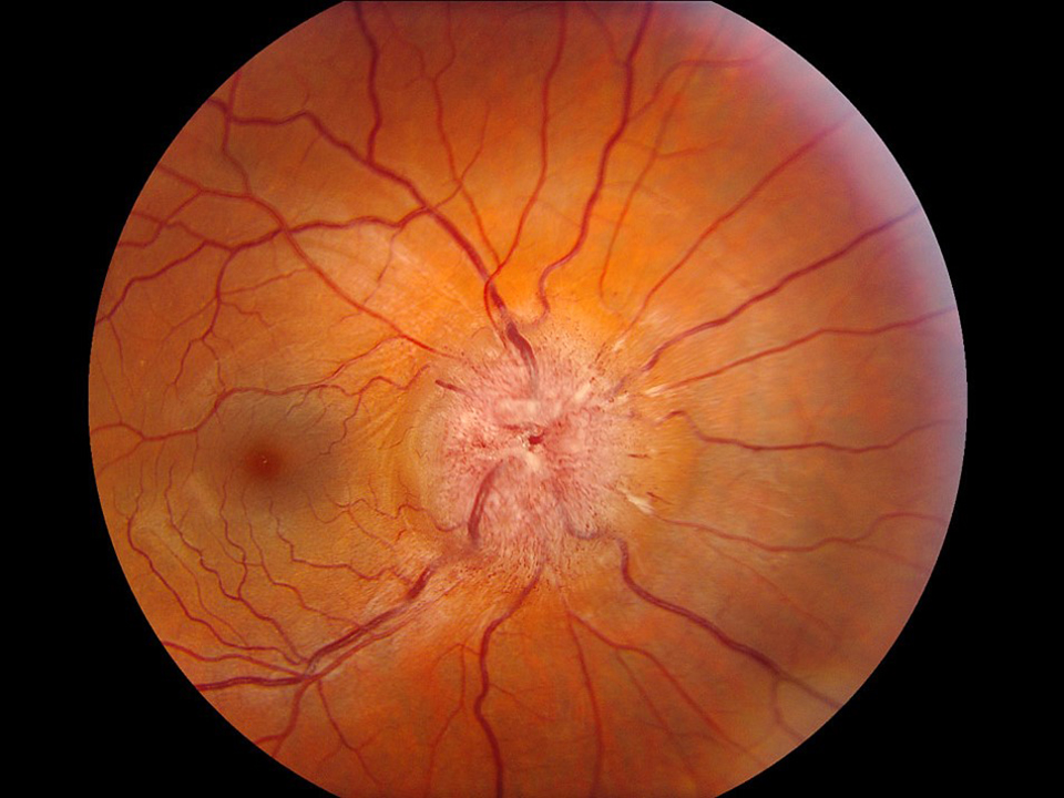
Grade 2
Features of grade 1 but with the temporal gap filled in and no obscuration of vessels; partial obscuration of major vessels in permitted.
Click the large image to highlight areas of importance. Change large image by clicking on a smaller examples below the main image.
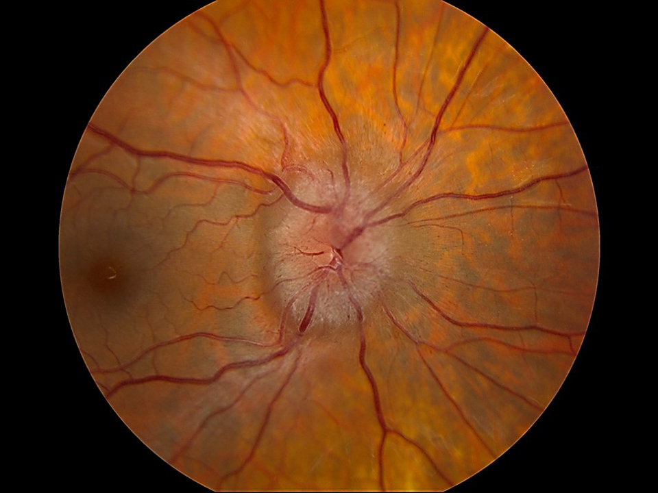
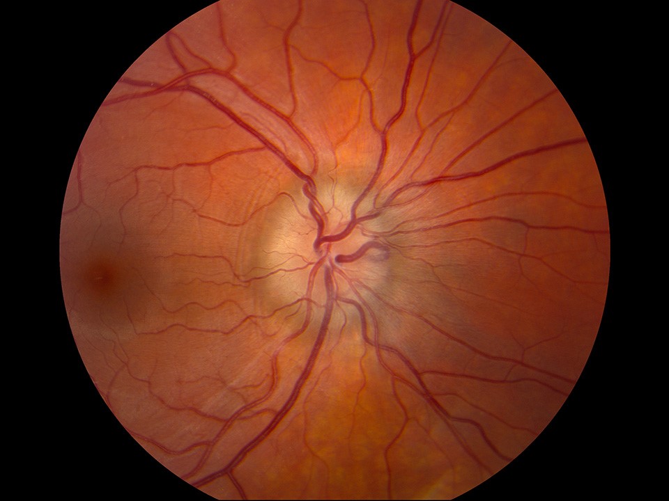
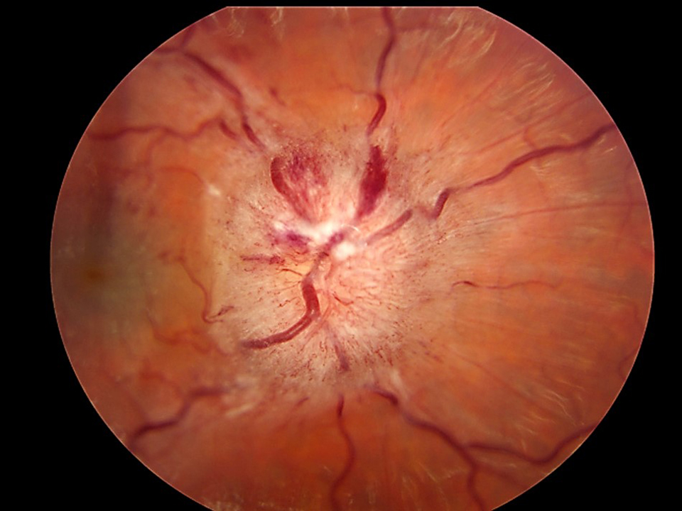
Grade 3
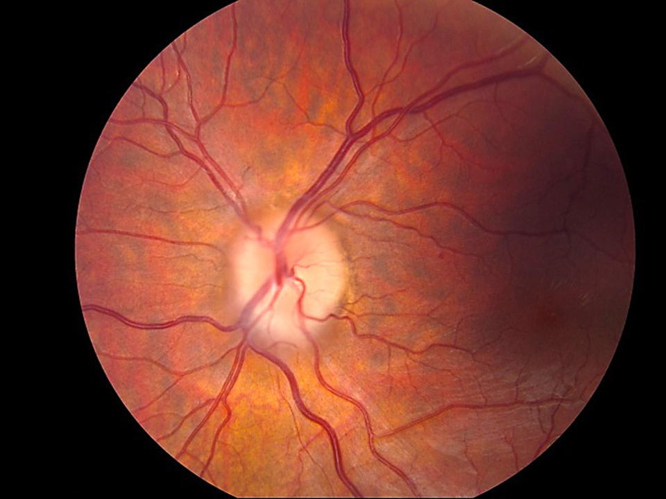
Features of grade 2 plus total obscuration of a portion of at least one major vessel as it leaves the disc.
Click the large image to highlight areas of importance. Change large image by clicking on a smaller examples below the main image.
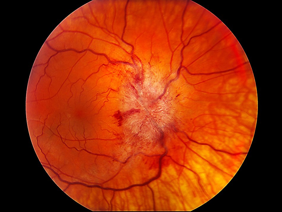
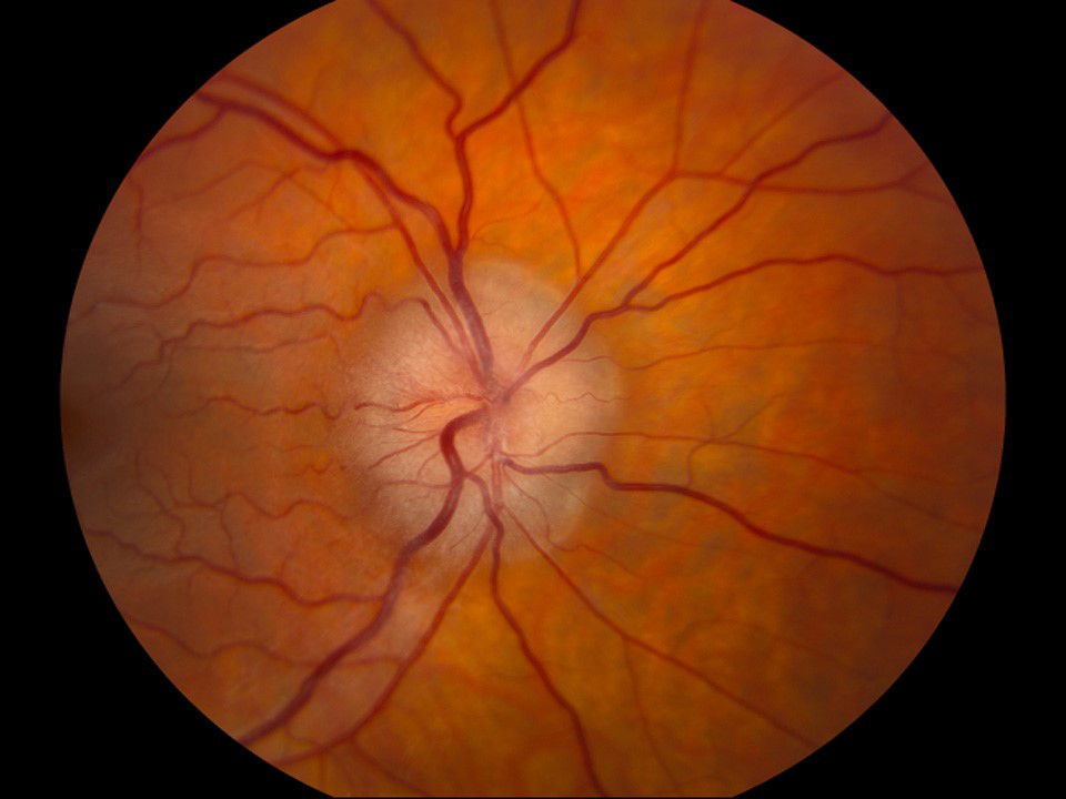
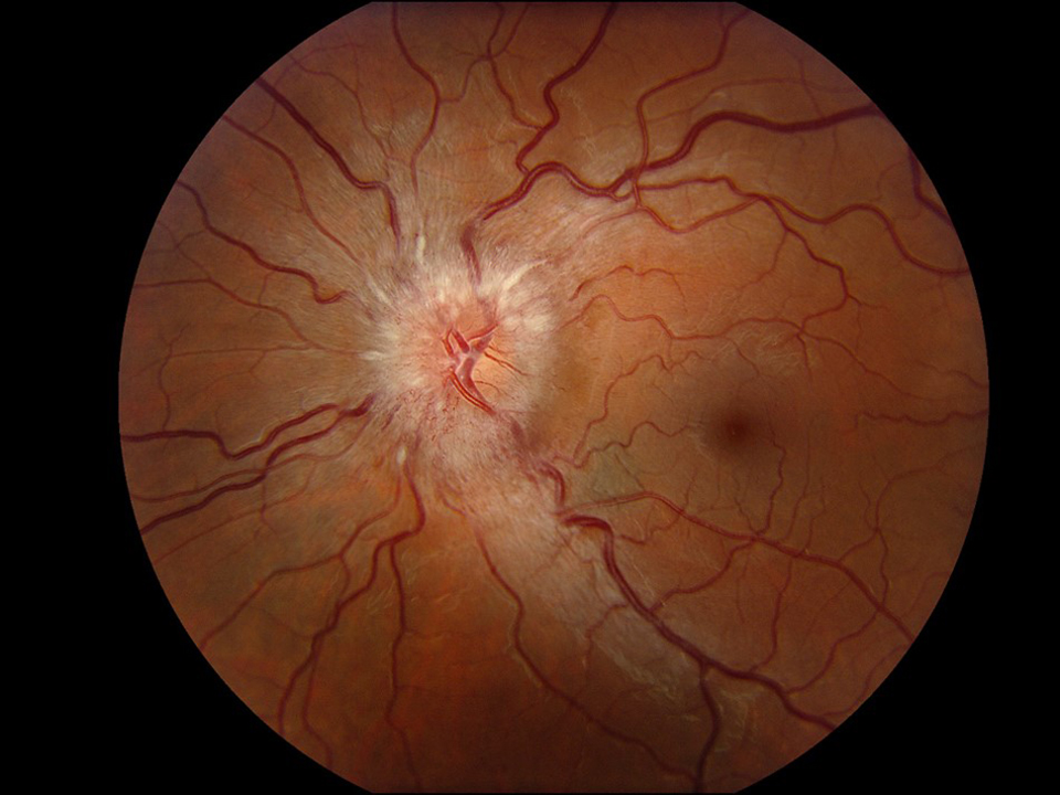
Grade 4
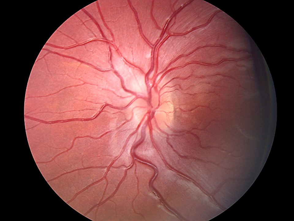
Features of grade 3 with the addition of total obscuration of a portion of a major vessel(s) on the disc. At least one major vessel on the disc must be spared and not obscured.
Click the large image to highlight areas of importance. Change large image by clicking on a smaller examples below the main image.
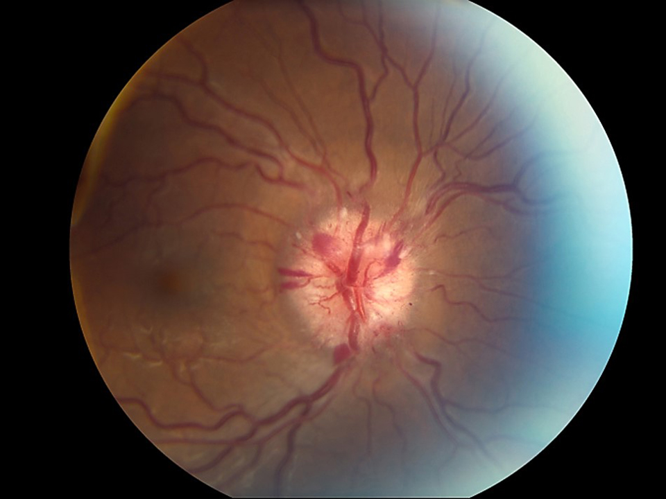
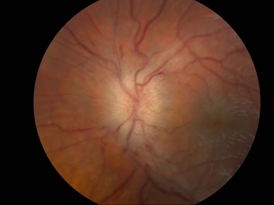
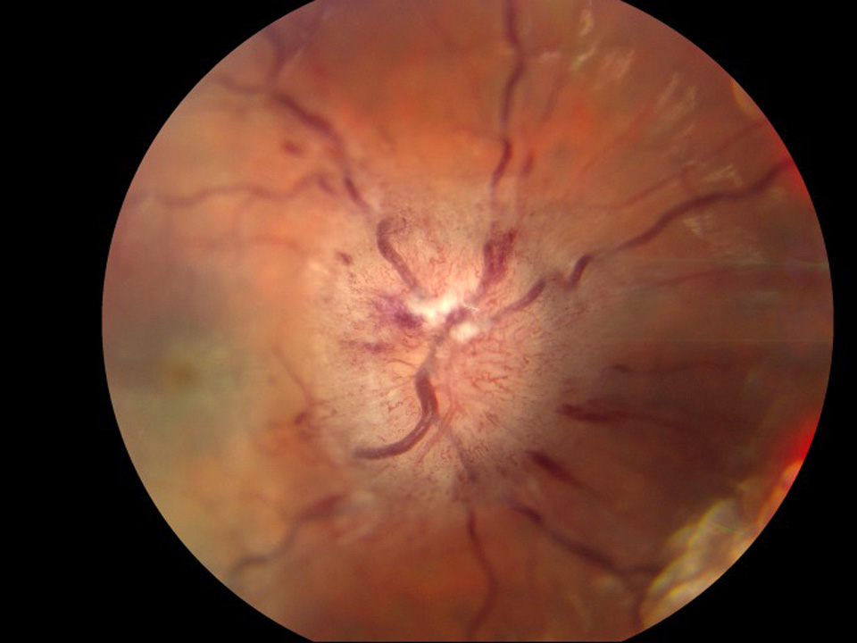
Grade 5
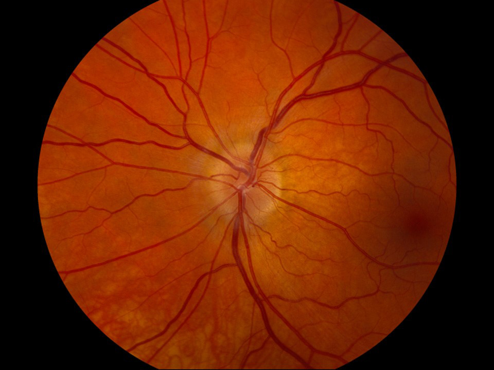
Features of grade 4 with total obscuration of a segment of every major vessel on the optic.
Click the large image to highlight areas of importance. Change large image by clicking on a smaller examples below the main image.
Results from Pretest
Legend: Correct - Incorrect -
Legend: Correct - Incorrect -
Select your career level:
Select your specialty:
Detailed Primer - Grading Papilledema
Grade 1
C-shaped halo with scalloped or feathered borders with a temporal gap that obscures underlying retinal details.
Disruption or thickening of normal retinal nerve fiber layer anatomy (striations seen with higher magnification).
Click the large image to highlight areas of importance. Change large image by clicking on a smaller examples below the main image.
Grade 2
Features of grade 1 but with the temporal gap filled in and no obscuration of vessels; partial obscuration of major vessels in permitted.
Click the large image to highlight areas of importance. Change large image by clicking on a smaller examples below the main image.
Grade 3
Features of grade 2 plus total obscuration of a portion of at least one major vessel as it leaves the disc.
Click the large image to highlight areas of importance. Change large image by clicking on a smaller examples below the main image.
Grade 4
Features of grade 3 with the addition of total obscuration of a portion of a major vessel(s) on the disc. At least one major vessel on the disc must be spared and not obscured.
Click the large image to highlight areas of importance. Change large image by clicking on a smaller examples below the main image.
Grade 5
Features of grade 4 with total obscuration of a segment of every major vessel on the optic.
Click the large image to highlight areas of importance. Change large image by clicking on a smaller examples below the main image.
Results from Pretest
Legend: Correct - Incorrect -
Legend: Correct - Incorrect -
Pressing the Browser "Back" will Reset the Quiz
Results from Post-Test
Legend: Correct - Incorrect -
Legend: Correct - Incorrect -
Results from Post-Test
Legend: Correct - Incorrect -
Legend: Correct - Incorrect -
Kardon R, Wall M. Grading of Papilledema – Self Test. EyeRounds.org. Posted March 1, 2023; Available from: https://eyerounds.org/cases/interactive_quizzes/grading_papilledema/grading_papilledema.htm

Ophthalmic Atlas Images by EyeRounds.org, The University of Iowa are licensed under a Creative Commons Attribution-NonCommercial-NoDerivs 3.0 Unported License.

Address
University of IowaLegal
Related Links
Grading and recognition of the features of papilledema have clinical importance for diagnosis, differentiating papilledema from pseudopapilledema and other causes of optic disc edema, and for monitoring a patient over time, especially with respect to their treatment. Optical coherence tomography (OCT) has advantages of being quantitative and provides insight into features that may or may not be obvious on clinical examination and has become an important adjunct to the clinical exam. Appearance of the optic nerve with fundoscopy is still the mainstay of clinical diagnosis and monitoring of papilledema. Many points of care do not have OCT capabilities or fundus photography and clinicians must rely on their clinical exam of the fundus.
Therefore, it is very important to be able to recognize features of papilledema as they appear on fundus exam and to have a standardized grading scale of its severity. The Frisen grading scale has become the standard for evaluating the severity of papilledema. Standardized grading also helps assess the risk of visual loss, which is higher as the severity of papilledema increases. With the advent of semi-automated fundus photography, grading of papilledema from photographs is needed. There is intense clinical research and interest into automated approaches to evaluate papilledema from fundus photographs and OCT and these goals have become bolstered by recent investigations into the use of different forms of artificial intelligence to evaluate image features.
This module will help clinicians and researchers to accurately and consistently grade papilledema. Future modules are being developed for differentiating papilledema, pseudopapilledema and optic disc drusen and for the use of OCT to diagnose and monitor these entities.
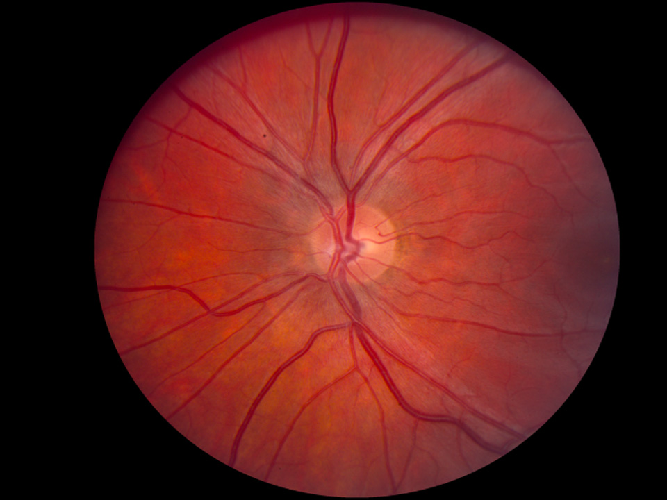
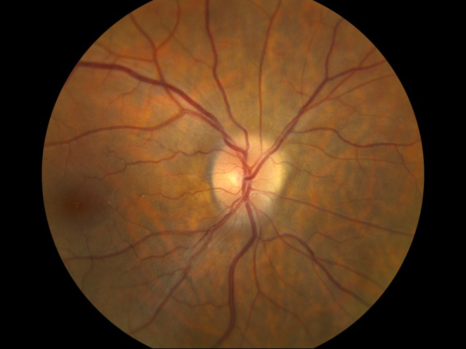
No C-shaped halo or obscuration of the peripapillary nerve fiber layer as observed in grade 1
C-shaped halo with scalloped or feathered borders with a temporal gap that obscures underlying retinal details.
Disruption or thickening of normal retinal nerve fiber layer anatomy (striations seen with higher magnification).
Features of grade 1 but with the temporal gap filled in and no obscuration of vessels; partial obscuration of major vessels in permitted.
Features of grade 2 plus total obscuration of a portion of at least one major vessel as it leaves the disc.
Features of grade 3 with the addition of total obscuration of a portion of a major vessel(s) on the disc. At least one major vessel on the disc must be spared and not obscured.
Features of grade 4 with total obscuration of a segment of every major vessel on the optic.
Grading Papilledema - Examples
Grade 1
C-shaped halo with scalloped or feathered borders with a temporal gap that obscures underlying retinal details.
Disruption or thickening of normal retinal nerve fiber layer anatomy (striations seen with higher magnification).
Click the large image to highlight areas of importance. Change large image by clicking on a smaller examples below the main image.
Grade 2
Features of grade 1 but with the temporal gap filled in and no obscuration of vessels; partial obscuration of major vessels in permitted.
Click the large image to highlight areas of importance. Change large image by clicking on a smaller examples below the main image.
Grade 3
Features of grade 2 plus total obscuration of a portion of at least one major vessel as it leaves the disc.
Click the large image to highlight areas of importance. Change large image by clicking on a smaller examples below the main image.
Grade 4
Features of grade 3 with the addition of total obscuration of a portion of a major vessel(s) on the disc. At least one major vessel on the disc must be spared and not obscured.
Click the large image to highlight areas of importance. Change large image by clicking on a smaller examples below the main image.
Grade 5
Features of grade 4 with total obscuration of a segment of every major vessel on the optic.
Click the large image to highlight areas of importance. Change large image by clicking on a smaller examples below the main image.