

EyeRounds Online Atlas of Ophthalmology
Contributor: William Charles Caccamise, Sr, MD, Retired Clinical Assistant Professor of Ophthalmology, University of Rochester School of Medicine and Dentistry
*Dr. Caccamise has very generously shared his images of patients taken while operating during the "eye season" in rural India as well as those from his private practice during the 1960's and 1970's. Many of his images are significant for their historical perspective and for techniques and conditions seen in settings in undeveloped areas.
Category: Cataract
Intracapsular cataract extraction that failed-ending up as an extracapsular extraction with retention of significant lens material
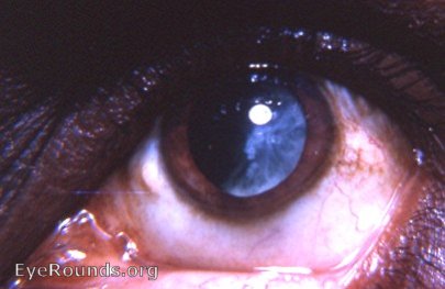
The intracapsular cataract surgeon was never happy with an extracapsular result like the one in the photo. The retained lens material could effectively interfere with the visual axis. Inflammation induced by the lens material could effectively block the pupil. The formation of Elschnig's pearls (detectable with the slit-lamp on the retained lens material near the pupil at 3 o'clock) could further occlude the pupil. Treatment usually involved needling of the aftercataract once the eye was quiet. A few surgeons would attempt to extract the aftercataract with forceps after introducing alpha chymotrypsin in an effort to weaken the zonular fibers. Adhesions of the aftercataract to the vitreous face made this a hazardous maneuver. It must be remembered that there was no laser surgery at that time.

Ophthalmic Atlas Images by EyeRounds.org, The University of Iowa are licensed under a Creative Commons Attribution-NonCommercial-NoDerivs 3.0 Unported License.