

EyeRounds Online Atlas of Ophthalmology
Contributor: William Charles Caccamise, Sr, MD, Retired Clinical Assistant Professor of Ophthalmology, University of Rochester School of Medicine and Dentistry
Expanded text by Brendan K. Penaluna, May 5, 2017
*Dr. Caccamise has very generously shared his images of patients taken while operating during the "eye season" in rural India as well as those from his private practice during the 1960's and 1970's. While the quality of his images are not up to today's higher web standards, many of his images are significant for their historical perspective and for techniques and conditions seen in settings in undeveloped areas.
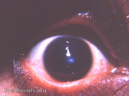
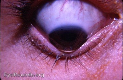
Cornea: keratoconus with apical scarring
Keratoconus is a bilateral degenerative disease that can lead to high astigmatism, thinning of the cornea and can result in apical or scarring of the cornea. Keratoconus has displayed both autosomal dominant and autosomal recessive inheritance patterns. In the top picture, apical scarring can be appreciated as a white opacity over the black of the pupil. In the bottom picture, Munson's sign, a protrusion of the lower lid caused by the ectatic cornea in downgaze, can be seen.
Reference
Biswell R. Chapter 6. Cornea. In: Riordan-Eva P, Cunningham ET, Jr. eds. Vaughan & Asbury's General Ophthalmology, 18e New York, NY: McGraw-Hill; 2011. http://accessmedicine.mhmedical.com/content.aspx?bookid=387§ionid=40229323. Accessed April 15, 2017.
originally posted 2/8/2008

Ophthalmic Atlas Images by EyeRounds.org, The University of Iowa are licensed under a Creative Commons Attribution-NonCommercial-NoDerivs 3.0 Unported License.