

EyeRounds Online Atlas of Ophthalmology
Contributor: William Charles Caccamise, Sr, MD, Retired Clinical Professor of Ophthalmology, University of Rochester School of Medicine and Dentistry
*Dr. Caccamise has very generously shared his images of patients taken while operating during the "eye season" in rural India as well as those from his private practice during the 1960's and 1970's. Many of his images are significant for their historical perspective and for techniques and conditions seen in settings in undeveloped areas.
Category: Cataract
Differential diagnosis of the white cataract in older patients
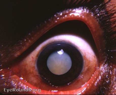
In the intracapsular era prior to Kelman's return to extracapsular surgery, great attention was placed on diagnosing the stage of a senile cataract. With the pupil in its undilated state, the situation could be quite perplexing. If the pupil was dilatable, the diagnosis was somewhat simplified. With a dilated pupil, a very white pupil was most likely a hypermature Morgagnian cataract. Slit-lamp examination would reveal no anterior cortical markings with perhaps a few scattered white dots behind the anterior capsule. There would be no radii or sectors as demonstrated in the photograph. Thus, the photograph is not of a hypermature Morgagnian cataract.A mature cataract has a dull-gray or brownish color. Radial cortical markings usually are still evident. The photograph is not of a mature senile cataract.The photograph shows the early stage of an intumescent cataract. Under the slit-lamp,it is beginning to show a cortex that is taking on a bluish-white color and acquiring a sily luster. Radial markings are evident in the anterior cortex. Please see the other Atlas photographs of full-blown intumescent cataracts. In both the intumescet cataract and the hypermature Morgagnian cataract, the anterior chamber may be shallower than normal.

Ophthalmic Atlas Images by EyeRounds.org, The University of Iowa are licensed under a Creative Commons Attribution-NonCommercial-NoDerivs 3.0 Unported License.