This site uses tracking information. Visit our privacy policy. Click to agree to this policy and not see this again.

Ophthalmology and Visual Sciences

Thyroid Eye Disease (TED)
An Introductory Tutorial and Overview of Disease

Thyroid Eye Disease (TED)
An Introductory Tutorial and Overview of Disease

Contributors: Chase A Liaboe, BA; Thomas J Clark, MD; Keith D Carter, MD; Erin M Shriver, MD

The University of Iowa
Department of Neurology and Department of Ophthalmology and Visual Sciences

Updated: November 18, 2016


Introduction

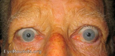
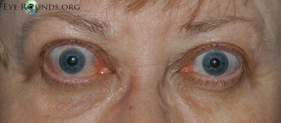
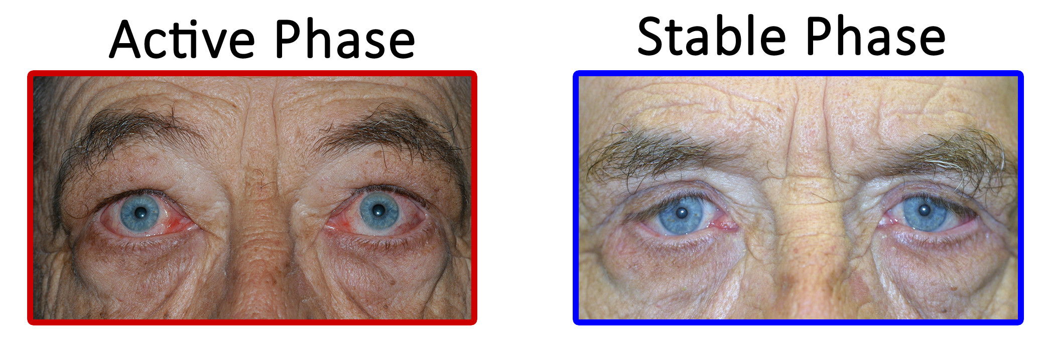
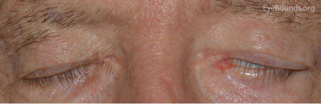
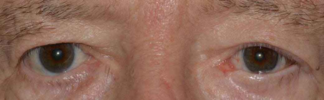
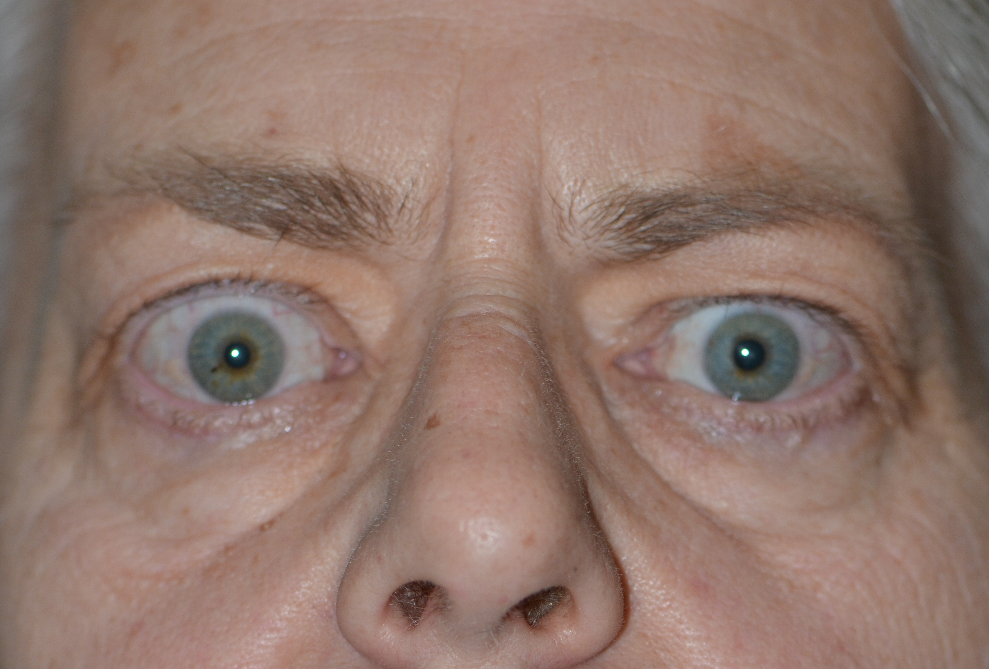
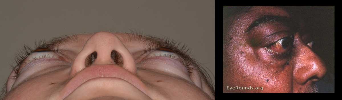
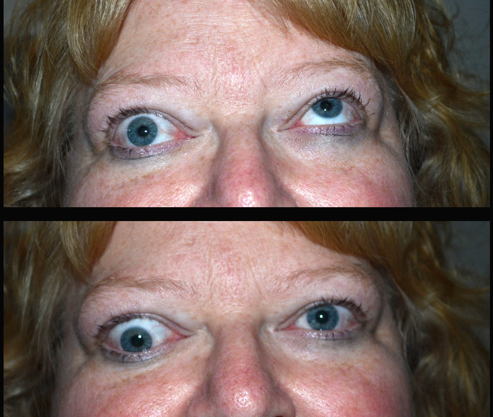
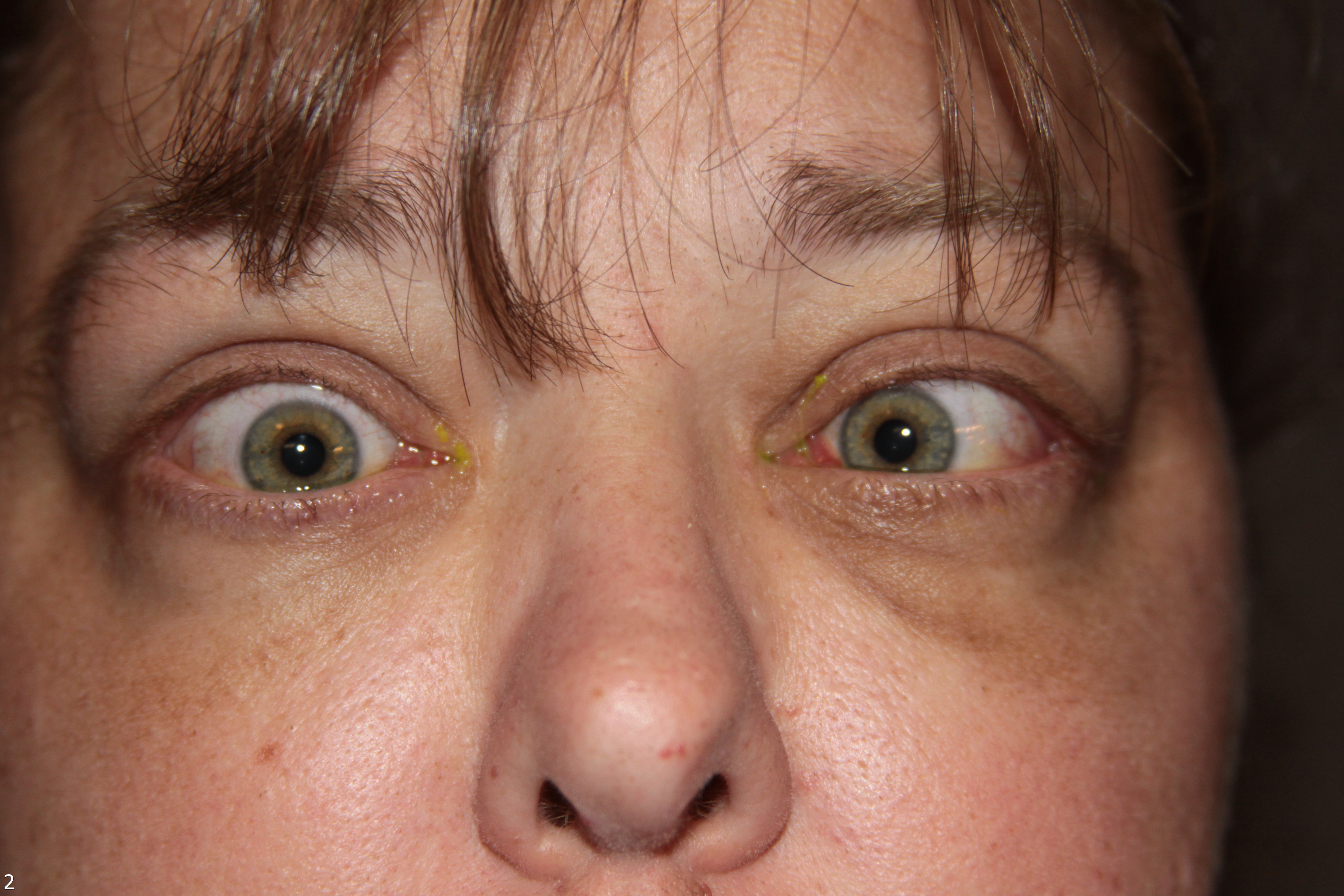
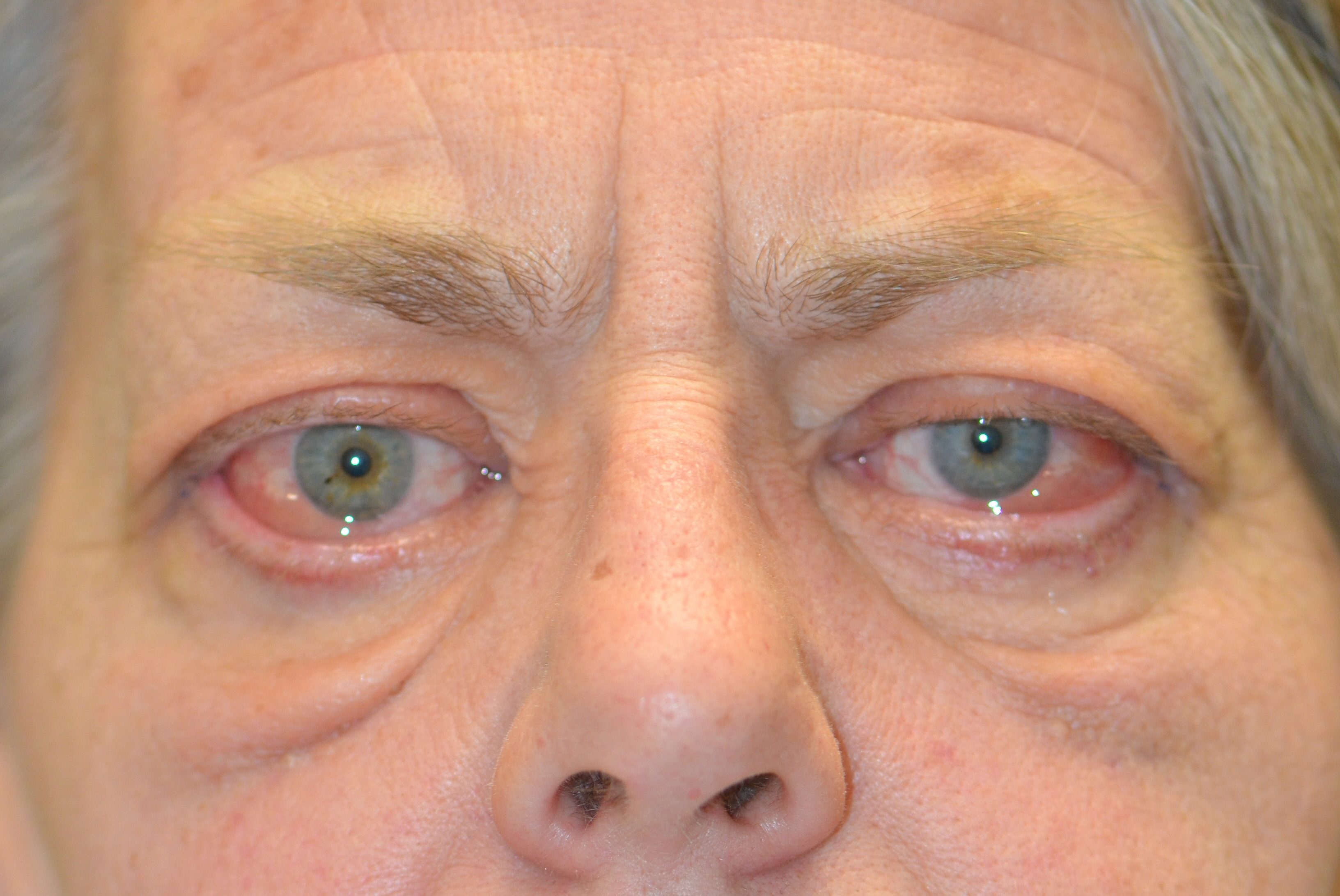
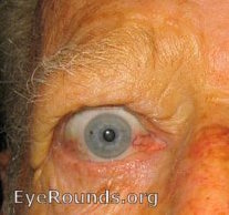
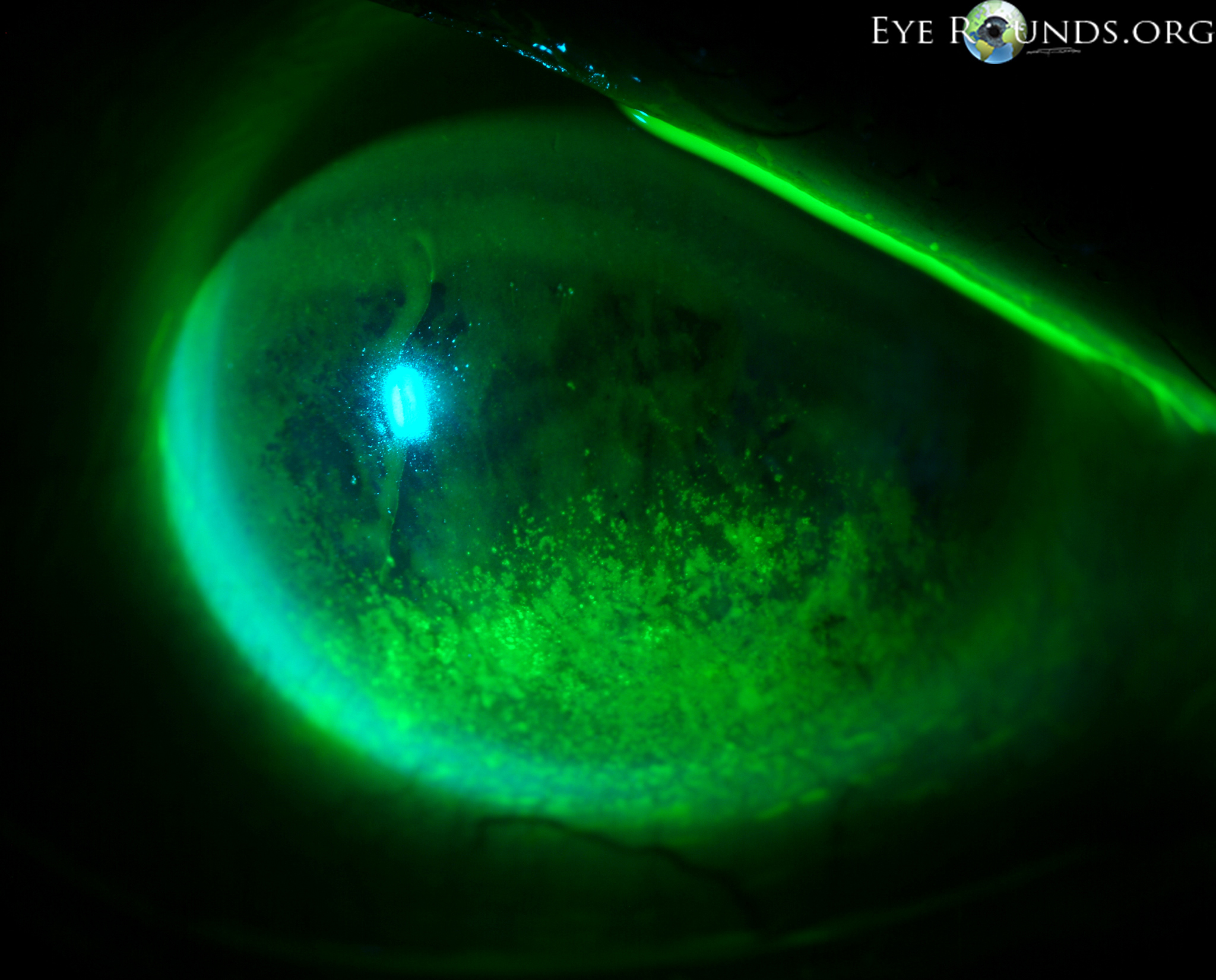
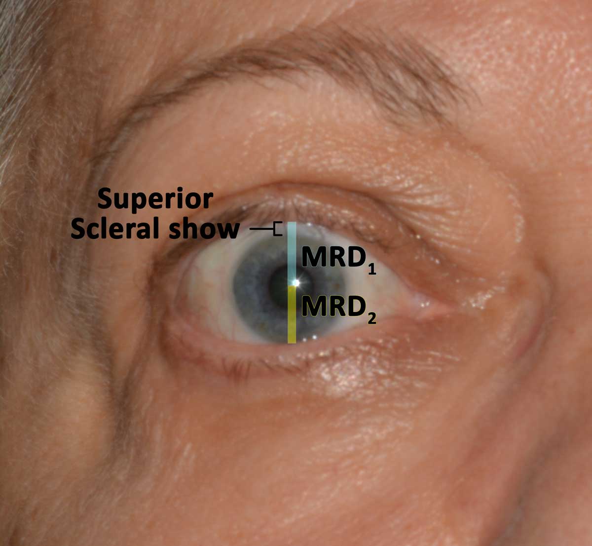
Thyroid eye disease (TED) is an autoimmune inflammatory disease of the eye and surrounding tissues. It is also recognized in the literature as Graves' ophthalmopathy, Graves' orbitopathy, thyroid-associated ophthalmopathy, and thyroid orbitopathy. TED was originally associated strictly with the Graves' triad of hyperthyroidism, pretibial myxedema, and eye disease. More recently, TED has also been noted in Hashimoto's thyroiditis as well as in the absence of a thyroid dysfunction. While symptoms are typically bilateral, they are often asymmetric. The most common presenting signs are orbital and periorbital edema, eyelid retraction, eyelid lag in downgaze, restrictive strabismus, compressive optic neuropathy, and exposure keratopathy with common symptoms of ocular irritation and dryness (Figures 1 and 2) [1]. The disease course of TED does not always coincide with thyroid activity or the treatment of underlying thyroid dysfunction.

 Thyroid Eye Disease. Paitent exhibits classic signs of the diesase.
Figure 1. These patients have some of the classic signs and symptoms of TED. Note the periorbital edema, eyelid retraction, scleral show, and conjunctival injection.
 Thyroid Eye Disease. Paitent exhibits classic signs of the diesase.
Figure 2. These patients have some of the classic signs and symptoms of TED. Note the periorbital edema, eyelid retraction, scleral show, and conjunctival injection.

Epidemiology

TED is the most common cause of orbital disease in North America and Europe and of both unilateral and bilateral exophthalmos. While TED is most commonly associated with Graves' disease, it can also occur in association with other thyroid states, pathologic or non-pathologic. TED has a higher prevalence in women than men (16 per 100,000 vs. 3 per 100,000, respectively). Both men and women demonstrate a bimodal pattern of age of diagnosis (40-44 and 60-64 years in women; 45-49 and 65-69 years in men). The median age of diagnosis is 43 years for all patients, with a range from 8-88 years. Patients diagnosed over the age of 50 years have a worse prognosis overall. Risk factors for TED include age, gender, ethnicity, and family history. A positive family history of TED is noted in 61% of TED patients [2].

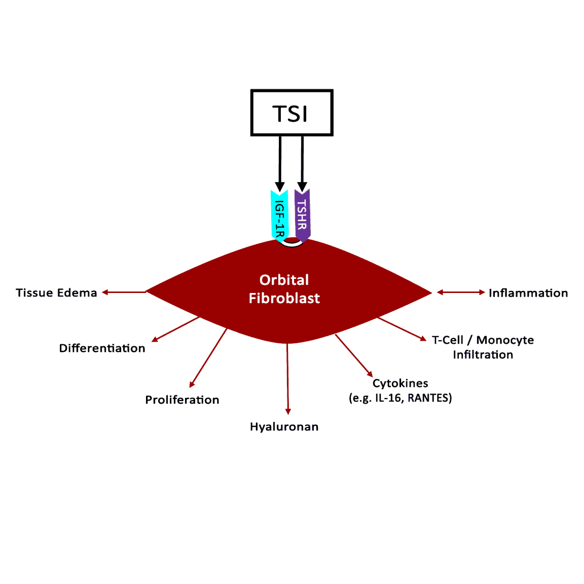
TED exacerbation is thought to be associated with both genetic and environmental factors, such as cigarette smoking, low selenium levels, and stress [3]. Smoking has been shown to adversely affect the development, progression, and response to treatment of TED. Smokers are twice as likely to develop Graves' disease, and smokers who have Graves' disease are 7.7 times more likely to develop TED compared to nonsmokers with Graves' disease.


Pathophysiology

  • Development of TED is centered on inflammation of orbital tissue via stimulation of orbital fibroblasts (Figure 3).
  • Orbital fibroblasts are unlike other fibroblasts in the body, in that they express CD40 receptors (CD40-R), which are normally found on B-cells [4]. When T-cells interact with CD40-R on orbital fibroblasts, the orbital fibroblasts produce pro-inflammatory cytokines. This leads to the synthesis of glycosaminoglycans (GAGs) and hyaluronic acid.
  • Up-regulation of GAG synthesis and deposition of GAGs results in congestion and edema of orbital tissue (Figure 4).
 Diagram demosntrating TSI activation of IGF-1R and TSHR recceptors in orbital fibroblasts.
Figure 3. Orbital fibroblast activation. Stimulation of orbital fibroblasts activates pro-inflammatory genes, leading to the synthesis of glycosaminoglycans (GAGs) and hyaluronic acid.
 Close-up of pateint showing orbital congestion.
Figure 4. Orbital congestion. Up-regulation of GAG production and deposition results in congestion of orbital tissue.
  • Orbital fibroblasts originate from neural crest cells and can differentiate into adipocytes or myofibroblasts [4].
    • Fibroblast-to-adipocyte differentiation explains the fatty hypertrophy of orbital tissue found in TED characterized by extensive orbital adipose tissue proliferation and deposition, which more commonly leads to compressive optic neuropathy. [5]
    • Fibroblast-to-myofibroblast differentiation explains another variant of TED that manifests primarily with muscle enlargement and more commonly leads to restrictive myopathy [5].
  • Two additional immunologic processes characterize the orbital fibroblast's role in TED
  • Antigen-dependent autoimmune response [1]
    • Expression of both insulin-like growth factor 1 receptor (IGF-1-R) and thyroid-stimulating hormone receptor (TSH-R) directly correlate with TED [6].
    • Both IGF-1-R and TSH-R are present on most types of human cells and are activated by their respective autoantibodies.
    • Activation of IGF-1-R stimulates synthesis of GAGs and secretions of chemoattractants by orbital fibroblasts, leading to orbital inflammation and congestion [6].
    • Activation of IGF-1-R and TSH-R cause orbital cytokine production, leading to GAG deposition in orbital tissues (Figure 5).
      • TSH-R activation upregulates the synthesis of TSH-R mRNA [7].
      • TSH-R mRNA signals for additional fibroblast differentiation in the orbit.
    • Three types of TSH-R antibodies (TRA) are known, but only one type, thyroid-stimulating immunoglobulin (TSI), is associated with hyperthyroidism and TED [8].
      • A recent study found TSI levels correlate directly with the activity and severity of TED, and all TED patients with diplopia were positive for TSI.
      • TSI is the closest "functional biomarker" of TED that we have at this time.
      • While the amount of TSI produced is directly correlated with TED severity, TED can present without Graves' disease or autoantibody formation [1].
 Thyroid Eye Disease Activation Model.
Figure 5: Antibody interaction. IGF-1-R and TSH-R are activated by TSI (GD-IgG). Activation of these receptors produces orbital cytokines, leading to GAG deposition and orbital inflammation and congestion.

Clinical Presentation

The thyroid state of a patient presenting with TED is quite variable: 90% hyperthyroid, 6% euthyroid, 3% with Hashimoto's thyroiditis, and 1% hypothyroid [2]. Patients are simultaneously diagnosed with TED and thyroid dysfunction 20% of the time, and 60% present within 1 year of onset of thyroid disease [9]. However, TED can present long before (up to 10 years) or long after (up to 20 years) the initial presentation of thyroid disease [2].

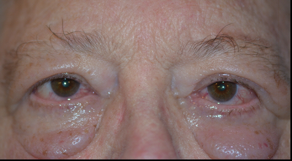
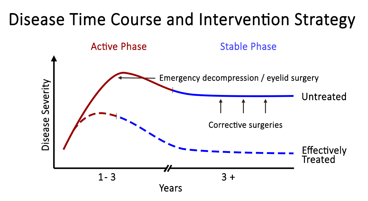
TED is a self-limiting disease and may present in one of two stages: active or quiescent (Figure 6). In the active stage, there is active inflammation, which can lead to orbital muscle enlargement, conjunctival injection and chemosis, ocular pain, and swelling of the periocular tissues and eyelids. This stage typically involves waxing and waning TED symptoms and can last months to years. On average, the active phase lasts for 1 year in non-smokers and 2-3 years in smokers. The quiescent phase follows spontaneous resolution of the active phase (Figure 7). Active TED has a recurrence rate of 5-10% but is less likely to recur after 18 months of quiescence [10].

 Active vs. Stable TED.
Figure 6: Active vs. Stable TED. Active TED is characterized by signs of inflammation (orbital muscle enlargement, conjunctival injection, swelling of periocular tissue, and chemosis). TED activity waxes and wanes, and usually transitions to stable TED within 1-3 years.
 Rundle's curve.
Figure 7: Rundle's curve. As seen in the representation of TED activity over time in Rundle's curve, early initiation of therapy is crucial in diminishing the final severity of disease manifestations.
  • Upper eyelid retraction – the most common presenting sign of TED (Figure 8)
    • Up to 90% of patients affected (bilateral or unilateral) [2]
    • Multifactorial cause [2]
      • Increased sympathetic tone acting on Müller's muscle
      • Contraction of the levator palpebrae superioris
      • Proptosis
      • Scarring between the lacrimal gland and the levator palpebrae superioris
    • Physical exam
      • Dalrymple's sign (Figure 8): widening of the palpebral fissure with inferior and superior scleral show
      • Temporal flare (Figure 9): elevation of the temporal upper eyelid compared to its normal anatomical location
      • Lagophthalmos (Figure 10)
        • Inability to close the eyes completely
        • Manifests as dry eye, tearing, foreign body sensation, blurred vision, and eventually exposure keratopathy, especially with a poor Bell's reflex
    • see Treatment options
  Eyelid retraction is the most common presenting sign of TED.
Figure 8: Eyelid retraction is the most common presenting sign of TED, and is the result of many factors associated with TED. This image also shows Dalrymple's sign, characterized by the widening of the palpebral fissure. Note the superior and inferior scleral show.
 Lagophthalmos.  Lagophthalmos.
Figure 9: Lagophthalmos. Lagophthalmos typically presents as dry eye, tearing, foreign body sensation, and blurred vision.
 Temporal flare.
Figure 10: Temporal flare. Note the elevation of the temporal portion of the upper eyelid.
 Exophthalmos.
Figure 11: Exophthalmos. The globe is displaced anteriorly out of the orbit. This is an ophthalmologic emergency – the cornea is at risk for exposure, and the optic nerve is at risk of irreversible damage.
  • Exophthalmos – the second most common sign associated with TED (Figure 11)
    • 60% of patients are affected [2]li>
    • Physical exam
      • Exposure keratopathy: characteristic punctate epithelial erosions
      • Globe subluxation: anterior displacement of the globe [11]
        • Globe equator protrudes anteriorly in relation to the lids
        • Ophthalmologic emergency
          • Decreased perfusion of the optic nerve and the retina
          • Anoxic destruction of the optic nerve can cause irreversible visual loss
    • see Treatment options
  • Other common signs and symptoms associated with TED: eyelid lag, extraocular myopathy, pain with eye movement, optic neuropathy, chemosis, and conjunctival injection
    • Eyelid lag
      • 50% of patients affecte
      • Static dysfunction in which the upper eyelid is elevated in relation to the globe while in downgaze
    • Von Graefe's sign
      • Dynamic form of eyelid lag associated with TED
      • Delayed descent of the upper eyelid during downgaz
 Chemosis
Figure 12: Hypotropia. Note the vertical misalignment of the eyes in primary gaze and the restrictive movement in upward gaze. This is due to an enlarged and restricted inferior rectus muscle.
 Conjunctival Injection.
Figure 13: Esotropia. Note the horizontal misalignment of the eyes in primary gaze. This is due to an enlarged and restricted medial rectus muscle.figcaption>
    • Restrictive extraocular myopathy (Figures 13 and 14)
      • 40% of patients affected
      • Inferior and medial rectus muscles most commonly affected, leading to hypotropia and esotropia, respectively
      • Corneal light reflex –
        • Clinical examination in which position of light reflex relative to pupil and limbus is used to evaluate degree of duction in the four cardinal directions
        • Shown to be the best method of evaluating restrictive extraocular myopathy [12]
    • Pain with eye movement [2]li>
      • 30% of patients affected
      • Characterized as dull, deep orbital pain
    • Optic nerve dysfunction from compressive optic neuropathy (Figure 14) [2]
      • 6% of patients affected
      • Compression of the optic nerve due to enlargement of the rectus muscles and increased volume of periorbital tissue within the confines of the bony orbit
      • Presents with dyschromatopsia, decreased vision, and/or visual field defects
      • Ophthalmic emergency requiring immediate treatment
Compressive optic neuropathy
Figure 14: Compressive optic neuropathy. This sequence shows a series of CT scans from patients with compressive optic neuropathy, with their associated visual fields.
    • Chemosis and conjunctival injection (Figures 15 and 16)
      • Sign of active inflammation cause by congestion of the orbital tissue
      • More pronounced at the site of the rectus muscle insertion
       Chemosis
      Figure 15: Chemosis. Note the swelling within the conjunctiva.
       Conjunctival Injection.
      Figure 16: Conjunctival Injection. Note the dilation of the nasal and temporal conjunctival vessels.
    • Exposure keratopathy (Figure 17)
      • Secondary sign due to lagophthalmos from eyelid retraction and/or exophthalmos
      • Predisposes the cornea to bacterial infection (keratitis), which can lead to ulceration, endophthalmitis, and perforation
      • Multimodal management
       Exposure keratopathy.
      Figure 17: Exposure keratopathy. Punctate epithelial erosions (PEE) secondary to exposure keratopathy.

Workup and Diagnosis

Differential Diagnosis

When examining a patient with suspected TED, it is important to have a working differential. The following diagnoses share some similarities to the clinical presentation of TED

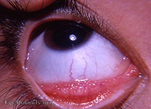
  • Allergic conjunctivitis– While both can cause excess tearing and conjunctivitis, allergic conjunctivitis tends to be acute in onset from a new exposure, causes itching, can have papillary conjunctival reaction, and is not associated with eyelid retraction or exophthalmos (Figure 18).

  • Allergic conjunctivitis.
    Figure 18: Allergic conjunctivitis. Note the presence of giant papillae.(larger image not available)
  • Myasthenia gravis (MG) – Like TED, MG patients can present with diplopia. However, MG tends to worsen throughout the day and improves after rest while diplopia in TED is not typically variable. Also, MG patients may present with ptosis, which is not associated with TED. Diplopia associated with TED is restrictive in nature, which can be determined by forced duction testing.

  • Orbital myositis (OM)OM causes enlargement and inflammation of the muscle body and tendon insertion, rather than just the muscle body, as is the case in TED patients. Orbital myositis is not generally associated with eyelid retraction. OM is usually unilateral. A bilateral presentation would be unusual for OM, whereas TED can present either way.

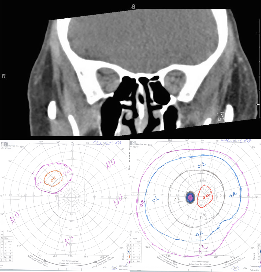
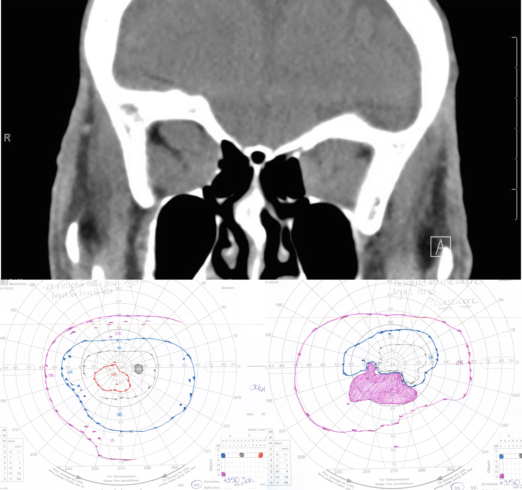
  • Orbital tumors – Orbital tumors are typically unilateral in presentation and can cause proptosis and a wide variety of motility disturbances depending on location. Orbital tumors are unlikely to cause eyelid retraction or lid lag. (see Eye Rounds case on Cavernous Hemangioma)

  • Carotid-cavernous fistula (CCF)– Patients may hear pulse-synchronous tinnitus. Presentation may include proptosis, pulsatile exophthalmos, dilated conjunctival and episcleral vessels, elevated intraocular pressure, or enlarged EOM depending on the amount of flow through the fistula and the degree of congestion. A CCF would not cause eyelid retraction or temporal flare. (see Eye Rounds case on CCF)

  • Chronic progressive external ophthalmoplegia (CPEO)– CPEO slowly progresses over 5-15 years with most patients presenting with ptosis. All cardinal directions of gaze are affected, with downgaze most likely spared. TED, conversely, typically affects downward and nasal gaze.

  • Inflammatory orbitopathy, such as granulomatosis with polyangitis (GPA, formerly known as Wegener's granulomatosis) – GPA typically presents with a mix of upper airway, lower airway, and renal pathologies. Patients may have conjunctivitis, episcleritis, scleritis, and/or uveitis. Other than conjunctivitis, these findings are uncommon in TED patients.

  • IgG4 disease – Tumefactive lesions and fibrosis affecting one or more organs characterize this fibro-inflammatory disorder. It is most commonly present in the biliary tree, retroperitoneum, salivary glands, orbit, and lymph nodes. It is thought to involve both humoral and cell-mediated immunity. Orbital IgG4 disease often involves painless swelling of the extraocular muscles, lacrimal glands, and infraorbital nerves in combination with paranasal sinus disease. IgG4 disease can also present as an inflammatory orbital mass lesion.

Clinical Requirements for Diagnosis

In diagnosing TED, two of the following three clinical requirements must be met [13 ,14]

  • Laboratory evidence (current or recently-treated immune-related thyroid dysfunction)
    • Graves' disease
    • Hashimoto's thyroiditis
  • Presence of thyroid antibodies without a dysthyroid state: TRA, thyroid-binding inhibitory antibodies, TSI, antimicrosomal antibody
  • Exam findings (1 or more of the following)
    • Unilateral/bilateral eyelid retraction with temporal flare
    • Unilateral/bilateral proptosis
    • Restrictive strabismus
    • Compressive optic neuropathy
    • Fluctuating eyelid edema/erythema
    • Chemosis/caruncular edema
  • Radiographic evidence: unilateral/bilateral fusiform enlargement of inferior rectus, medial rectus, super rectus/levator complex, or lateral rectus

Disease Stratification

If TED is suspected, one must determine disease activity and severity in order to assess the urgency of treatment.

Disease Activity

In assessing the activity level of TED in a patient, the clinical activity score (CAS) can be used [15].

  • At the initial visit, patients are given a CAS score of 1-7, one point for each sign or symptom (Figure 19):
  • Spontaneous orbital pain in the last 4 weeks
  • Gaze evoked orbital pain in the last 4 weeks
  • Eyelid swelling that is considered to be due to active TED
  • Eyelid erythema
  • Conjunctival injection considered to be due to active TED
  • Chemosis
  • Inflammation of caruncle or plica semilunaris
  • At follow-up visits, add the 3 following criteria for a potential CAS score of 10 (one point for each sign or symptom)
  • Increase of ≥ 2 mm in proptosis
  • Decrease in uniocular motility in any one direction of ≥ 8 degrees
  • Decrease in visual acuity equivalent to 1 Snellen line
  • TED is considered "active" if the CAS ≥ 3 at the initial visit or ≥ 4 at follow-up visits

Figure 19. Clinical Activity Score

Initial Visit( 1 point each)

  1. spontaneous orbital pain in last 4 weeks
  2. Gaze-evoked orbital pain in last 4 weeks
  3. Eyelid swelling
  4. Eyelid erythema
  5. Conjunctival injection
  6. Chemosis
  7. Inflammation of caruncle or plica semilunaris

CAS ≥ 3 → "Active"

Follow-up Visit (1 point each)

- Criteria 1-7

  1. Increase ≥ 2mm proptosis
  2. Decrease in uniocular motility in any one direction of ≥ 8°
  3. Decrease in visual acuity equivalent to 1 Snellen line

CAS ≥ 4 → "Active"

Disease Severity

In classifying the severity of TED, 3 indices are typically used

  • The Werner's NOSPECS measures clinical severity based on presenting features [17]
    • Class 0: No signs or symptoms
    • Class 1: Only signs (upper lid retraction and stare, ± lid lag)
    • Class 2: Soft tissue involvement (edema of conjunctiva and lids, conjunctival injection)
    • Class 3: Proptosis
    • Class 4: Extraocular muscle involvement (usually with diplopia)
    • Class 5: Corneal involvement (primarily lagophthalmos)
    • Class 6: Sight loss (due to optic nerve involvement)
  • The European Group Of Graves' Orbitopathy (EUGOGO) reports that TED severity can be classified by the following measurements [16]
    • Assess for an RAPD
    • demonstration of the MRD1 and MRD2 calculations .
      Figure 20: External eye measurements. This patient has lid retraction and superior scleral show. A demonstration of the MRD1 and MRD2 calculations is shown here.
    • Record lid retraction (Figure 20)
      • MRD 1 (note superior scleral show)
      • MRD 2 (note inferior scleral show)
      • Note if temporal flare is present
    • Hertel exophthalmometer with intercanthal distance
    • Risk of cornea ulceration
      • Lagophthalmos
      • Bell's Phenomenon (if absent, eye will not rotate up and out with lid closure)
  • EUGOGO has also proposed a classification scheme that grades TED as mild, moderate-to-severe, and sight-threatening based on the following criteria [16]
    • Mild
    • Mild impact on daily life
    • Insufficient to justify immunosuppressive/surgical treatment
    • One or more of the following
      • Minor lid retraction (< 2 mm)
      • Mild soft tissue involvement
      • Exophthalmos < 3 mm above normal for race and gender (~18 mm for Asians, 20 mm for Caucasians, and 22 mm for African Americans)
      • Transient or no diplopia
      • Corneal exposure responsive to lubricants
    • Moderate-to-severe
      • Non-sight-threatening but sufficient impact on life to justify immunosuppression or surgical intervention
      • One or more of the following
        • Lid retraction ≥ 2 mm
        • Moderate or severe soft tissue involvement
        • Exophthalmos ≥ 3mm above normal for race and gender
        • Transient or constant diplopia
    • Sight-threatening
      • TED patients with resultant optic neuropathy and/or corneal breakdown
      • Warrants immediate intervention
  • The VISA analysis (Vision, Inflammation, Strabismus, and Appearance) subjectively and objectively evaluates each category, which is based on the presence and severity of signs and symptoms (Figure 21).
VISA Chart.
Figure 21: VISA chart

Treatment

Introduction to Treatment

Management goals include maintenance of general health and well-being, achieving a euthyroid state (without post-treatment hypothyroidism), and promotion of smoking cessation. Both smoking cessation and euthyroidism help prevent further exacerbation and decrease the duration of active disease. From an ophthalmologist's perspective, the primary goal is to preserve visual function, while also preventing exposure keratopathy, correcting diplopia, and improving blink dynamics and cosmesis.

  • Smoking cessation is a key part of treatment.
    • Cigarette smoking
      • Increases severity of disease
      • Decreases the effectiveness of core treatment methods [3]
      • Cyanide, contained in cigarette smoke, is converted in the body to thiocyanate, an anti-thyroid agent
        • inhibits iodine uptake
        • increases iodine excretion
        • inhibits thyroid hormone synthesis
    • Smokers are twice as likely to develop Graves' disease when compared to nonsmokers.
    • Patients with Graves' disease who smoke are 7.7 times more likely to develop TED when compared to nonsmokers.
    • Smoking reduces the effectiveness of TED treatments such as corticosteroids and RAI.

TED is a self-limiting disease, with patients moving from the active to quiescent phase within 1-3 years with a 5-10% risk of recurrence [10] Treatment for TED should start at the time of the diagnosis, as treatment becomes less effective as the disease progresses from the early, acute, active phase to the chronic quiescent phase.

  • Rundle's Curve (Figure 22)
    • Early initiation of therapy is crucial in diminishing the final severity of disease manifestations.
    • Treatment initiation
      • Treatment initiated during the early months of the active inflammatory phase has been shown to be most effective.
      • Initiation of therapy during the final months of active inflammatory phase has little effect on the final outcome of disease.
    • Once the chronic fibrotic stage has set in, treatment options become more limited, i.e. primarily surgical.
    Rundle Curve.
    Figure 22: Rundle's curve. As seen in the representation of TED activity over time in the Rundle's curve, early initiation of therapy is crucial in diminishing the final severity of disease manifestations.
  • The majority of patients with TED (~75%) have mild to moderate disease and require primarily supportive care as a means of symptom management.
    • Ocular lubrication, by way of daytime eyedrops and nighttime ophthalmic ointment, is the mainstay of treatment for mild to moderate disease.
    • Topical cyclosporine has been shown to be beneficial in reducing symptoms of ocular surface irritation [10].
    • The following lifestyle modifications are also helpful adjuncts to ocular lubrication
      • Smoking cessation
      • Sodium restriction to reduce water retention and tissue edema
      • Sleeping with the head of the bed elevated to decrease orbital edema
      • Sunglasses to decrease photophobia and feelings of dryness
    • Oral NSAIDs can be used if periocular pain is a prominent complaint [10].
    • In the presence of diplopia, temporary press-on prism lenses (e.g. Fresnel) can be utilized.
      • Once stability in diplopia is achieved, the prism can be ground into glasses.
      • Alternatively, for stable diplopia, the patient may undergo strabismus surgery, assuming the patient has been in the stable phase of TED for several months.
    • Mineral supplementation with selenium, when taken regularly, has been shown to exert a significant benefit in European patients with mild, non-inflammatory orbitopathy, but the benefit of this supplementation in other regions is debated [18].
  • Approximately 20% of patients with TED undergo some type of surgical intervention [19].
    • In one study, 13% of patients with TED had eyelid surgery, 9% strabismus surgery, and 7% orbital decompression [19].
    • As a general guideline, surgery is not advised until a euthyroid state is maintained and the TED has been in the stable phase for at least 6-9 months.
      • Exceptions include visual loss from compressive optic neuropathy or corneal exposure, in which cases urgent surgical intervention is warranted.
      • In order to prevent repeat surgery following recovery from subsequent procedures, surgery for TED occurs in the following order, whenever possible
        • Orbital decompression
        • Strabismus surgery
        • Eyelid surgery

Aside from threatening vision and causing ocular and orbital pain, TED can be disfiguring and emotionally and psychologically taxing for many patients. Waxing and waning symptoms can be frustrating for both patient and provider. Education and reassurance are integral components of patient care. Peer support groups are invaluable for many patients.

Management of Systemic Hyperthyroidism

The following section starts with an overview of managing hyperthyroidism, followed by the different treatment options used in TED. It concludes with a discussion about therapeutic modalities specific to each sign or symptom associated with TED.

  • While the course of TED does not parallel the status of systemic thyroid disease, achieving a euthyroid state is an important part of management.
    • Hyper- or hypothyroidism has been associated with a greater severity score than euthyroid patients.
    • Restoration of a euthyroid state by antithyroid drugs has been associated with improvement of TED over several months [20].
  • Oral beta-blockers can be used for symptom control (Figure 23).
    • They decrease conversion of free T4 to T3.
    • They decrease heart rate, palpitations, anxiety, and heat intolerance.
  • Propylthiouracil and methimazole, thiourea derivatives, are used for thyroid hormone suppression.
    • Remission rates are 30-50% at 12-24 months [21].
    • Relapse and/or hypothyroidism can occur.
    • Thyroidectomy can successfully treat the hyperthyroid state.
    • Nearly half of patients will become hypothyroid following surgery so close monitoring is needed [21].
    • Many studies have shown that post-thyroidectomy hypothyroidism results in worsening progression of TED [22].
    • Consider thyroidectomy in patients who are high risk for severe exacerbations of hyperthyroidism and are refractory to other treatment modalities [10].
      • Smokers
      • Severe, active TED
      • Elevated T3 concentrations
  • Radioactive Iodine (RAI)
    • 80% of patients achieve a hypothyroid state at 6-12 months [21].
    • RAI therapy is known to exacerbate TED in nearly 1/3 of patients undergoing treatment [23].
      • This may be caused by an increased release of TSH-R antigens from the thyroid cells, which enhances the immune response [8].
      • Smokers are more likely to demonstrate worsening following RAI [24].
      • Closely monitor thyroid labs following RAI therapy .
      • "Block and replace" therapy (RAI + methimazole + thyroxine) has been shown to limit post-RAI hypothyroidism [10].
      • RAI with moderate-dose oral prednisone is indicated when the risks of worsening TED outweigh the potential risks of systemic side effects from glucocorticoid therapy [22].
  • Thyroid Storm [22]
    • This hypermetabolic state is triggered by release of excessive thyroid hormone, which can lead to severe hypotension, heart failure, and shock.
    • It is inevitably fatal if untreated.
    • Triggers include stress (i.e. surgery, anesthesia), thyroid surgery, or RAI.
    • To prevent thyrotoxicosis, avoid operating on patients who are currently hyperthyroid.
    Anti-thyroid drug mechanisms Diagram.
    Figure 23: Anti-thyroid drug mechanisms. This diagram depicts the formation of thyroid hormone, as well as the mechanism of action of anti-thyroid drugs to decrease levels of circulating thyroid hormone.

Overview of Treatment Options

Corticosteroid Therapy

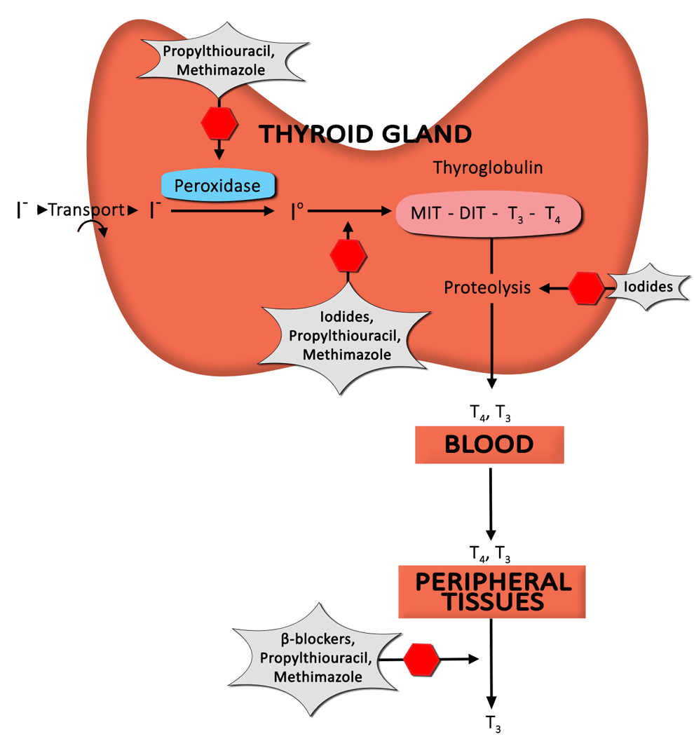
Corticosteroids are a mainstay of treatment in TED. The benefit derived from corticosteroid administration is due to anti-inflammatory and immunosuppressive effects. Unfortunately, a significant percentage of patients respond only partially (or not at all), and recurrences upon dose reduction or cessation are not infrequent [19].

  • Oral prednisone [19]
    • This is typically started in high doses (60-100 mg daily).
    • Most effective in treating soft tissue changes and optic neuropathy
    • Usually tapered slowly over a course of several months
    • Multiple studies show a mean effectiveness rate of ~ 60%.
    • Drawbacks include
      • High rate of ineffectiveness (~ 40%)
      • Need for high doses for an extended period of time
      • Frequent relapses upon dose reduction/therapy cessation
      • Multiple potential side effects, including but not limited to
      • Cushing's syndrome
      • Weight gain
      • Increased risk of infection
      • Exacerbation of diabetes mellitus, hypertension, and/or osteoporosis
  • Intravenous (IV) corticosteroids [15 ,25]
    • Compared to oral administration, IV is usually better tolerated and more effective (mean effectiveness ~ 70%).
    • Compared to months of treatment with oral prednisone, IV treatment usually lasts 12 weeks (6 weeks – 500 mg once weekly; 6 weeks – 250 mg once weekly), which allows for easy and early detection of "non-responders."
    • IV decreases the need for additional medical therapy.
    • Additional considerations
      • If prolonged high-dose treatment is anticipated, treat with calcium, vitamin D, and a proton pump inhibitor (for patients at high risk for gastric ulceration).
      • Incorporate frequent monitoring of serum electrolytes, blood glucose, liver function tests (LFTs), and blood pressure.
      • Patients receiving cumulative doses exceeding 8 g are at risk for hepatic toxicity, electrolyte disturbances, and cardiac arrhythmias

Orbital Radiotherapy

Orbital radiotherapy (ORT) has been used in the management of TED for nearly a century and can be used alone or in conjunction with corticosteroids [26 ,27].

  • Mechanisms of action [28]
    • Lymphocytes are temporarily sterilized.
    • Terminal differentiation in orbital fibroblasts is induced.
      • Adipocyte differentiation and fatty hypertrophy are limited.
      • This is more effective in patients < 40 years old who have more orbital fat hypertrophy.
    • Radiation induces death of tissue-bound monocytes.
      • Radiation blunts the immune response at the orbit, reducing inflammation of both the ocular surface and eyelids in ~ 60% of patients [10].
      • ORT has not been shown to have notable effects on proptosis or eyelid retraction.
  • ORT can be used to treat the following sequelae of TED
    • Compressive optic neuropathy
    • Ocular surface and periorbital tissue inflammation
    • Orbital congestion
    • Strabismus and ocular motility deficits
  • Treatment regimen and effects
    • Use 2000 cGy, administered over 10 treatment sessions, during a 2-week time course [26].
    • A maximum effect occurs around 6 months post-treatment [29].
    • Treatment is associated with a transient exacerbation of periorbital edema, conjunctival injection, and chemosis [29].
  • The role of ORT monotherapy has been controversial due to highly irregular results [26].
    • Effectiveness rates range from 20% - 90% in previously published studies.
    • The most convincing evidence for benefit is found in the treatment of restrictive vertical strabismus.
  • A recent Cochrane review concluded that the combination of corticosteroids + ORT has a more sustained anti-inflammatory effect, leading to a decreased risk of compressive optic neuropathy in active TED patients, when compared to treatment with either corticosteroids or ORT monotherapy [27].
  • Contraindications [29]
    • Patients with underlying microvascular retinopathy (e.g. diabetic or hypertensive retinopathy) have an increased incidence of radiation retinopathy (1-2%).
    • Patients may have increased risk of cataracts.
    • There is a theoretical concern for tumorigenesis, and thus, ORT is typically avoided in patients younger than 35 years.

Selenium [18]

  • When taken regularly for one year, selenium has been shown to exert significant benefits in patients with mild, non-inflammatory orbitopathy.
  • One study showed a benefit from selenium supplementation (100 µg twice daily) in Europe where the soil was selenium deficient.
  • The benefit of selenium supplementation from non-selenium-deficient populations is not known.

Emerging Therapies

  • Azathioprine [30]
    • This is a chemotherapeutic agent that inhibits DNA synthesis.
    • It is currently being studied in a Combined Immunosuppression and Radiotherapy in TED (CIRTED) trial [31].
    • Sporadic case reports have shown a benefit with use of the anti-TNF alpha biologics infliximab and etanercept.
  • Rituximab [32]
    • This targets CD-20 on B-cells, which leads to B-cell depletion in the thyroid gland and decreased TSI production.
    • A recent study showed TED patients with CAS ≥ 4 that received rituximab (1000 mg, IV, twice over a two week interval) showed an average decrease in CAS that ranged from 2.3 to 4.7, with a median decrease ranging from 2.5 to 4.5.
    • The overall efficacy of rituximab therapy is complicated by the variable presentations and course of TED.
  • Teprotumumab
    • This is a human monoclonal antibody (Graves' Disease IgG – GD-IgG) against the IGF-1-R recently investigated (phase II clinical trial) in patients with active, moderate to severe TED.
    • IGF-1-R has mitogenic and anti-apoptotic functions.
    • GD-IgG interacts directly with IGF-1-R.
    • IGF-1-R is upregulated in TED.
    • Anti-IGF-1-R therapy may interfere with this abnormal signaling pathway present in TED.

Treatment of Emergent Conditions

Optic Neuropathy

Overview (Figure 24)

Compressive optic neuropathy
Figure 24: Compressive optic neuropathy. This sequence shows a series of CT scans from patients with compressive optic neuropathy, with their associated visual fields.
  • Optic neuropathy occurs in about 6% of TED patients.
    • Mechanism
      • Inflammation and congestion at the orbital apex are severe enough that the optic nerve and its blood supply become compressed in the confines of the bony orbit.
      • Progressive expansion and congestion of the orbital tissues can lead to further stretching of the optic nerve.
  • Presentation
    • In most cases, vision loss is insidious, progressive, and typically bilateral (usually asymmetric).
    • Dyschromotopsia is an early sign of optic nerve dysfunction.
    • Common visual field deficits are loss of vision or central or diffuse visual field depression.
    • The appearance of the optic disc is often unremarkable but can be edematous or atrophic.
    • If disease is asymmetric, an RAPD may be present.

Treatment Modalities

  • Corticosteroid therapy (Oral or IV)
    • This is considered first-line therapy for compressive optic neuropathy [33]
      • This may be primary treatment, or a temporizing measure, until surgical decompression can be performed.
      • In non-emergent cases, steroid treatment (100 mg oral, tapering by 10 mg/week, for a total of 12 weeks) has been shown to be as effective as orbital decompression [15].
      • IV methylprednisolone can be used in management of both emergent and non-emergent cases of optic neuropathy.
        • High-dose IV methylprednisolone (830 mg weekly for 6 weeks, followed by 415 mg weekly for 6 weeks) in pulsatile administration is more efficacious than oral corticosteroids [25].
        • When cumulative doses exceed 8 g, measure liver enzymes and test liver function, as fatal hepatotoxicity has been reported [25].
  • Orbital radiotherapy (ORT)
    • This may be an alternative treatment to avoid extensive side effects associated with corticosteroid use [28].
    • Numerous studies have demonstrated the effectiveness of ORT [27].
      • Over a 2 week time course, 2000 cGy are administered over 10 treatments.
      • This reduces extraocular muscle width near the orbital apex and may avoid need for urgent surgical decompression.
    • There is concern for an initial inflammatory response with ORT, which can worsen compressive optic neuropathy [34].
    • Fat may become more fibrotic after radiation, making decompression less effective post-radiation.
  • Orbital Decompression
    • See videos
    • If vision loss is rapid and progressive, surgical orbital decompression may be required to relieve pressure on the optic nerve and its blood supply – typically after a failed trial of corticosteroids.
    • This invasive procedure involves removal of bone and sometimes adipose tissue to provide access to space outside the orbit, which allows excess tissue to decompress from the confined orbit.
    • Lateral wall decompression
      • Partial- or full-thickness inner aspects of the zygomatic bone and greater wing of the sphenoid bone are removed, which is typically performed through a canthotomy with inferior cantholysis.
      • Potential complications include cerebrospinal fluid leak secondary to dural tear, pulsatile exophthalmos, and oscillopsia [35].
    • Medial wall decompression
      • Segments of the ethmoid bone are removed allowing intraorbital contents to expand into the ethmoid air cells.
        • The lateral wall of the sphenoid bone may be removed as well.
        • The approach may be external or endoscopic.
        • When an external incision is made, it is typically transcaruncular and/or an extension of the orbital floor decompression incision, when applicable.
      • Potential complications
        • Diplopia secondary to alteration of globe position may occur.
        • Medial rectus prolapse into the ethmoid sinus can cause an abduction deficit.
        • Compression of the lesser wing of the sphenoid can cause internal carotid artery laceration or optic nerve damage.
    • Orbital floor decompression
      • This allows for expansion of orbital contents into the maxillary sinus.
      • Decompression may be performed just medial to the infraorbital nerve (largest area with the most benefit in decompressing the optic nerve) or may be both medial and lateral to the infraorbital nerve.
      • The optic strut is often left intact to minimize globe displacement.
      • Caution must be taken to avoid the infraorbital neurovascular bundle, which traverses the infraorbital canal.
      • Decompression is typically performed through a transconjunctival incision with a lateral canthotomy and inferior cantholysis, but a subciliary approach is used as well.
      • Potential complications include diplopia secondary to globe ptosis, supraduction deficit from inferior rectus prolapse, and CN V2 - distribution hypoesthesia.
    • Orbital fat decompression
      • The orbital fat may be removed alone or in conjunction with bony decomposition.
      • Orbital fat decompression has been shown to be especially beneficial in patients who tend to have more orbital fat hypertrophy than EOM involvement, which is more common in patients < 40 years old.
    • Pre-operative planning with computed tomography (CT) [36]
      • This may help confirm the diagnosis of TED.
      • CT allows for evaluation of the anatomy of the sinuses, cribiform plate, and lateral wall of the orbit.
      • CT also helps assess the potential benefit of fat and bone removal.
    • Considerations with orbital decompression surgery
      • Decompression may be of any wall alone or in combination with other walls.
      • The medial wall and orbital floor decompressions are of the most benefit in compressive optic neuropathy but have a slightly higher rate of diplopia post-operatively.
      • The decompression is called "balanced" when the medial wall and lateral wall are included.
      • Studies have shown that the balanced decompression has lower rates of diplopia as compared to decompressions involving the floor.

Globe Subluxation

  • Initial management with digital repositioning [37]
    • If digital repositioning fails, a Desmarres retractor (or if not available, a large-sized paperclip, bent to form a right angle (similar to a laryngoscope)) can be used to navigate between the upper lid margin and superior rectus to allow for proper repositioning.
    • Lateral tarsorrhaphy or orbital decompression surgery may be necessary, if initial managements fail.

Corneal Exposure

  • Patients at high risk for corneal exposure include those with a combination of proptosis, eyelid retraction, lagophthalmos, neurotrophic cornea, and poor Bell's reflex.
  • Pathogenesis is centered on prolonged exposure of corneal surface, leading to corneal drying, which decreases vision and threatens barrier to infection.
  • Treatment of non-emergent cases includes
    • Environmental modifications
    • Ocular surface lubrication
    • Increasing tear production via immunosuppression (cyclosporine, loteprednol, flurometholone)
    • Decreasing tear evaporation
      • Increasing oil content of tears
        • Omega-3 fatty acids
        • Warm compresses
      • Decreasing surface area for exposure and evaporation with eyelid surgery
        • Lid retraction repair
        • Orbital decompression
        • Lid tarsorrhaphy
    • Decreasing tear outflow (punctal plugs or cautery)
  • Treat emergent cases when corneal integrity is threatened.
    • The cause of the exposure must be corrected, and more aggressive therapy is pursued to provide protection and moisture in the meantime.
    • A bandage contact lens (BCL) or scleral contact lens with concomitant topical antibiotic will protect the cornea from further drying, while also preventing infection.
    • A temporary tarsorrhaphy may be indicated while pursuing other treatment modalities to decrease factors that exacerbate exposure, including
      • Chemosis
      • Proptosis
      • Eyelid retraction
      • Ocular surface inflammation

Treatment of Non-Emergent Conditions

Proptosis (Figure 25)

  • Orbital decompression increases the volume of the bony orbit through removal of orbital bone and adipose tissue and allows a proptotic globe to recess back into its normal confines.
    • It is considered first-line therapy for cases of severe optic nerve compression.
    • Other indications include
      • Globe subluxation
      • Exposure keratopathy
      • Restoration of pre-morbid appearance in patients with residual proptosis following the active stage of TED
    • Because decompression surgery can alter globe positioning, decrease eyelid retraction, and affect extraocular motility, it should precede any extraocular muscle or eyelid surgery.
    • Orbital decompression can reduce proptosis and eyelid retraction [38].
    • See the "Emergent Treatment" section above for more information on orbital decompression.
 Exophthalmos.
Figure 25: Exophthalmos. The globe is displaced anteriorly out of the orbit. This is an ophthalmologic emergency – the cornea is at risk for exposure, and the optic nerve is at risk of irreversible damage.

Strabismus

  • TED affects extraocular muscles in a predictable manner [39].
    • The inferior rectus and medial rectus are most commonly involved.
    • This presents as hypotropia and/or esotropia.
  • Most TED patients with diplopia due to strabismus will not require surgical intervention, as most can be effectively managed with prism spectacles [39].
  • Indications for strabismus surgery [39]
    • Patients may have intractable diplopia in primary gaze or with reading.
    • Abnormal head positioning may be present.
    • The position of the globe may be cosmetically unacceptable.
  • Surgical approach [39]
    • Delay strabismus surgery until disease stability has been demonstrated.
    • Recession of the affected muscles is the most commonly used surgical method.
    • Adjustable sutures are helpful in especially difficult cases.
    • Although diplopia is frequently improved post-operatively, normal ocular motility is infrequently achieved for the following reason
    • With recession of the inferior rectus muscle, infraduction deficits may result, making the use of bifocals challenging.
      • Restrictive nature of myopathy
      • Large muscle recessions
      • Ongoing chronic disease
  • Extraocular muscle recession can worsen proptosis [40].
    • If orbital decompression is foreseeable, it should be performed prior to strabismus surgery, as orbital decompression can also alter strabismus.
  • Extraocular muscle recession can effect eyelid position [40].
  • Large inferior rectus muscle recession can result in lower eyelid retraction, which is largely due to adherence between the inferior rectus muscle and the capsulopalpebral fascia of the lower eyelid.
    • With superior rectus recession, connection points between the superior rectus and upper eyelid elevators may worsen upper eyelid retraction.
    • Strabismus surgery should be undertaken prior to any corrective eyelid procedures.
  • Botulinum toxin [41]
    • Poor surgical candidates that cannot be treated with prisms may benefit.
    • Restricted extraocular muscles are injected, which temporarily relieves restrictive strabismus and may have some lasting effects.
    • Consider alternative treatment if there is need for recurrent injections, difficulty in precisely delivering the agent within the orbit, or variability of effect on fibrotic extraocular muscles.

Eyelid Retraction

  • The etiology is multifactorial and may include (Figure 26)
    • Increased sympathetic tone stimulating Müller's muscle
    • Contraction and/or fibrosis of the levator palpebrae superioris in the upper lid and lower lid retractors in the lower lid
    • Proptosis
    • Scarring between the lacrimal gland and the levator palpebrae superioris
  • Treatment
    • Non-surgical
      • Injection of hyaluronic acid gel fillers
      • Botulinum toxin to levator palpebrae superioris and/or Müller's muscle [41]
        • A temporary measure injected into the levator palpebrae superioris to lower the upper eyelid position
        • Good for poor surgical candidates, patients in the active phase, or patients awaiting stability
    • Surgical
      • Implantation of an eyelid weight into the upper lid (e.g. gold or titanium)
      • Incision and/or recession of one or more of the eyelid retractors
        • Levator palpebrae superioris
        • Müller's muscle
        • Capsulopalpebral fascia
        • Inferior tarsal muscle
      • Full-thickness blepharotomy
      • Insertion of "spacer" material to lengthen the eyelid
        • Ear cartilage
        • Hard palate
        • Sclera
        • Synthetic material
        • Porcine
    • Eyelid contouring [40]
      • Aimed at restoring the natural appearance of the eyelid , while minimizing temporal flare
      • Upper eyelid peak: medial edge of the pupil
      • Lower eyelid trough: lateral limbus
 Eyelid retraction.
Figure 26: Eyelid retraction. Eyelid retraction is the most common presenting sign of TED, and is the result of many factors associated with TED.

References

  1. Douglas RS, Gupta S. The pathophysiology of thyroid eye disease: implications for immunotherapy. Curr Opin Ophthalmol 2011;22(5):385-390. [PMID 21730841]
  2. Bartley GB, Fatourechi V, Kadrmas EF, Jacobsen SJ, Ilstrup DM, Garrity JA, Gorman CA. Clinical features of Graves' ophthalmopathy in an incidence cohort. Am J Ophthalmol 1996;121(3):284-290. [PMID 8597271]
  3. Krassas GE, Wiersinga W. Smoking and autoimmune thyroid disease: the plot thickens. Eur J Endocrinol 2006;154(6):777-780. [PMID 16728534]
  4. Kazim M, Goldberg RA, Smith TJ. Insights into the pathogenesis of thyroid-associated orbitopathy: evolving rationale for therapy. Arch Ophthalmol 2002;120(3):380-386. [PMID 11879144]
  5. Smith TJ, Koumas L, Gagnon A, Bell A, Sempowski GD, Phipps RP, Sorisky A. Orbital fibroblast heterogeneity may determine the clinical presentation of thyroid-associated ophthalmopathy. J Clin Endocrinol Metab 2002;87(1):385-392. [PMID 11788681]
  6. Tsui S, Naik V, Hoa N, Hwang CJ, Afifiyan NF, Sinha Hikim A, Gianoukakis AG, Douglas RS, Smith TJ. Evidence for an association between thyroid-stimulating hormone and insulin-like growth factor 1 receptors: a tale of two antigens implicated in Graves' disease. J Immunol 2008;181(6):4397-4405. [PMID 18768899]
  7. Naik V, Khadavi N, Naik MN, Hwang C, Goldberg RA, Tsirbas A, Smith TJ, Douglas RS. Biologic therapeutics in thyroid-associated ophthalmopathy: translating disease mechanism into therapy. Thyroid 2008;18(9):967-971. [PMID 18713027]
  8. Ponto KA, Zang S, Kahaly GJ. The tale of radioiodine and Graves' orbitopathy. Thyroid 2010;20(7):785-793. [PMID 20578895]
  9. Holds JB, Buchanan AG. Graves orbitopathy. Focal Points: Clinical Modules for Ophthalmologists. Module 11. San Francisco: American Academy of Ophthalmology; 2010.
  10. Bartalena L, Marcocci C, Bogazzi F, Manetti L, Tanda ML, Dell'Unto E, Bruno-Bossio G, Nardi M, Bartolomei MP, Lepri A, Rossi G, Martino E, Pinchera A. Relation between therapy for hyperthyroidism and the course of Graves' ophthalmopathy. N Engl J Med 1998;338(2):73-78. [PMID 9420337]
  11. Talke LM, Murchison AP. Globe Subluxation: Review and Management. Review of Ophthalmology 2007;14 (https://www.reviewofophthalmology.com/article/globe-subluxation-review-and-management)
  12. Dolman PJ, Cahill K, Czyz CN, Douglas RS, Elner VM, Feldon S, Kazim M, Lucarelli M, Sivak-Collcott J, Stacey AW, Strianese D, Uddin J. Reliability of estimating ductions in thyroid eye disease: an International Thyroid Eye Disease Society multicenter study. Ophthalmology 2012;119(2):382-389. [PMID 21959369]
  13. Mourits MP, Prummel MF, Wiersinga WM, Koornneef L. Clinical activity score as a guide in the management of patients with Graves' ophthalmopathy. Clin Endocrinol (Oxf) 1997;47(1):9-14. [PMID 9302365]
  14. Gerding MN, van der Meer JW, Broenink M, Bakker O, Wiersinga WM, Prummel MF. Association of thyrotrophin receptor antibodies with the clinical features of Graves' ophthalmopathy. Clin Endocrinol (Oxf) 2000;52(3):267-271. [PMID 10718823]
  15. Bartalena L, Baldeschi L, Dickinson A, Eckstein A, Kendall-Taylor P, Marcocci C, Mourits M, Perros P, Boboridis K, Boschi A, Curro N, Daumerie C, Kahaly GJ, Krassas GE, Lane CM, Lazarus JH, Marino M, Nardi M, Neoh C, Orgiazzi J, Pearce S, Pinchera A, Pitz S, Salvi M, Sivelli P, Stahl M, von Arx G, Wiersinga WM, European Group on Graves O. Consensus statement of the European Group on Graves' orbitopathy (EUGOGO) on management of GO. Eur J Endocrinol 2008;158(3):273-285. [PMID 18299459]
  16. Stan MN, Garrity JA, Bahn RS. The evaluation and treatment of graves ophthalmopathy. Med Clin North Am 2012;96(2):311-328. [PMID 22443978]
  17. Cawood T, Moriarty P, O'Shea D. Recent developments in thyroid eye disease. BMJ 2004;329(7462):385-390. [PMID 15310608]
  18. Marcocci C, Kahaly GJ, Krassas GE, Bartalena L, Prummel M, Stahl M, Altea MA, Nardi M, Pitz S, Boboridis K, Sivelli P, von Arx G, Mourits MP, Baldeschi L, Bencivelli W, Wiersinga W, European Group on Graves O. Selenium and the course of mild Graves' orbitopathy. N Engl J Med 2011;364(20):1920-1931. [PMID 21591944]
  19. Bartalena L, Pinchera A, Marcocci C. Management of Graves' ophthalmopathy: reality and perspectives. Endocr Rev 2000;21(2):168-199. [PMID 10782363]
  20. Prummel MF, et al. Amelioration of eye changes of Graves' ophthalmolopathy by achieving euthyroidism. Acta Endocrinology (Copenhagen) 1989;121 (suppl 2):185-189
  21. Kronenberg HM, Melmed S, Polonsky KS, Larsen PR, editors. Williams Textbook of Endocrinology. 11th ed. Philadelphia: Elsevier/Saunders, 2008.
  22. Bahn RS, Burch HB, Cooper DS, Garber JR, Greenlee MC, Klein I, Laurberg P, McDougall IR, Montori VM, Rivkees SA, Ross DS, Sosa JA, Stan MN, American Thyroid Association, American Association of Clinical Endocrinologists. Hyperthyroidism and other causes of thyrotoxicosis: management guidelines of the American Thyroid Association and American Association of Clinical Endocrinologists. Thyroid 2011;21(6):593-646. [PMID 21510801]
  23. Stan MN, Durski JM, Brito JP, Bhagra S, Thapa P, Bahn RS. Cohort study on radioactive iodine-induced hypothyroidism: implications for Graves' ophthalmopathy and optimal timing for thyroid hormone assessment. Thyroid 2013;23(5):620-625. [PMID 23205939]
  24. Eckstein A, Quadbeck B, Mueller G, Rettenmeier AW, Hoermann R, Mann K, Steuhl P, Esser J. Impact of smoking on the response to treatment of thyroid associated ophthalmopathy. Br J Ophthalmol 2003;87(6):773-776. [PMID 12770979]
  25. Vannucchi G, Covelli D, Campi I, Origo D, Curro N, Cirello V, Dazzi D, Beck-Peccoz P, Salvi M. The therapeutic outcome to intravenous steroid therapy for active Graves' orbitopathy is influenced by the time of response but not polymorphisms of the glucocorticoid receptor. Eur J Endocrinol 2014;170(1):55-61. [PMID 24128430]
  26. Rajendram R, Bunce C, Lee RW, Morley AM. Orbital radiotherapy for adult thyroid eye disease. Cochrane Database Syst Rev 2012;10.1002/14651858.CD007114.pub2(7):CD007114. [PMID 22786503]
  27. Shams PN, Ma R, Pickles T, Rootman J, Dolman PJ. Reduced risk of compressive optic neuropathy using orbital radiotherapy in patients with active thyroid eye disease. Am J Ophthalmol 2014;157(6):1299-1305. [PMID 24582992]
  28. Kazim M. Perspective--Part II: radiotherapy for Graves Orbitopathy: the Columbia University experience. Ophthal Plast Reconstr Surg 2002;18(3):173-174. [PMID 12021645]
  29. Wakelkamp IM, Tan H, Saeed P, Schlingemann RO, Verbraak FD, Blank LE, Prummel MF, Wiersinga WM. Orbital irradiation for Graves' ophthalmopathy: Is it safe? A long-term follow-up study. Ophthalmology 2004;111(8):1557-1562. [PMID 15288988]
  30. Stan MN, Garrity JA, Carranza Leon BG, Prabin T, Bradley EA, Bahn RS. Randomized controlled trial of rituximab in patients with Graves' orbitopathy. J Clin Endocrinol Metab 2015;100(2):432-441. [PMID 25343233]
  31. Rajendram R, Lee RW, Potts MJ, Rose GE, Jain R, Olver JM, Bremner F, Hurel S, Cook A, Gattamaneni R, Tomlinson M, Plowman N, Bunce C, Hollinghurst SP, Kingston L, Jackson S, Dick AD, Rumsey N, Morris OC, Dayan CM, Uddin JM. Protocol for the combined immunosuppression & radiotherapy in thyroid eye disease (CIRTED) trial: a multi-centre, double-masked, factorial randomised controlled trial. Trials 2008;9:6. [PMID 18237441]
  32. Silkiss RZ, Reier A, Coleman M, Lauer SA. Rituximab for thyroid eye disease. Ophthal Plast Reconstr Surg 2010;26(5):310-314. [PMID 20562667]
  33. Wakelkamp IM, Baldeschi L, Saeed P, Mourits MP, Prummel MF, Wiersinga WM. Surgical or medical decompression as a first-line treatment of optic neuropathy in Graves' ophthalmopathy? A randomized controlled trial. Clin Endocrinol (Oxf) 2005;63(3):323-328. [PMID 16117821]
  34. Prummel MF, Mourits MP, Blank L, Berghout A, Koornneef L, Wiersinga WM. Randomized double-blind trial of prednisone versus radiotherapy in Graves' ophthalmopathy. Lancet 1993;342(8877):949-954. [PMID 8105213]
  35. Fayers T, Barker LE, Verity DH, Rose GE. Oscillopsia after lateral wall orbital decompression. Ophthalmology 2013;120(9):1920-1923. [PMID 23618229]
  36. Trokel S, Kazim M, Moore S. Orbital fat removal. Decompression for Graves orbitopathy. Ophthalmology 1993;100(5):674-682. [PMID 8493010]
  37. Tse DT. A simple maneuver to reposit a subluxed globe. Arch Ophthalmol 2000;118(3):410-411. [PMID 10721966]
  38. Chang EL, Bernardino CR, Rubin PA. Normalization of upper eyelid height and contour after bony decompression in thyroid-related ophthalmopathy: a digital image analysis. Arch Ophthalmol 2004;122(12):1882-1885. [PMID 15596598]
  39. Thomas SM, Cruz OA. Comparison of two different surgical techniques for the treatment of strabismus in dysthyroid ophthalmopathy. J AAPOS 2007;11(3):258-261. [PMID 17572340]
  40. Nerad JA. Oculoplastic Surgery: The Requisites in Ophthalmology. St. Louis, MO: Mosby, 2001.
  41. Morgenstern KE, Evanchan J, Foster JA, Cahill KV, Burns JA, Holck DE, Perry JD, Wulc AE. Botulinum toxin type a for dysthyroid upper eyelid retraction. Ophthal Plast Reconstr Surg 2004;20(3):181-185. [PMID 15167723]

Suggested citation format

Liaboe, CA, Clark TJ, Carter KD, Shriver EM. Thyroid Eye Disease (TED): An Introductory Tutorial and Overview of Disease. EyeRounds.org. November 18, 2016; available from https://eyerounds.org/tutorials/thyroid-eye-disease/index.htm


Quiz


Select your current training level: (optional)

Question 1

What is NOT a typical symptom of IIH?

The correct answer is "No of the above, all are typical of IIH"

The symptoms most commonly reported by IIH patients followed by their frequency are:

  • headache (94%)
  • transient visual obscurations or blurring (68%)
  • pulse synchronous tinnitus or "wooshing noise" in the ear (58%)
  • pain behind the eye (44%)
  • double vision (38%)
  • visual loss (30%)
  • pain with eye movement (22%)
Question 2

Does a diagnosis of IIH require a high spinal fluid pressure (measured during a spinal tap)?

The correct answer is "True"

The diagnosis of IIH is made by identifying the typical symptoms of the disease along with documentation of a high spinal fluid pressure (measured during a spinal tap). The neurologic examination is normal except for the presence of swollen optic nerves called papilledema (seen by examining the back of the eye).

Question 3

Based on the diagnosis of IIH iwth mild visual loss, which treatment strategy would be most appropriate?

The correct answer is

  • Low Na+ diet
  • 10% weight loss
  • Acetazolamide

Answer: low sodium diet, wieght loss and acetazolamide