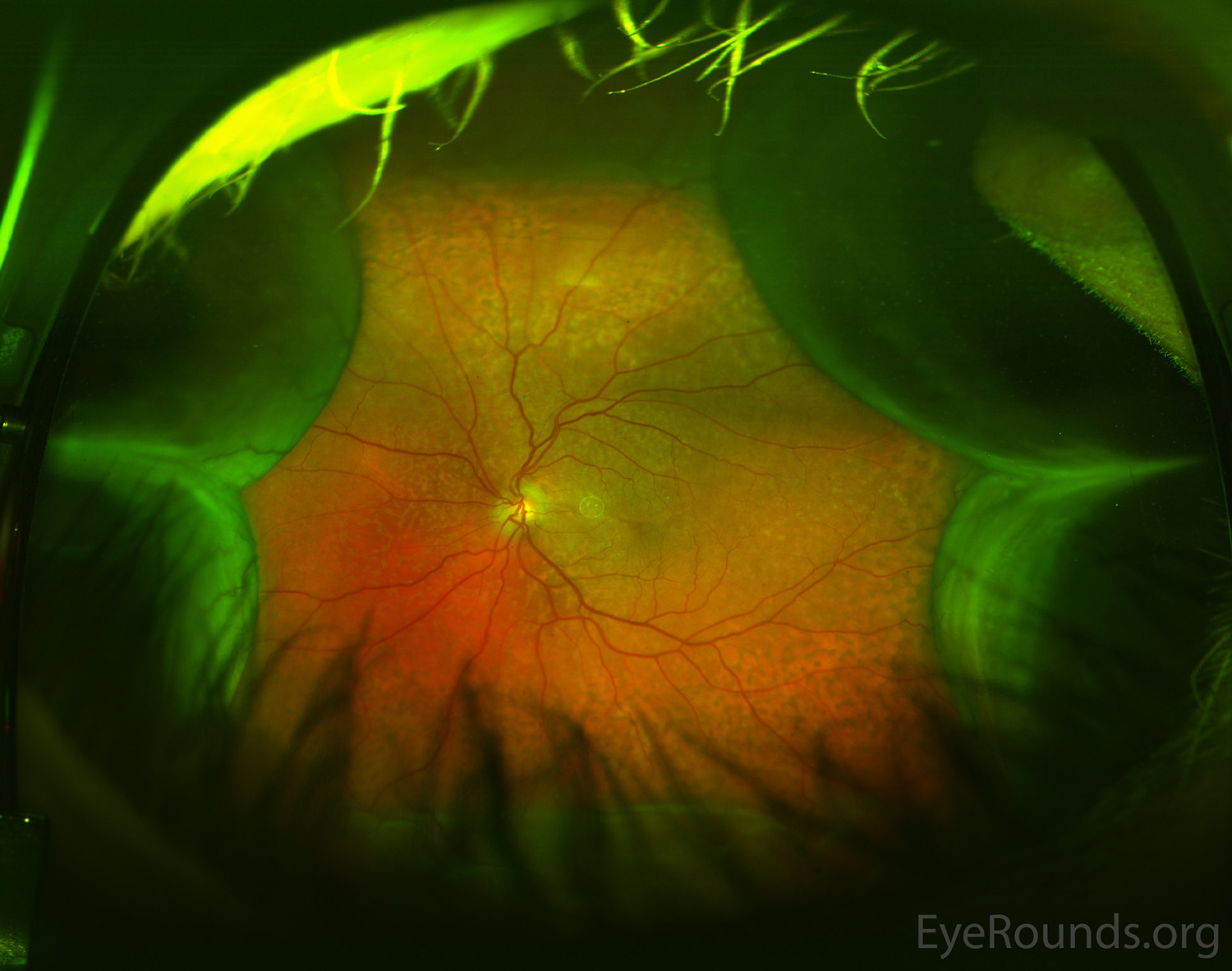
Uveal effusion syndrome is a rare entity involving the idiopathic collection of serous fluid in the suprachoroidal space. The condition is more common in middle-aged men. Serous retinal detachment and suprachoroidal collection of serous fluid may be seen and confirmed with transillumination of the globe or ultrasonography. Other examination findings may include leopard-spotted changes in the retinal pigment epithelium (indicating chronicity), normal intraocular pressure (as opposed to hypotonous choroidal effusion), vitreous cell, dilation of episcleral blood vessels, and blood in Schlemm's canal. Due to the idiopathic nature, the diagnosis is made after excluding other causes of effusion such as inflammation or infection, neoplasm, or trauma. Effective treatment often involves sclerotomy, quadrantic sclerectomy, and/or decompression of vortex veins. Medical management with steroids and surgical management with scleral bucking and pars plana vitrectomy are less effective in reducing the effusion and treating the retinal detachment. Visual decline and often severe vision loss usually occurs without treatment.

Ophthalmic Atlas Images by EyeRounds.org, The University of Iowa are licensed under a Creative Commons Attribution-NonCommercial-NoDerivs 3.0 Unported License.


Address
University of IowaLegal
Related Links