Terrien marginal degeneration is a painless, slowly progressive thinning of the peripheral corneal stroma. It is usually bilateral but may be asymmetric. As opposed to other causes of peripheral corneal thinning, there is typically no inflammation and the corneal epithelium remains intact. Characteristic features of this disorder include peripheral stromal thinning that typically begins superiorly with intact epithelium, overlying pannus, and a leading edge of lipid. The thinning classically has a steep central edge and a gradually sloping peripheral edge. The thinning may create against-the-rule astigmatism due to its relaxing effect on the corneal curvature and patients may experience corneal perforation with mild trauma.
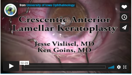
See the related video to view a crescentic lamellar keratoplasty for Terrien marginal degeneration.
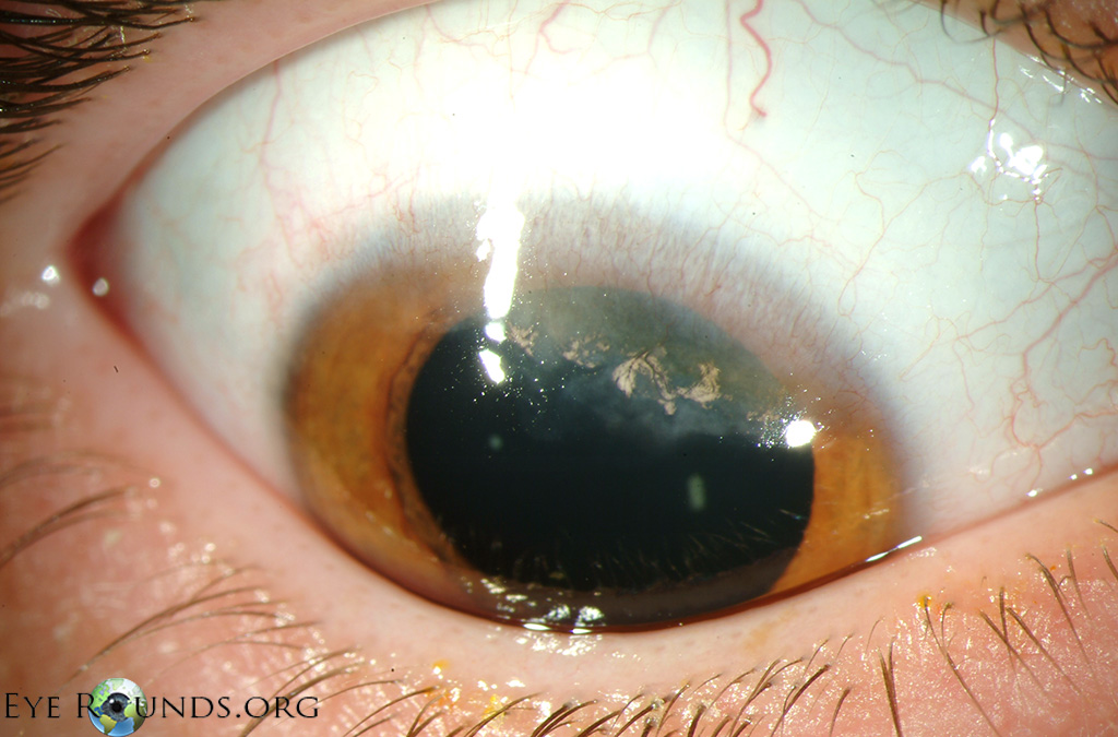
Patient with Terrien marginal degeneration displaying superior stromal thinning with intact epithelium, overlying pannus, and lipid at the leading edge.
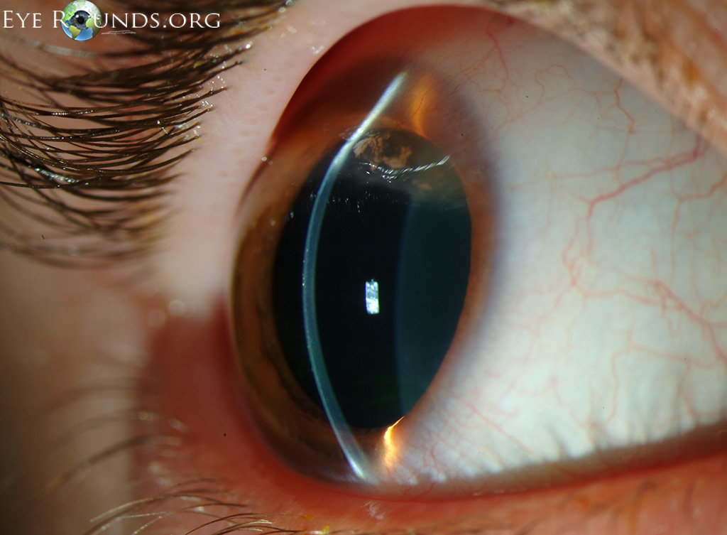
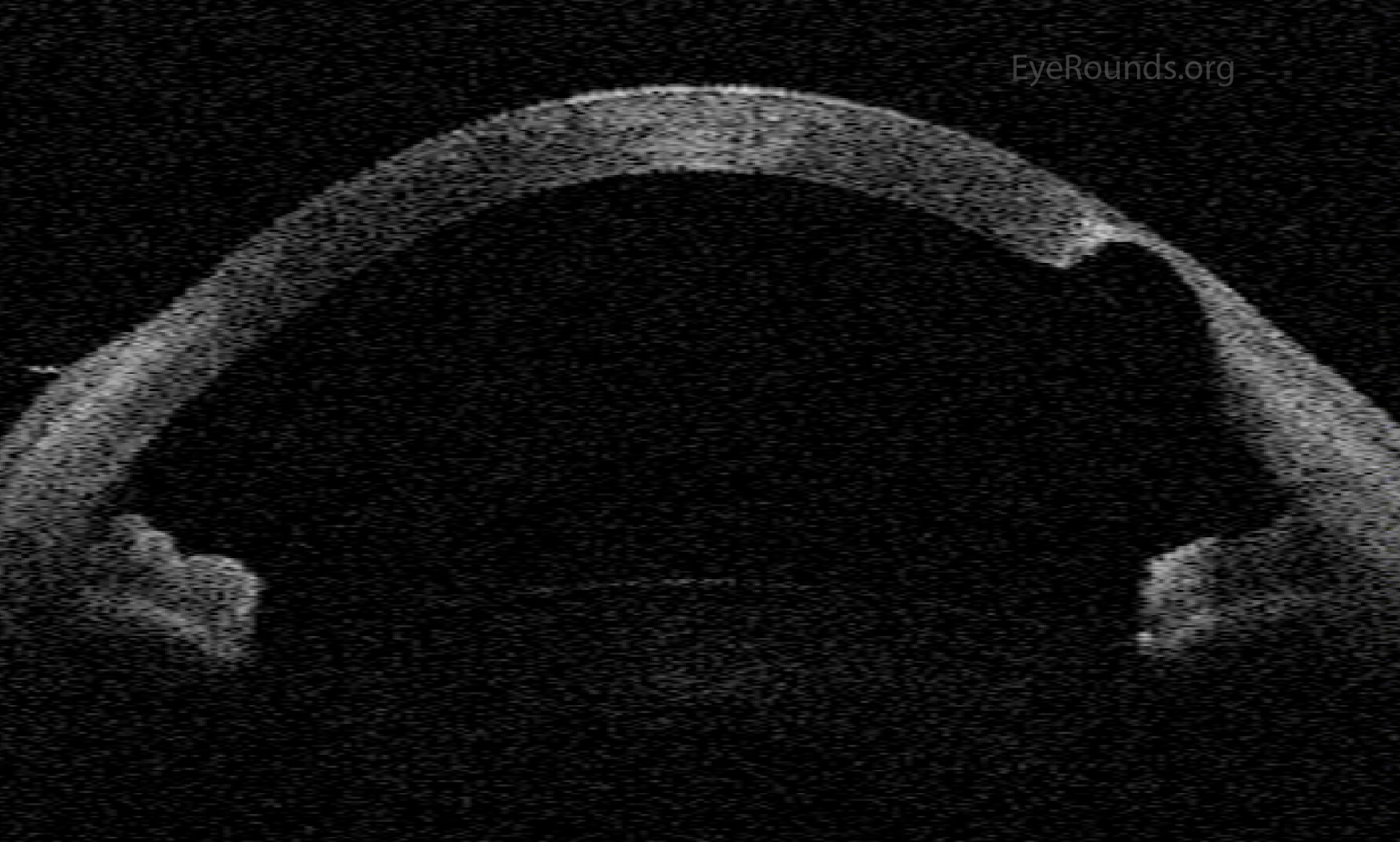
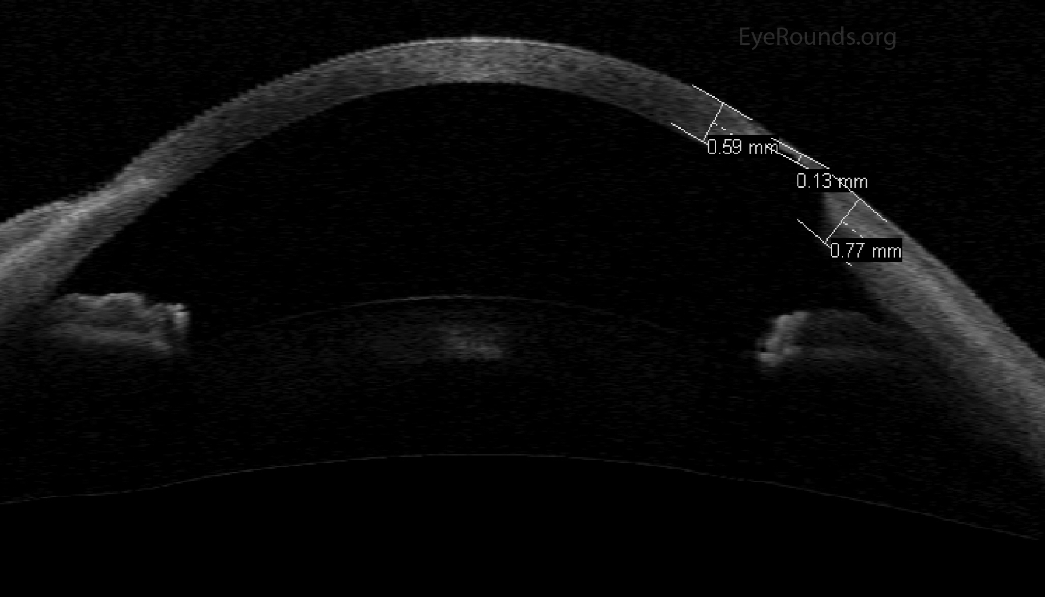
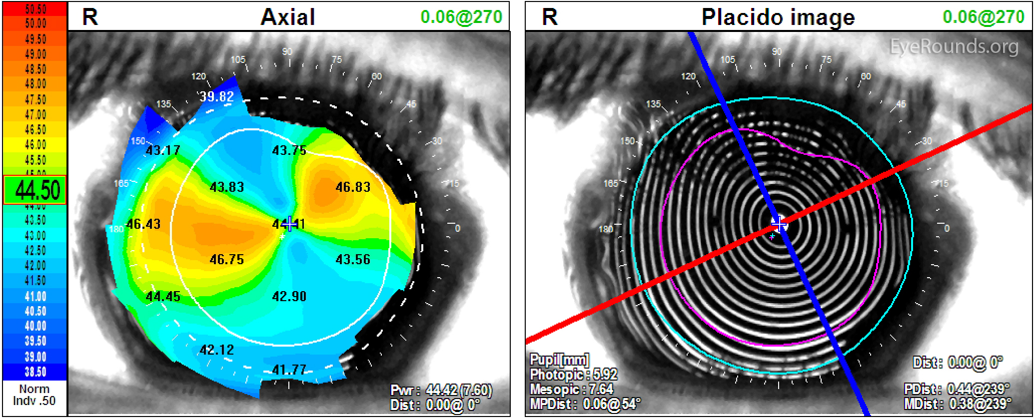
This patient with Terrien marginal degeneration has characteristic findings in the right eye. The severe superior stromal thinning is visible on anterior segment OCT and is creating a characteristic against-the-rule pattern of astigmatism. The patient was filtering aqueous through the area of superior thinning resulting in positive Seidel testing and creation of an adjacent conjunctival bleb. He underwent a crescentic lamellar keratoplasty to repair the defect (section below)
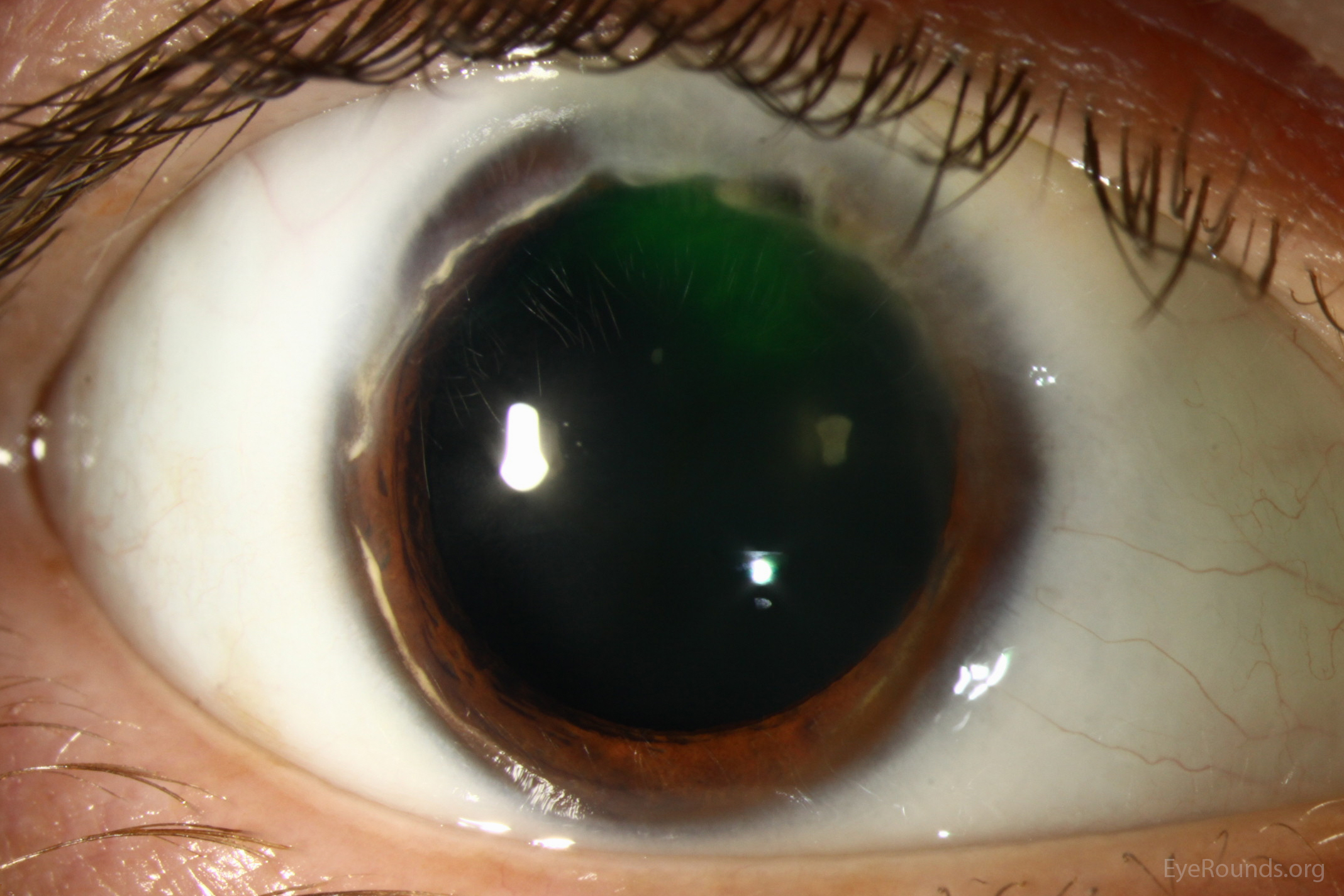
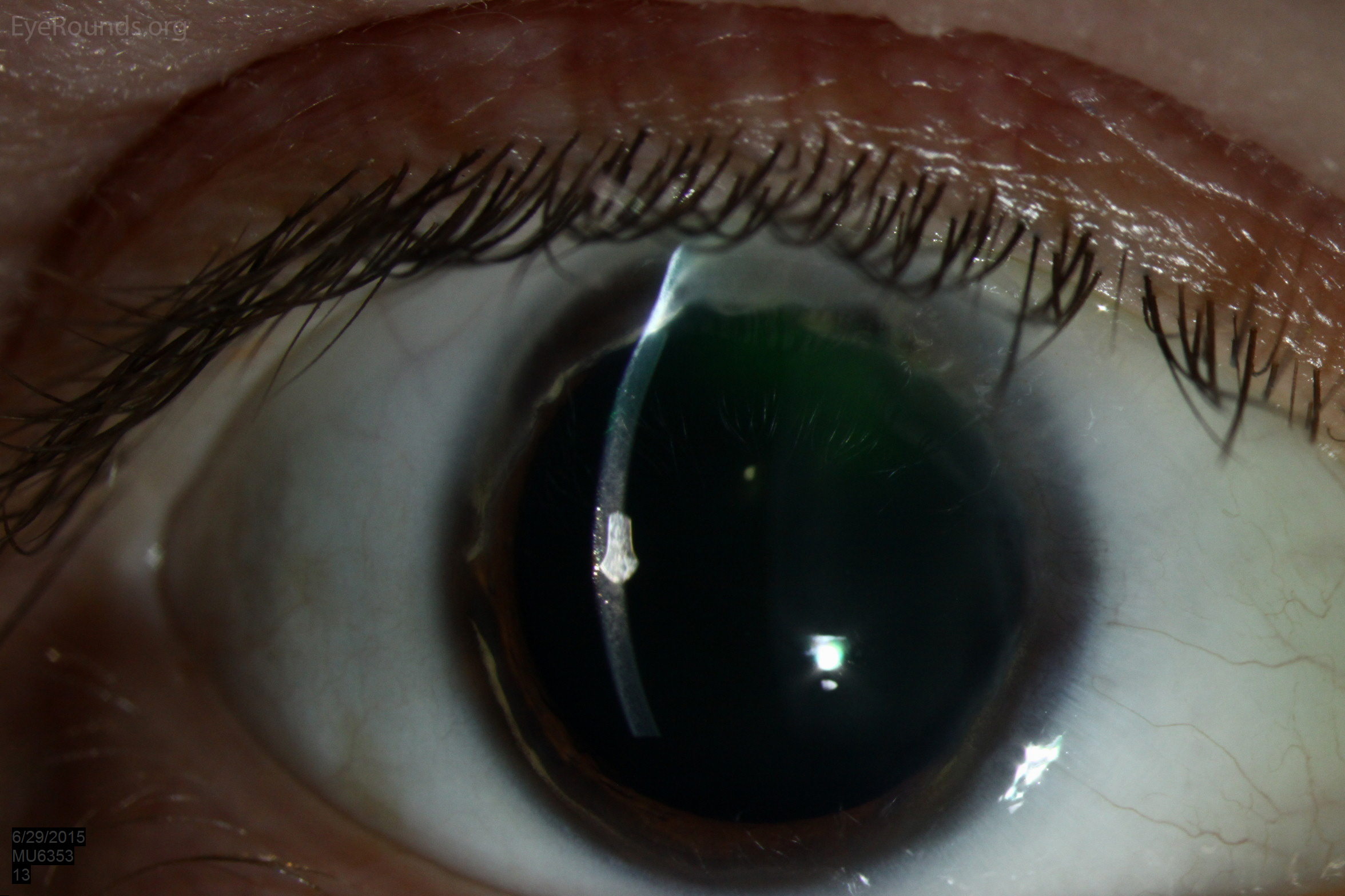
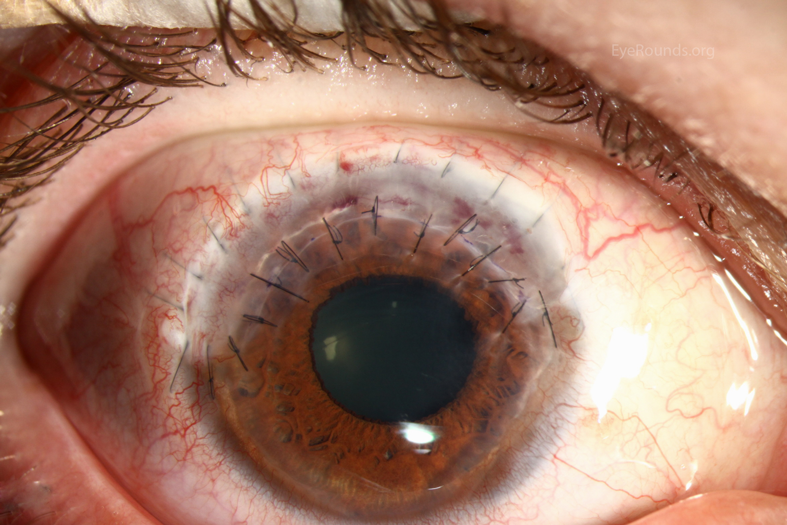
Post-operative appearance of the patient from previous images after undergoing crescentic lamellar keratoplasty at 1 week and 1 month after the procedure. See the related video to view the procedure.

Ophthalmic Atlas Images by EyeRounds.org, The University of Iowa are licensed under a Creative Commons Attribution-NonCommercial-NoDerivs 3.0 Unported License.

Address
University of IowaLegal
Related Links