This site uses tracking information. Visit our privacy policy. Click to agree to this policy and not see this again.

Ophthalmology and Visual Sciences

Retinitis pigmentosa (RP)

Retinitis pigmentosa (RP)

Category(ies): Inherited Eye Diseases and Retina / Vitreous
Contributor: Aaron M. Ricca MD
Photographer: Susan J. Wright

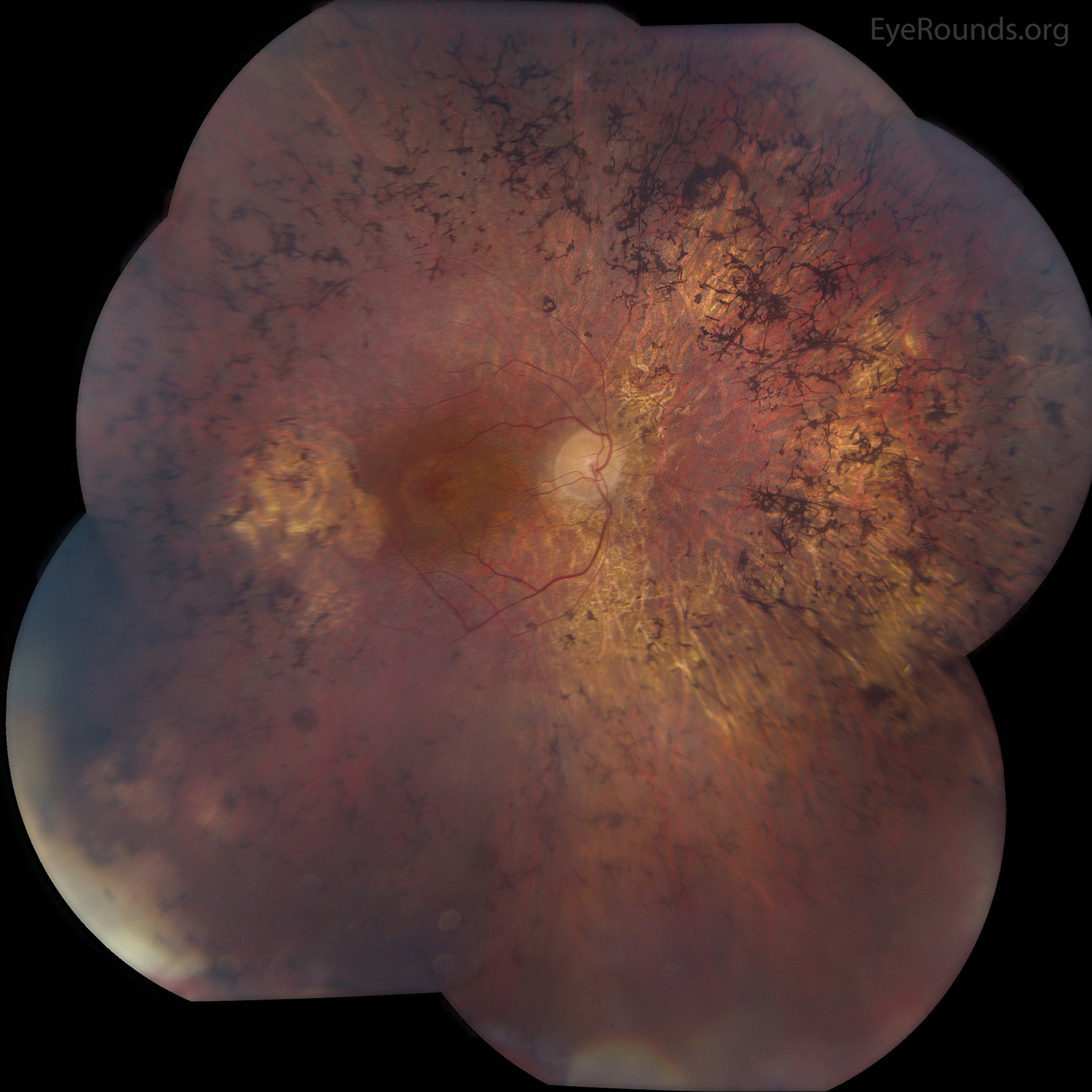
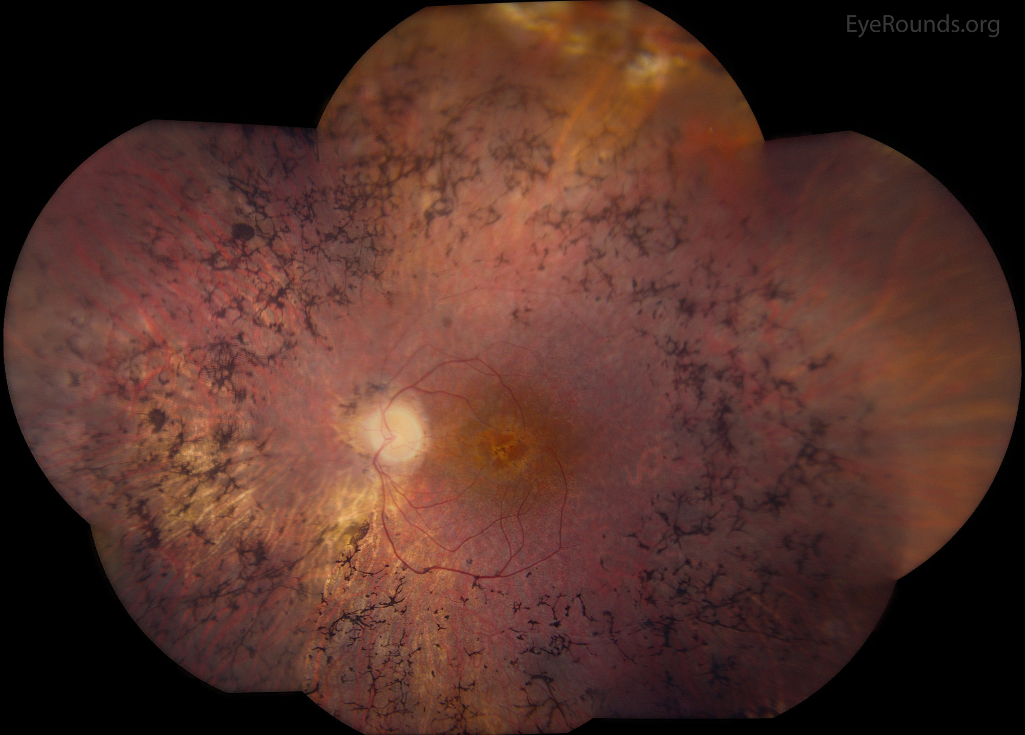
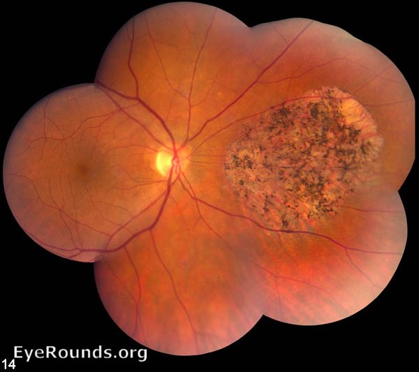
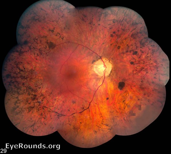
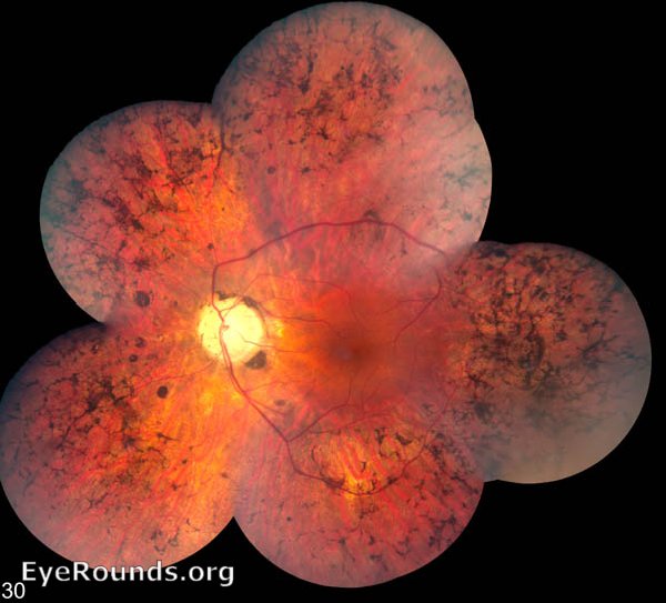
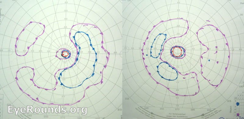
The montage color fundus photographs are from a 57-year-old male with molecularly confirmed retinitis pigmentosa (RP). He has a Pro347Ala change in the rhodopsin gene, which is consistent with autosomal dominant inheritance pattern. RP disease is rod-predominant photoreceptor degeneration, with late-stage atrophy of the cones and retinal pigment epithelium. It presents early in life with nyctalopia as a common first symptom. Visual field loss classically occurs in a ring-shaped scotoma with sparing of the central macula until late in the disease. Classic funduscopic findings include waxy-pallor of the optic disc, severe arteriolar attenuation, and bone-spicule-like pigmentation in the mid-peripheral retina. Routine dilated fundus examination and optical coherence tomography are important, as patients may develop cystoid macular edema, which may be respond to treatment with topical carbonic anhydrase inhibitors.


Category(ies): Retina
Contributor: Andrew Doan, MD, PhD
montage color fundus photograph of the right eye in a patient with autosomal recessive RP
1. This is a montage color fundus photograph of the right eye in a patient with autosomal recessive RP. The photograph depicts waxy disc pallor, arteriolar attenuation, and bone-spicule like pigmentation of the peripheral retina (distributed along the microvascular pattern of the retina).
montage color fundus photograph of the left eye in a patient with autosomal recessive RP
2. This is a montage color fundus photograph of the left eye in the same patient as above. Findings are similar to the right eye.
Goldmann visual perimetry  demonstrating bilateral ring-pattern scotomas
3. Goldmann visual perimetry of the right and left eye in the same patient as above demonstrating bilateral ring-pattern scotomas.
 montage color fundus photograph of the right eye in a female with x-linked RP in the carrier state
4. This is a montage color fundus photograph of the right eye in a female with x-linked RP in the carrier state. The photograph depicts unilateral, patchy pigmentation representing lyonization/x-inactivation. REFERENCE(S
References:
  1. Ryan SJ. Retina. 5th ed. London: Saunders/Elsevier; 2013.

Image Permissions:
Creative Commons

Ophthalmic Atlas Images by EyeRounds.org, The University of Iowa are licensed under a Creative Commons Attribution-NonCommercial-NoDerivs 3.0 Unported License.