
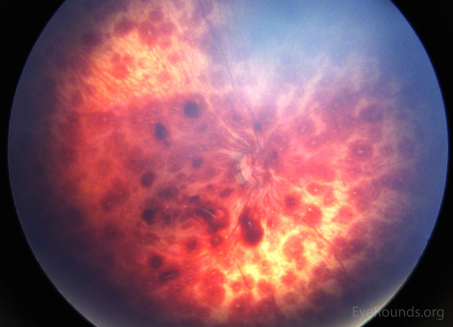
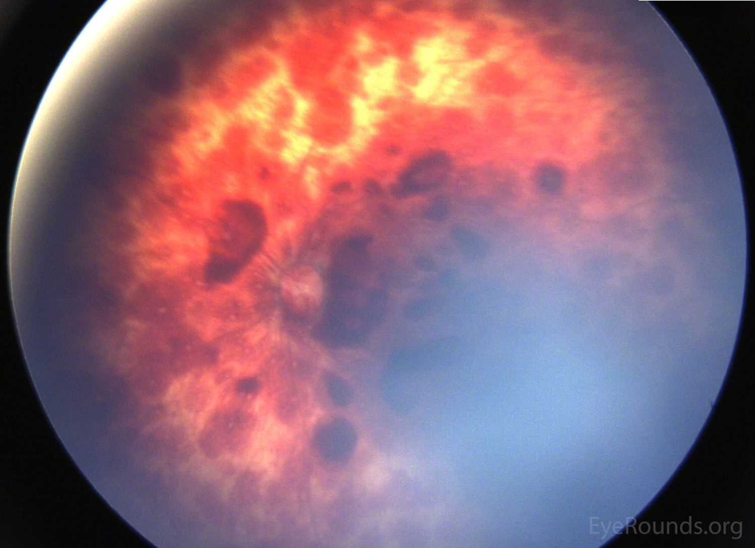
Retinal hemorrhages (RHs) are present throughout the macula and periphery of both eyes in this 4-month-old child. The differential diagnosis for retinal hemorrhages in an infant includes non-accidental trauma, sepsis, coagulopathy, leukemia, uncontrolled hypertension, cardiopulmonary resuscitation, and hyperviscosity syndromes, amongst others (1). It is prudent to rule out other causes by history, physical exam, and laboratory testing.
If intracranial pathology (e.g., subdural hematoma) is present without an identifiable etiology, one must suspect repetitive shaking or repetitive impact (resulting in repetitive rotational forces on the head) as the most likely cause of the RHs. Bilateral hemorrhages in multiple levels of the retina (i.e., pre, intra, and subretinal) and paramacular folds further raise the suspicion of child abuse. The number of hemorrhages and extent into the periphery (to the ora) also increase the likelihood that non-accidental trauma has occurred.
The primary mechanism of RHs in child abuse is mechanical with resultant shearing and damage to the retinal blood vessels, with or without direct blunt injury to the child. It is known that a correlation exists between the severity of ocular and intracranial injury and the presence of numerous RHs (2).
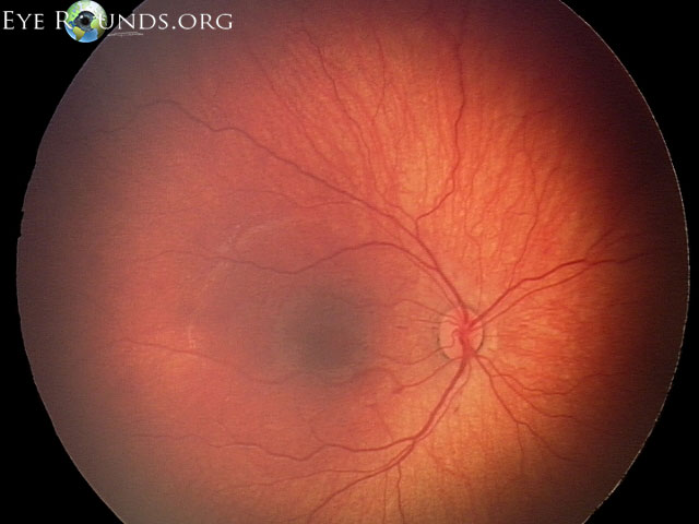
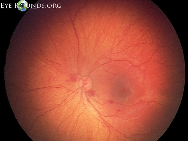
6 month-old girl found unresponsive and evaluated in the setting of a work-up of suspected non-accidental trauma. Fundus exam shows retinal nerve fiber layer hemorrhages OS > OD.

Ophthalmic Atlas Images by EyeRounds.org, The University of Iowa are licensed under a Creative Commons Attribution-NonCommercial-NoDerivs 3.0 Unported License.


Address
University of IowaLegal
Related Links
