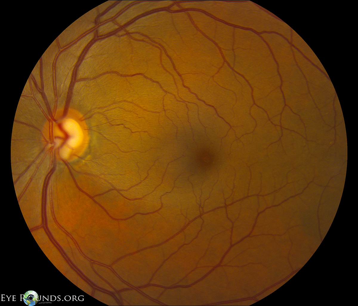
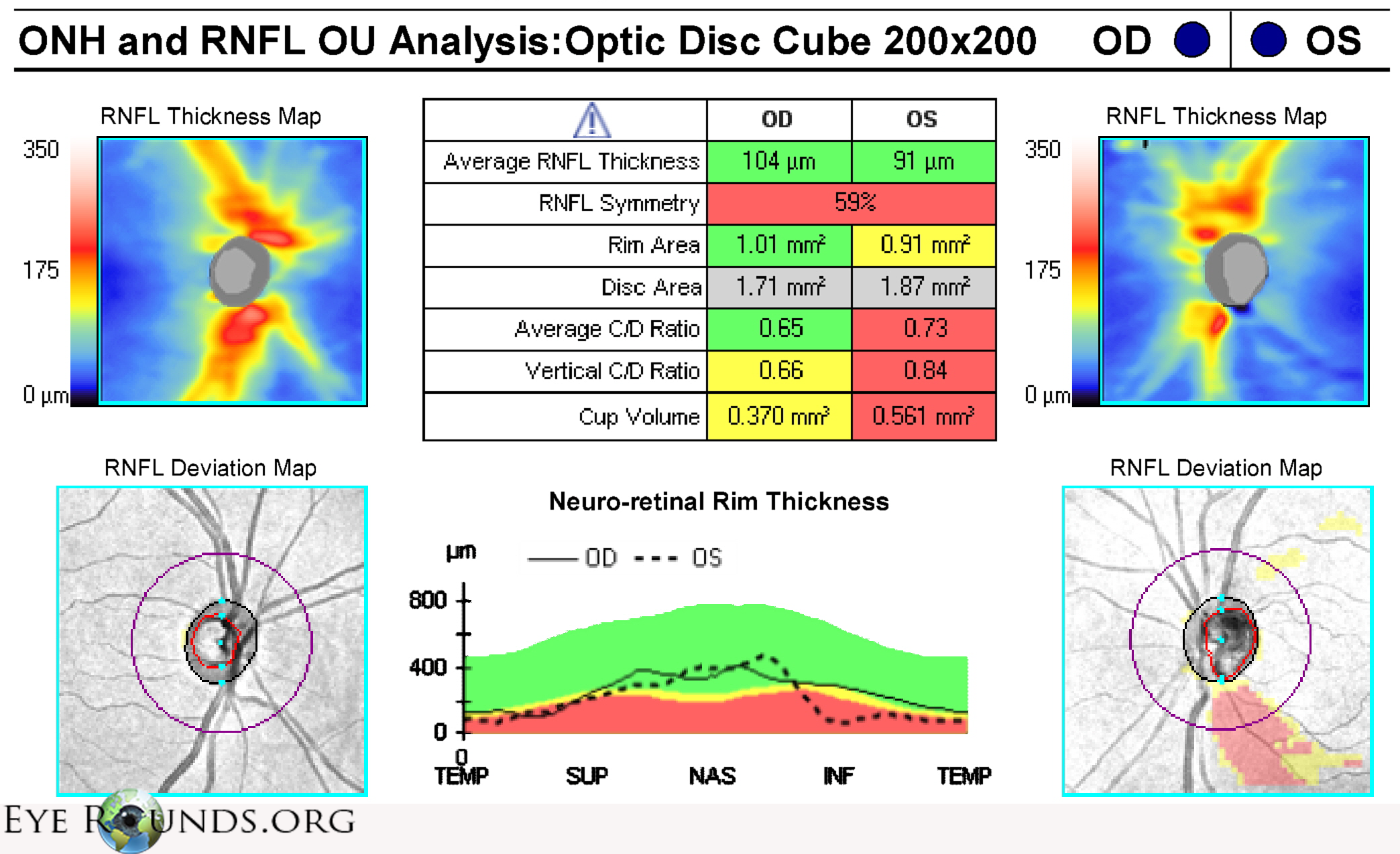
The optic nerve shows moderate cupping but there is a prominent inferior notch. The normal sheen of the nerve fiber layer is absent in a distribution radiating temporally from this notch due to cellular loss. The notch and nerve fiber layer defect can be more easily visualized on optical coherence tomography.
University of Iowa
Roy J. and Lucille A. Carver College of Medicine
Department of Ophthalmology and Visual Sciences
200 Hawkins Drive
Iowa City, IA 52242
University of Iowa
Roy J. and Lucille A. Carver College of Medicine
Department of Ophthalmology and Visual Sciences
200 Hawkins Drive
Iowa City, IA 52242