

EyeRounds Online Atlas of Ophthalmology
Contributor: William Charles Caccamise, Sr, MD, Retired Clinical Assistant Professor of Ophthalmology, University of Rochester School of Medicine and Dentistry
*Dr. Caccamise has very generously shared his images of patients taken while operating during the "eye season" in rural India as well as those from his private practice during the 1960's and 1970's. Many of his images are significant for their historical perspective and for techniques and conditions seen in settings in undeveloped areas.
Category: Cornea
Corneal macular scar with neovascularization of the superior cornea
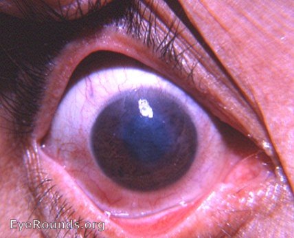
The corneal scar is too visible to be called a nebula. Therefore, it is a macula. Bloodvessels are growing into the cornea superiorly. This qualifies as neovascularization since the bloodvessels go beyond the normal limbal arcade. The term pannus does not apply to pure neovasdcularization. Pannus must include a granulation tissue invasion of the cornea - not just new bloodvessels but also young vascularized connective tissue, i.e. granulation tissue. The absence of trichiasis involving the upper lid suggests that the corneal scarring is the residual of a previous keratitis and not the result of trachoma.

Ophthalmic Atlas Images by EyeRounds.org, The University of Iowa are licensed under a Creative Commons Attribution-NonCommercial-NoDerivs 3.0 Unported License.