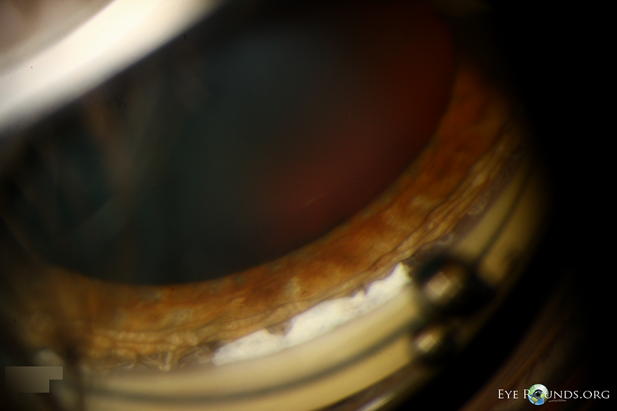
Traumatic angle recession on gonioscopic photos: This patient developed angle recession and a cyclodialysis cleft after racketball impact to the eye. Angle recession results from separation of the longitudinal and circular muscles of the ciliary body. Note the broad band of visible ciliary body face. He also had a cyclodialysis cleft, or separation of ciliary body from the scleral spur, which appears as a broad white band at the junction of the ciliary body and scleral spur.
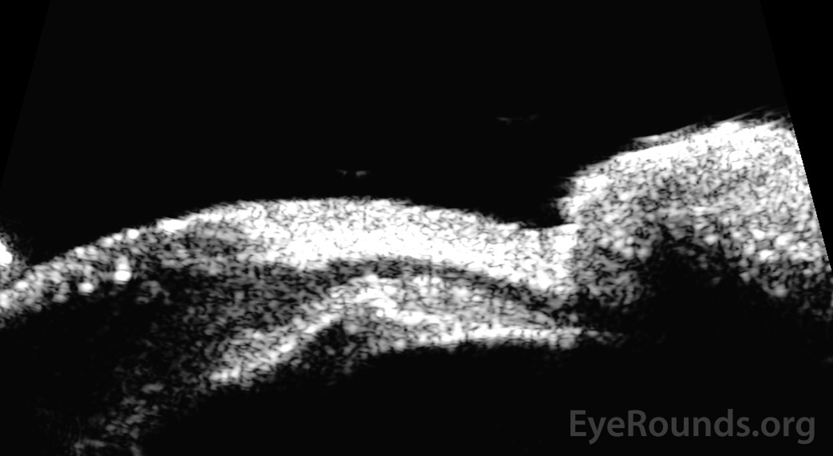
Traumatic cyclodialysis cleft on high frequency ultrasound biomicroscopy: This patient presented with hypotony after blunt trauma. Please note the separation of the ciliary body from the scleral spur, leading to a direct communication between the anterior chamber and the supraciliary space. Also note the blood in the angle which extends into the supraciliary space through the cleft.
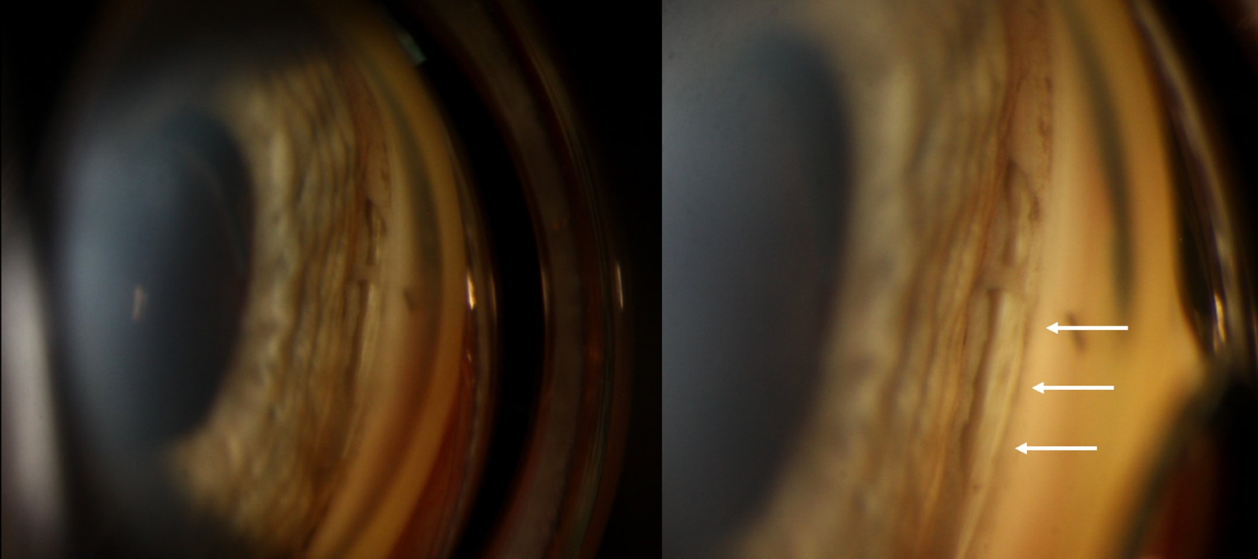
A 65-year-old woman presented for evaluation of hypotony after cataract surgery in her left eye. On examination, the patient’s intraocular pressure was 1-2 mmHg and on gonioscopy there was a large, mature cyclodialysis cleft nasally (white arrows). Cyclodialysis is the result of disinsertion of the scleral spur from the ciliary body, which creates a direct passageway from the anterior chamber to the suprachoroidal space.

Ophthalmic Atlas Images by EyeRounds.org, The University of Iowa are licensed under a Creative Commons Attribution-NonCommercial-NoDerivs 3.0 Unported License.


Address
University of IowaLegal
Related Links