In 1950, Dr. Phillips Thygeson reported a case series of 26 patients. These patients classically have a history of cyclical exacerbations and remissions with symptoms of foreign body sensation, photophobia, burning, tearing and occasionally blurring of vision. Symptoms are commonly bilateral but asymmetric. On exam, there are multiple slightly elevated corneal lesions which are round to oval conglomerates of gray, granular "crumblike" subepithelial opacities. These lesions normally negatively stain, but during exacerbations one can see staining with fluorescein dye. There is minimal conjunctival reaction. On average there are 15-20 lesions, with the greatest density being in the central cornea. These patients can present a difficult treatment conundrum. However, typical treatment consists of topical corticosteroids primarily with cyclosporine and bandaged contact lenses as secondary options. Visual prognosis is excellent.
As an interesting side note, Dr. Thygeson was a faculty member of the University of Iowa Department of Ophthalmology in the 1930's.
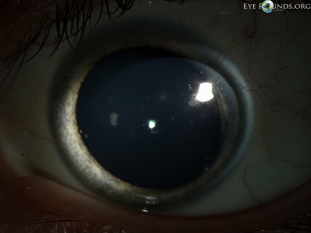
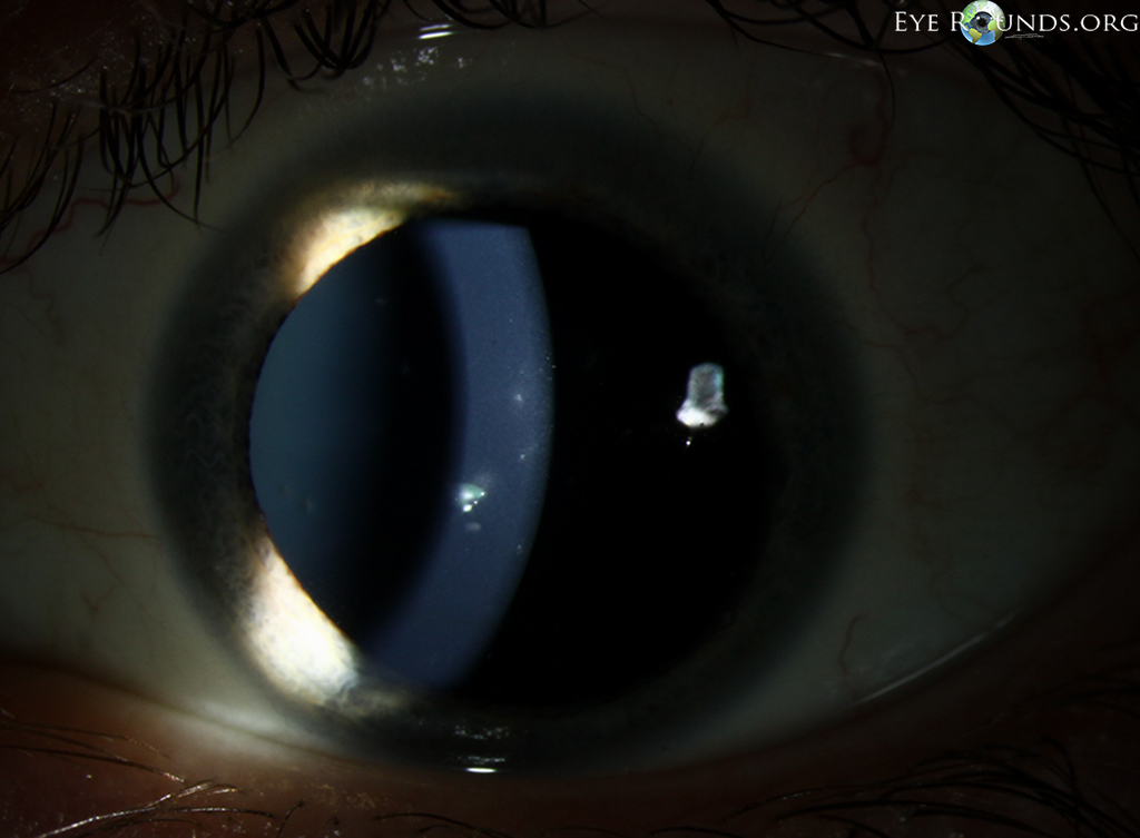
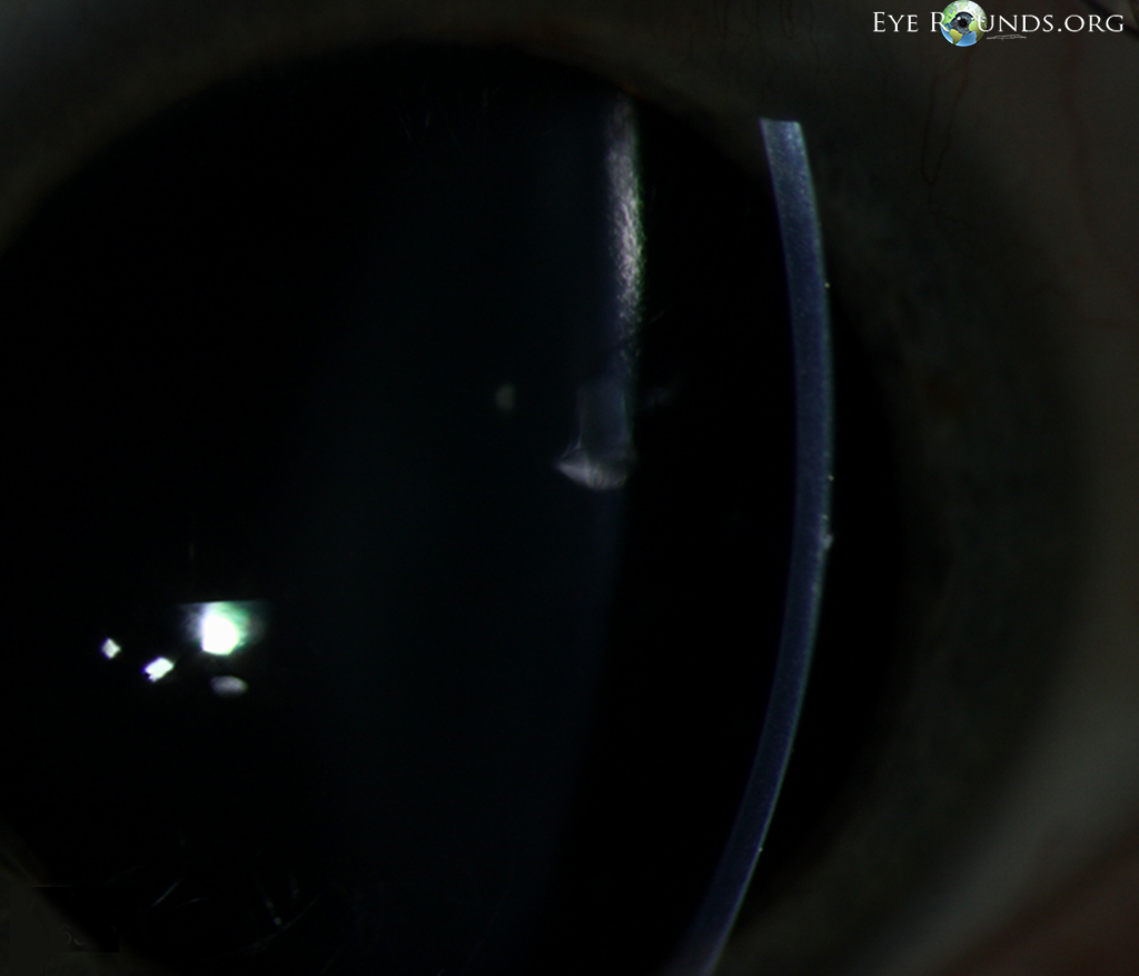
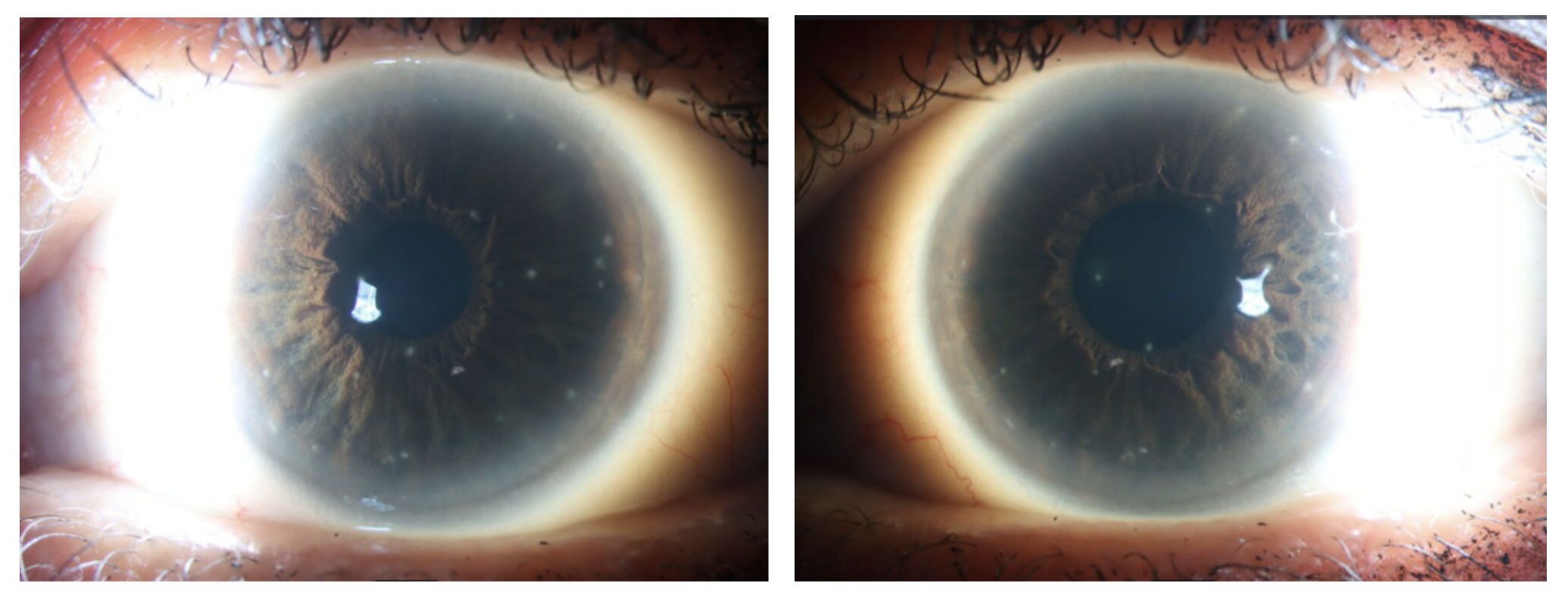
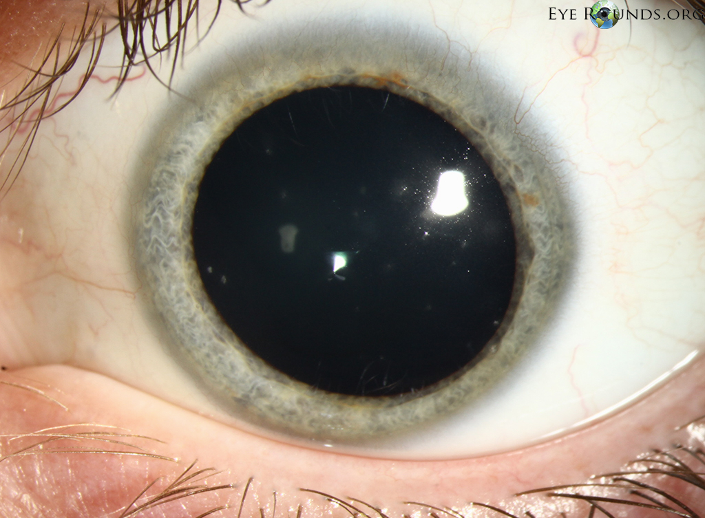
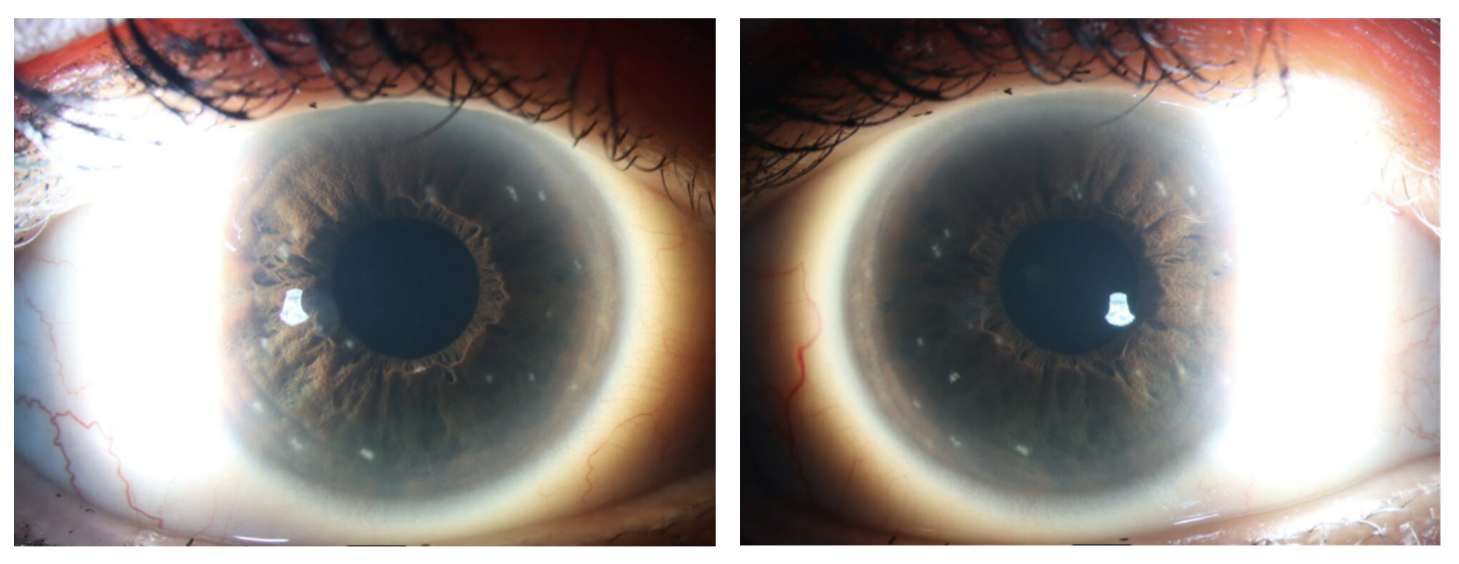
A woman in her 50s presented to the Cornea clinic with a 20-year history of recurrent episodes of eye pain, redness, and light sensitivity that improved with topical corticosteroid therapy and recurred after taper and cessation. Slit lamp exam of the cornea showed multiple intraepithelial opacities in both eyes that were small, circumscribed, slightly elevated, and grayish-white in color, as highlighted in these photos using sclerotic scatter. Findings are classic for Thygesons superficial punctate keratitis.

Ophthalmic Atlas Images by EyeRounds.org, The University of Iowa are licensed under a Creative Commons Attribution-NonCommercial-NoDerivs 3.0 Unported License.

Address
University of IowaLegal
Related Links