Contributor: Bethlehem Wole, BS; Ryan J. Diel, MD; Luis A. Leal, MD; Matthew J. Thurtell, MBBS, MSc, FRACP
Photographer: Nicole M. Radunzel
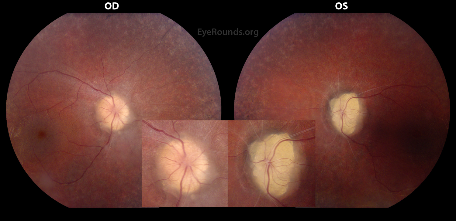
Figure 1: Color fundus photograph of the right and left eyes from a 15 year old boy with autosomal dominant retinitis pigmentosa and exposed optic disc drusen. The optic discs have a "lumpy bumpy" appearance with ill-defined margins suggesting optic disc drusen. The drusen appear as white-yellow excrescences that have joined together to form a "rocky candy" conglomerate (most marked in the left eye). Marked arteriolar attenuation secondary to retinitis pigmentosa is also evident in these photographs. Magnified views of the optic disc are displayed in the lower right and left hand corners.
Contributor: Ryan J. Diel, MD; Bethlehem Wole, BS; Alanna Tisdale, MD, MPH; Sophia M. Chung, MD
Photographer: Nicole M. Radunzel (photography) and Laura L. Warner (echography)
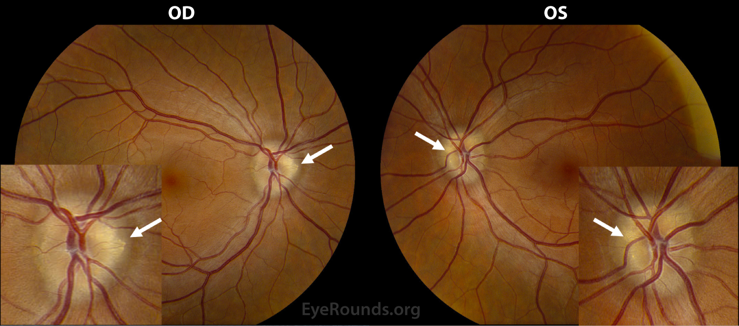
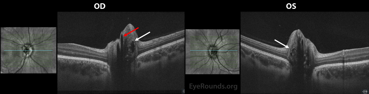
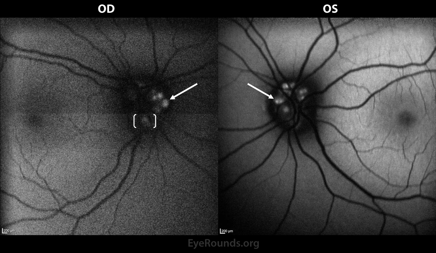
Color fundus photography (A), spectral domain optical coherence tomography (B), fundus autofluorescence (C), and ultrasound echography (D) of the right and left eyes in a 28 year-old female who presented for evaluation of pseudopapilledema and blurry vision
Figure 1A: Color fundus photography. Both optic discs are "lumpy bumpy" in appearance with highly refractile bodies seen extruding from the disc margins (denoted by white arrows) consistent with bilateral optic disc drusen.
Figure 1B: Spectral domain optical coherence tomography (SD-OCT). Buried optic disc drusen are denoted by the white arrows. Buried optic disc drusen are characterized as oval hyporeflective voids with overlying scattered hyperreflective dots [1]. Optic disc drusen should be differentiated from blood vessels seen in cross-section (red arrow) with underlying shadowing [2].
Figure 1C: Fundus autofluorescence. Exposed optic disc drusen are highly refractile (white arrows) when compared to deeper buried drusen which appear larger with a more diffuse signal (denoted by the white brackets) [2].
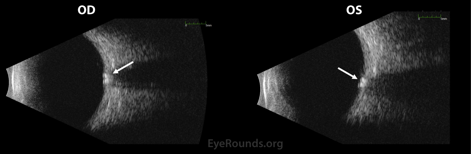
Figure 1D: Ultrasound echography. In cases of extruded/exposed optic disc drusen, the drusen are easily visible on fundus exam, SD-OCT, and/or autofluorescence as demonstrated in A-C. Ultrasound echography may be needed to idenitify very deep calcified drusen. Calcified drusen are seen on ultrasound as hyperechoic with posterior shadowing (white arrows) [1].
Contributor: Robert B Dinn, MD and Wallace L. M. Alward, MD , University of Iowa
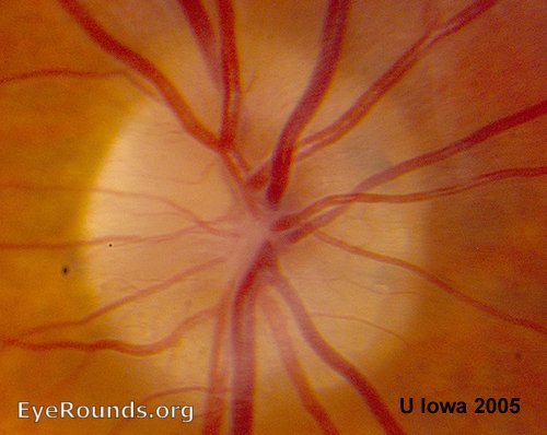
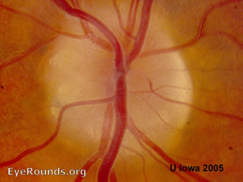
These disc photos are from an asymptomatic patient noted to have optic disc drusen. They are often bilateral and asymmetric. Optic disc drusen are refractile bodies frequently present at the nasal margin. Visual field defects are common in patients with optic disc drusen, and an RAPD may be noted in asymmetric cases.
These disc photos are from an asymptomatic patient noted to have optic disc drusen. They are often bilateral and asymmetric. Optic disc drusen are refractile bodies frequently present at the nasal margin. Visual field defects are common in patients with optic disc drusen, and an RAPD may be noted in asymmetric cases.
Caramoy A, Engel L, Koch KR, Kirchhof B, Cursiefen C, Heindl LM. Multiple imaging modalities for the detection of optic nerve head drusen: Is echography still mandatory? Acta Ophthalmol 2017;95(3):320-323. [PMID 27681817]
Malmqvist L, Bursztyn L, Costello F, Digre K, Fraser JA, Fraser C, Katz B, Lawlor M, Petzold A, Sibony P, Warner J, Wegener M, Wong S, Hamann S. The Optic Disc Drusen Studies Consortium Recommendations for Diagnosis of Optic Disc Drusen Using Optical Coherence Tomography. J Neuroophthalmol 2018;38(3):299-307. [PMID 29095768]
University of Iowa
Roy J. and Lucille A. Carver College of Medicine
Department of Ophthalmology and Visual Sciences
200 Hawkins Drive
Iowa City, IA 52242
University of Iowa
Roy J. and Lucille A. Carver College of Medicine
Department of Ophthalmology and Visual Sciences
200 Hawkins Drive
Iowa City, IA 52242