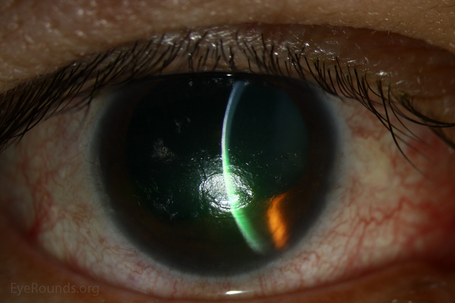
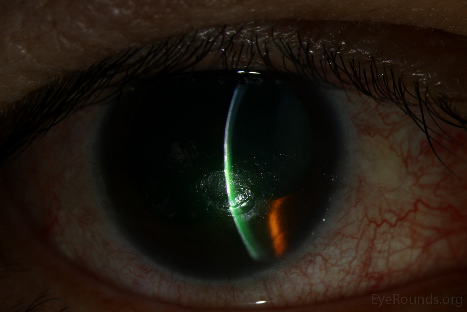
This is a 51-year-old woman was referred to the ophthalmology clinic from otolaryngology with progressive blurred vision in the right eye after resection of large petroclival meningioma 1 month prior. She had lagopthalmos and incomplete blink with significantly decreased corneal sensation. She was found to have a corneal ulcer with a gray, heaped-up rim, surrounding white blood cell recruitment, and mild corneal edema. She was started on aggressive lubrication and topical antibiotics to prevent secondary bacterial infection with plan for future tarsorrhaphy.
Round or oval-shaped ulcers with grayish, elevated edges are classic for neurotrophic ulcers. In this case the patient had a 5th cranial nerve palsy causing decreased sensation and was also predisposed to exposure given her 7th cranial nerve palsy. The differential for etiology of neurotrophic ulcer also includes cerebrovascular accidents, aneurysms, multiple sclerosis, herpes simplex or herpes zoster keratitis, or toxicity with topical medications such as anesthetics.

Ophthalmic Atlas Images by EyeRounds.org, The University of Iowa are licensed under a Creative Commons Attribution-NonCommercial-NoDerivs 3.0 Unported License.

Address
University of IowaLegal
Related Links