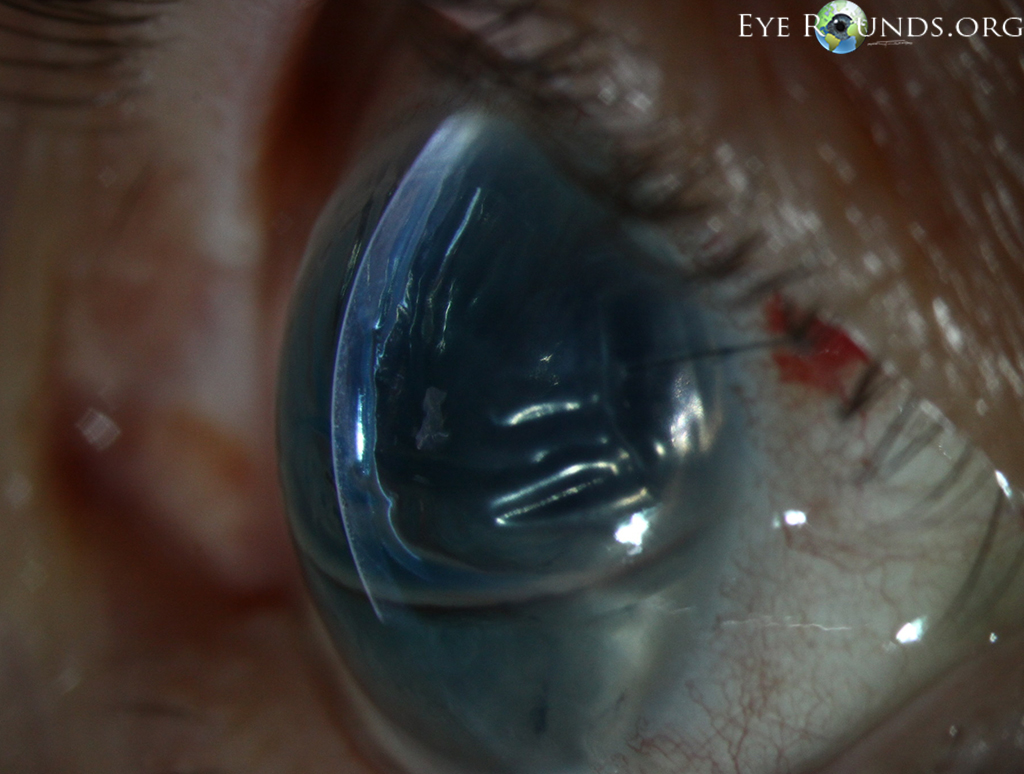
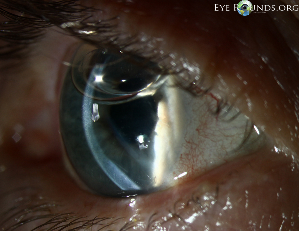
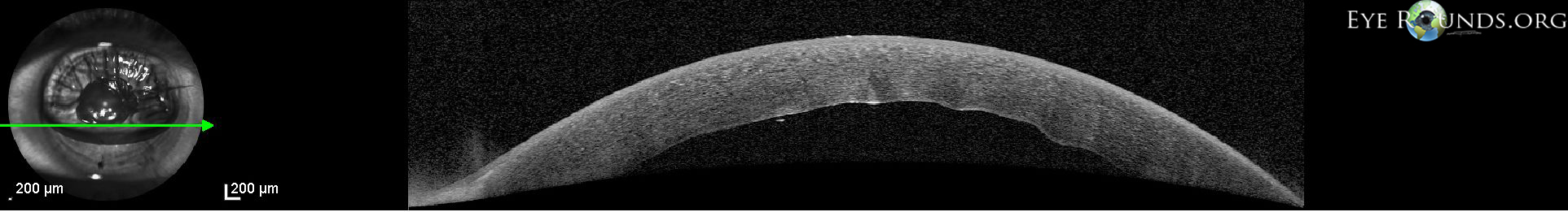
TThis patient had residual corneal edema with prominent Descemet's membrane folds visible on the posterior corneal surface on her first postoperative day after undergoing Descemet's membrane endothelial keratoplasty (DMEK). In the first photograph, the anterior chamber is 70% filled with SF6 gas and a superotemporal limbal suture can be seen. Anterior segment OCT verifies that the lenticule is in place and also shows the folds on the posterior corneal surface. After several days, the corneal edema eventually cleared and guttae were visible centrally in the graft tissue, consistent with Fuchs' endothelial dystrophy of the donor. These guttae are visible in the second photograph and this is likely the cause for delayed corneal clearing. Despite this, the patient still achieved 20/20 acuity.

Ophthalmic Atlas Images by EyeRounds.org, The University of Iowa are licensed under a Creative Commons Attribution-NonCommercial-NoDerivs 3.0 Unported License.

Address
University of IowaLegal
Related Links