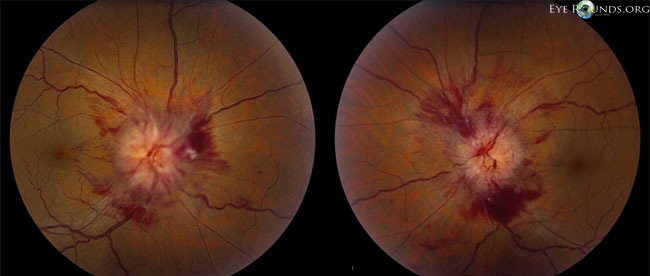
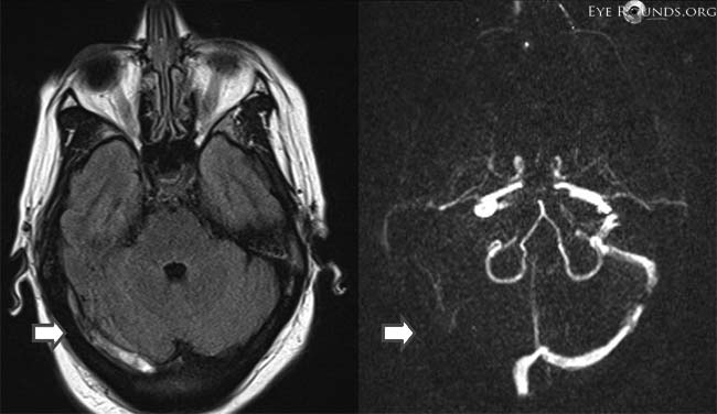
This patient presented with headaches, pulse synchronous tinnitus, and transient visual obscurations of 1 week duration, and was found to have grade 4 disc edema with prominent peripapillary hemorrhages. FLAIR MRI (left lower image) and MRV (right lower image) demonstrated a subacute right transverse sinus thrombosis (blue arrow) as the cause of the intracranial hypertension.
Cerebral venous sinus thrombosis must be evaluated for in patients with signs and symptoms of increased intracranial pressure. On MRI, an acute thrombus within 5 days will have intermediate signal on T1-weighted imaging and low signal on T2-weighted imaging. A subacute thrombus will have high signal on both T1- and T2-weighted imaging. A late thrombus will have progressive heterogeneous signal loss on T1- and T2-weighted imaging. MRV is helpful in demonstrating a lack of flow through the thrombosed venous sinus.

Ophthalmic Atlas Images by EyeRounds.org, The University of Iowa are licensed under a Creative Commons Attribution-NonCommercial-NoDerivs 3.0 Unported License.


Address
University of IowaLegal
Related Links