This site uses tracking information. Visit our privacy policy. Click to agree to this policy and not see this again.

Ophthalmology and Visual Sciences

Acute posterior multifocal placoid pigment epitheliopathy (APMPPE)

Acute posterior multifocal placoid pigment epitheliopathy (APMPPE)

Category(ies): Retina, Vitreous, Uveitis
Contributor: Angela McAllister, MD
Photographer: Brice Critser, CRA

APMPPE is one of the white dot syndromes that occurs in young healthy adults and similarly affects males and females. It is usually bilateral, although may be worse in one eye. APMPPE is generally a self-limited condition that requires no treatment and has a good prognosis.

A 47-year-old female presents with a four day history of fluttering bright spots in the right eye. She denies a viral prodrome or headaches.
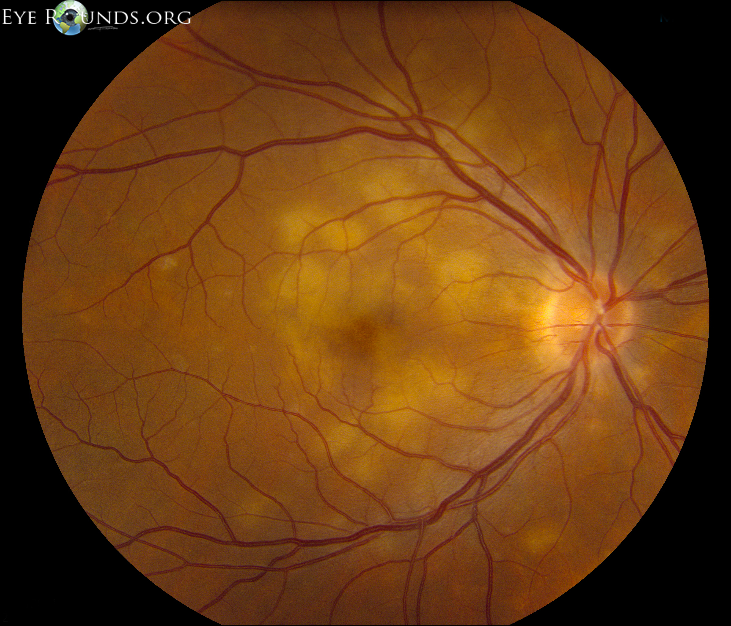
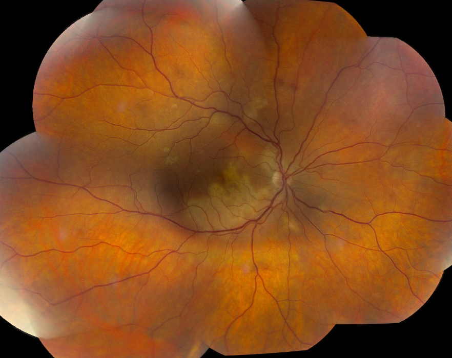
fundus image, yellow-white lesions throughout the macula
Color fundus photo, right eye (OD): Multiple creamy yellow-white lesions throughout the macula. There is mild vitritis on exam and mild disc edema.
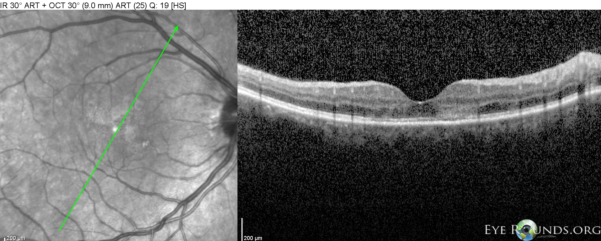
OCT
Optical coherence tomography OD: Disruption of the outer retinal layers.
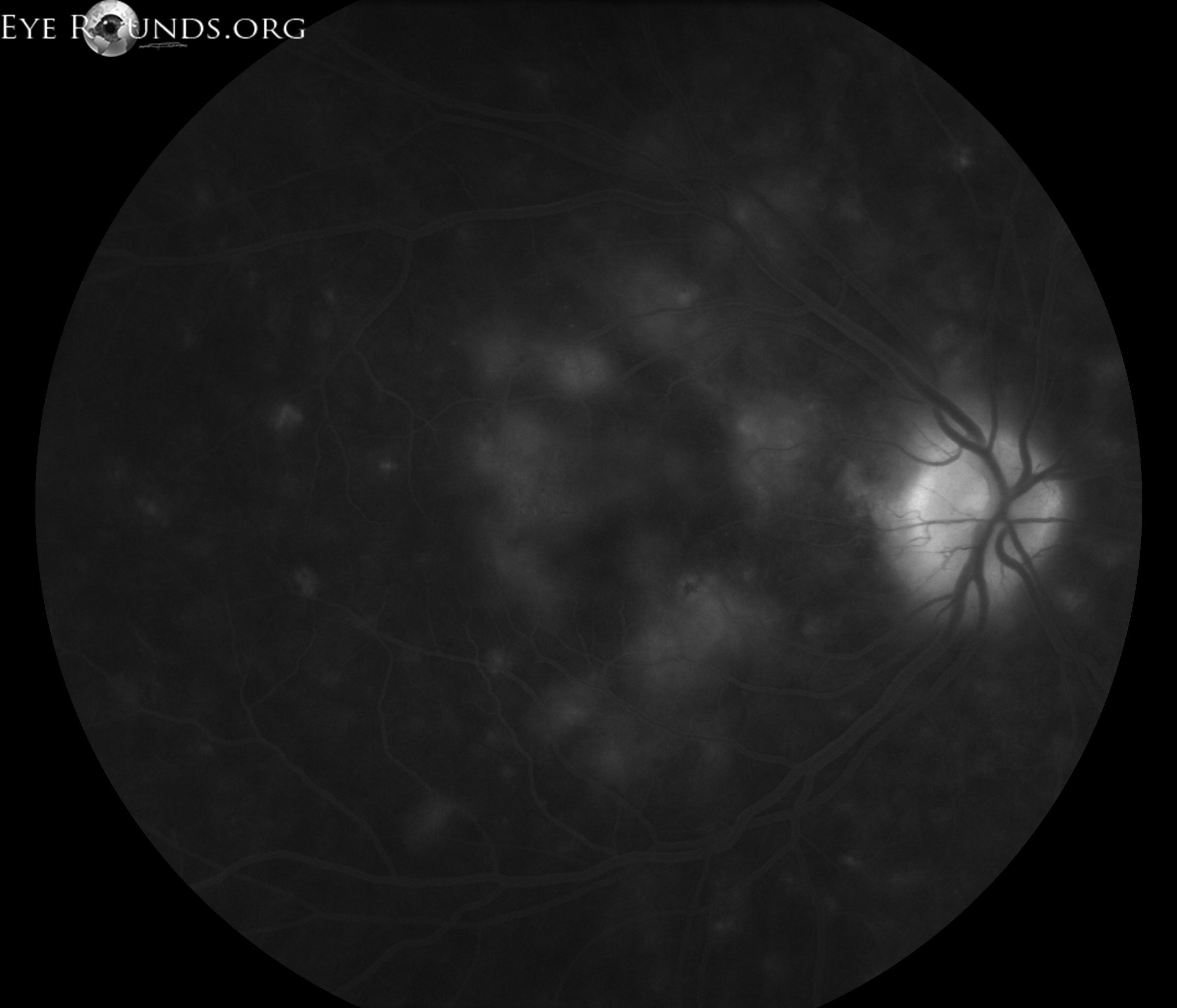
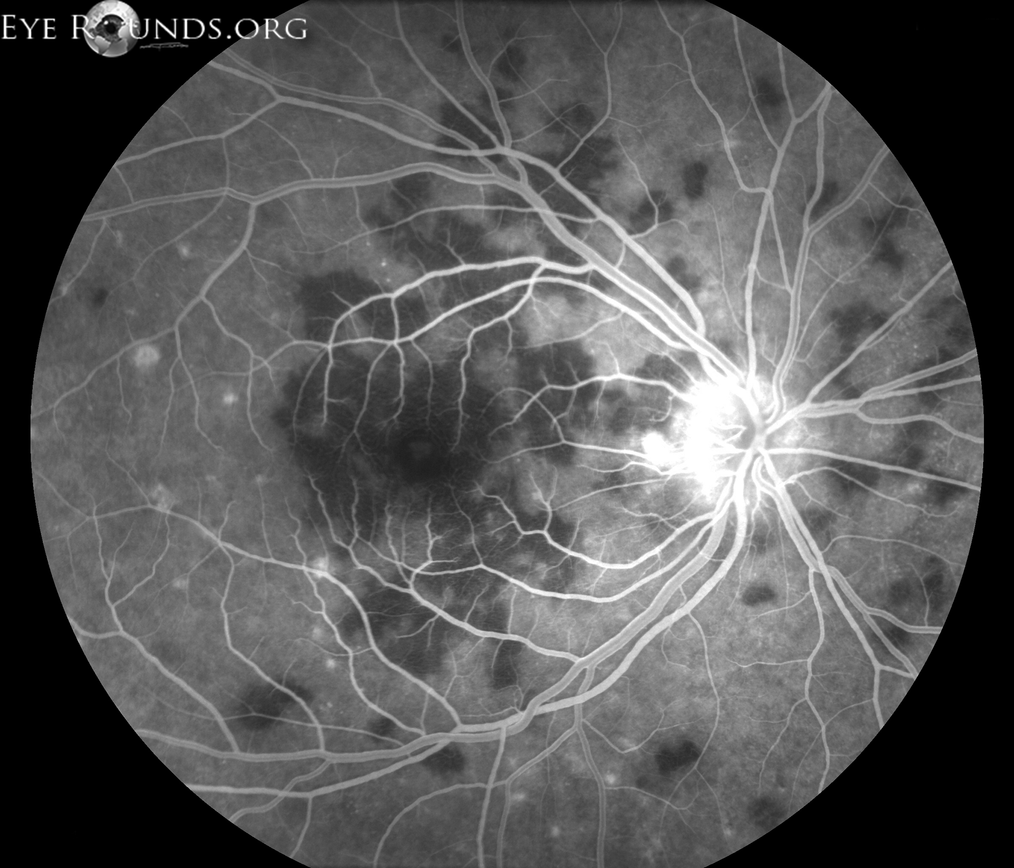
hypofluorescence of the lesions
Laminar phase of fluorescein angiogram demonstrating early hypofluorescence of the lesions
fluorescein angiogram demonstrating late staining of lesions
Late phase of fluorescein angiogram demonstrating late staining of lesions

Acute posterior multifocal placoid pigment epitheliopathy (APMPPE)

Acute posterior multifocal placoid pigment epitheliopathy (APMPPE)

Category(ies): Retina / Vitreous,Uveitis
Contributor: Mahsaw Mansoor, MD
Photographer: Jody Troyer, CRA
Posted: 06/04/2024

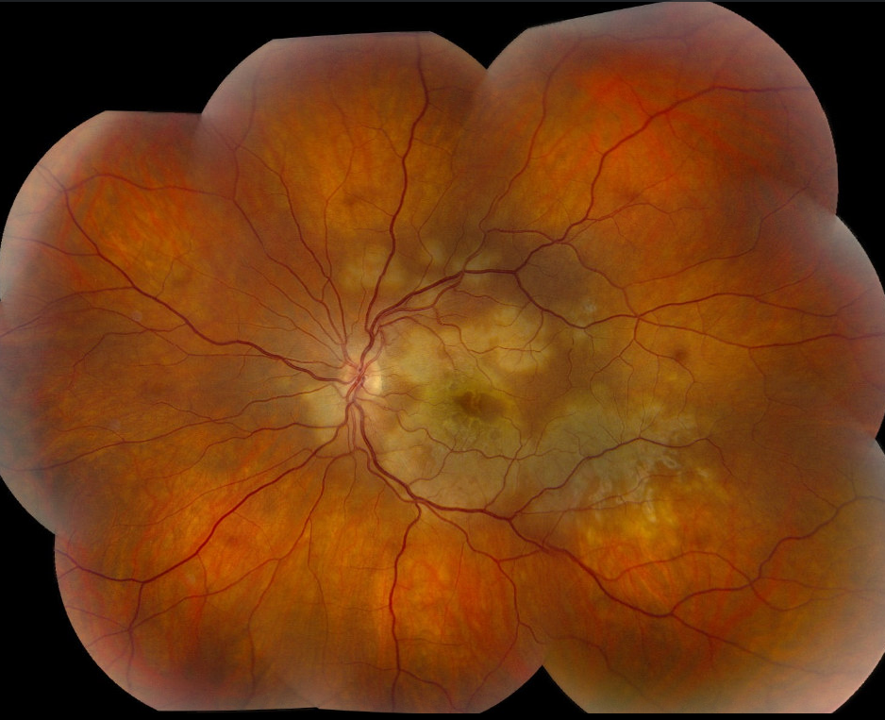
A 36-year-old woman presented with progressive vision loss in both eyes following a viral infection. On exam, there were placoid areas of whitening throughout the posterior pole of both eyes. On fluorescein angiography, the lesions on exam demonstrated early blocking defects and late staining.


Acute posterior multifocal placoid pigment epitheliopathy (APMPPE)

Acute posterior multifocal placoid pigment epitheliopathy (APMPPE)

Category(ies): Retina / Vitreous
Contributor: Alina Husain MD, Saloni Kapoor MD, Elliott H. Sohn MD, Timothy M. Boyce MD
Photographer: Madigan M. Peimann
Posted: 08/11/2026

A 39-year-old woman with a history of multiple sclerosis on ofatumumab (CD20 inhibitor) presented with progressive vision loss, left greater than right, for one week. The patient reported a history of a self-limited upper respiratory infection five weeks prior. An anterior chamber tap was performed and was negative for CMV, VZV, HSV-1 and HSV-2 PCR testing. Additional laboratory testing was notable for negative syphilis total antibody, negative quantiferon-TB gold and a mildly elevated platelet count. MRI/MRA of the Brain WWO Contrast and MRI Orbit WWO contrast demonstrated no evidence of CNS vasculitis or evidence of new or active demyelination. On exam, she demonstrated creamy placoid lesions in the posterior pole and periphery. The peripheral patches were subacute appearing with pigmentary changes and atrophy. She started on prednisone 60 mg daily after infectious testing returned negative and the patient was monitored for recurrence.

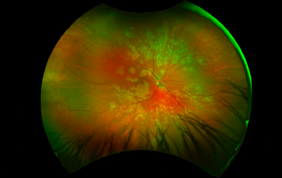
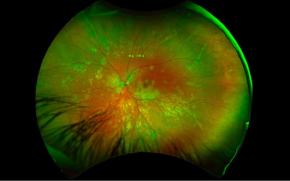
Fundus photograph, right eye. Creamy placoid lesions in the posterior pole with peripheral subacute placoid lesions with pigmentary changes and atrophic appearance
Figure 1. Fundus photograph, right eye. Creamy placoid lesions in the posterior pole with peripheral subacute placoid lesions with pigmentary changes and atrophic appearance.
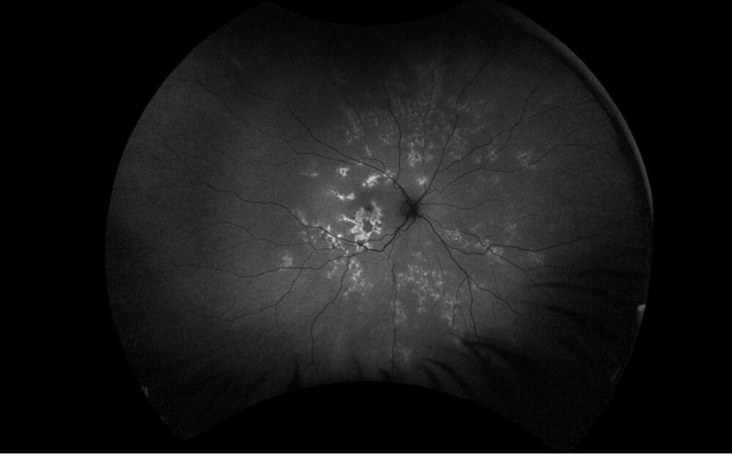
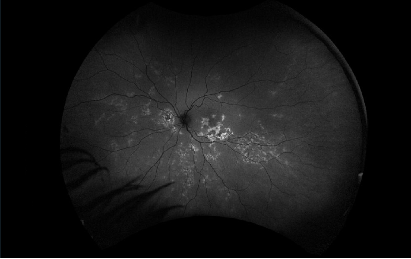
Fundus autofluorescence, right eye. There are multiple patches of hyperautofluorescence with granular hypoautofluorescence located in the macula and midperiphery
Figure 2. Fundus autofluorescence, right eye. There are multiple patches of hyperautofluorescence with granular hypoautofluorescence located in the macula and midperiphery.
Fundus photograph, left eye. Creamy placoid lesions in the macula with subacute placoid lesions in the periphery with pigmentary and atrophic appearance.
Figure 3. Fundus photograph, left eye. Creamy placoid lesions in the macula with subacute placoid lesions in the periphery with pigmentary and atrophic appearance.
Fundus autofluorescence, left eye. There are multiple patches of hyperautofluorescence with granular hypoautofluorescence located in the macula and midperiphery.
Figure 4. Fundus autofluorescence, left eye. There are multiple patches of hyperautofluorescence with granular hypoautofluorescence located in the macula and midperiphery.