This site uses tracking information. Visit our privacy policy. Click to agree to this policy and not see this again.

Ophthalmology and Visual Sciences

Paracentral Acute Middle Maculopathy (PAMM)

70-year-old woman with a history of cerebrovascular and cardiovascular disease presents with new scotomas in left eye.

Paracentral Acute Middle Maculopathy (PAMM)

70-year-old woman with a history of cerebrovascular and cardiovascular disease presents with new scotomas in left eye.
Kelly H. Yom, BA; Ryan J. Diel, MD; Heather Stiff, MD; A. Tim Johnson, MD; Ian C. Han, MD

Posted June 10, 2020

INITIAL PRESENTATION

Chief Complaint

"Splotchy" grey vision in the left eye

History of Present Illness

A 70-year-old Caucasian woman with an extensive cardiovascular history including multiple prior strokes, coronary artery disease, peripheral vascular disease, and hypertension presented to her local ophthalmologist for acute vision loss in her left eye. Two days prior, she had experienced a sudden onset of "blotchiness," like "dark grey paint splatter," in her left eye vision while driving. This blotchiness improved after 30 minutes but did not resolve completely, and she noted continued fluctuation in vision over the next two days. There was no associated pain, flashes, floaters, or diplopia; the right eye was unaffected.

Past Ocular History

  • None

Past Medical History

  • Multiple strokes:
    • Most significant stroke 21 years prior, with residual right-sided hemiparesis
    • Most recent stroke 13 years ago
  • Peripheral vascular disease
  • Coronary arteriosclerosis
  • Carotid artery stenosis
  • Hypertension
  • Hyperlipidemia
  • Osteoporosis
  • Mild intermittent asthma
  • Cervical spine degeneration

Pertinent Past Surgical History

  • No history of ocular or vascular procedures

Medications

  • No ocular medications
  • Albuterol inhaler, alendronate, aspirin, atorvastatin, cholecalciferol, cyanocobalamin, montelukast, sucralfate, ursodiol, warfarin

Allergies (Reaction)

  • NKDA

Family History

  • Non-contributory

Social History

  • Married, retired
  • Never smoked, no alcohol use, no illicit drug use 

Review of Systems

  • One week of pulsatile headache over left temple, improved with acetaminophen use
  • No fever, jaw claudication, or scalp tenderness
  • No photo/phonophobia, nausea, or vomiting
  • Otherwise negative except as described in HPI

OCULAR EXAMINATION

Visual Acuity with correction (Snellen)

  • Right eye (OD): 20/40, 20/25 -2 with pinhole
  • Left eye (OS): 20/100 -1, no improvement with pinhole

Ocular Motility/Alignment

  • Both eyes (OU): Full, orthophoric

Intraocular Pressure (IOP) by Tonopen

  • OD: 12 mmHg
  • OS: 16 mmHg

Pupils

  • OD: 4mm (dark) à 3mm (light), round, brisk, no APD
  • OS: 4mm (dark) à 3mm (light), round, brisk, no APD

Confrontation Visual Fields

  • Full OU

Ishihara Color Plates

  • OD: 14/14
  • OS: 5/14

External

  • Normal OU
  • No tenderness upon palpation of temporal artery bilaterally

Slit lamp exam

  • Lids/lashes: Normal OU
  • Conjunctiva/sclera: Clear and quiet OU
  • Cornea: Clear OU
  • Anterior chamber: Deep and quiet OU
  • Iris: Normal architecture OU
  • Lens: 2+ NS and 1+ anterior cortical cataract OU
  • Anterior vitreous: Normal OU

Dilated fundus examination (DFE)

  • Disc: Normal, well perfused OU; no disc edema or pallor
  • Cup to disc (C/D): 0.4 OU
  • Macula: Normal OD; subtle grayish perifoveal discoloration OS
  • Vessels: Mild tortuosity, no Hollenhorst plaques OU
  • Periphery: cotton wool spot along the superotemporal arcade OS, otherwise unremarkable OU; no intraretinal hemorrhages or areas of peripheral retinal whitening

Additional Testing

  • MRI brain with contrast: chronic small vessel ischemic disease but no acute intracranial findings
  • MRA head/neck with contrast: atherosclerotic involvement of bilateral carotid arteries but no hemodynamically significant stenosis
  • Color fundus photography (Figure 1)
  • Humphrey visual field testing (Figure 2)
  • Optical coherence tomography (OCT) of optic nerve head:
    • OD: average RNFL thickness 76 µm, superior quadrant thinning
    • OS: average RNFL thickness 76 µm, no thinning
  • Spectral domain OCT (SD-OCT) of macula (Figure 3)
  • OCT angiography (OCT-A) (Figures 4 and 5)

Color fundus photograph of (A) the right eye, which is unremarkable except for mild vascular tortuosity, and (B, inset: high magnification) the left eye, demonstrating a white lesion along the superior arcade representing a cotton wool spot (arrow) and faint grayish-white mottling in the temporal macula.

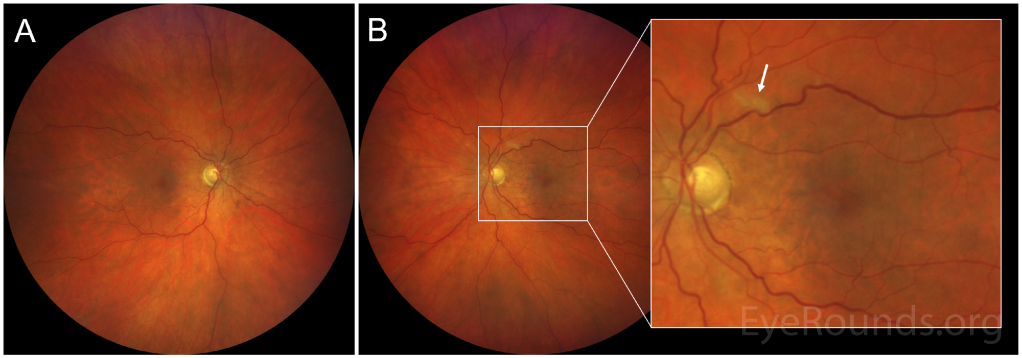
Figure 1: Color fundus photograph of (A) the right eye, which is unremarkable except for mild vascular tortuosity, and (B, inset: high magnification) the left eye, demonstrating a white lesion along the superior arcade representing a cotton wool spot (arrow) and faint grayish-white mottling in the temporal macula.

Central 24-2 Humphrey visual field testing of (A) the left eye, which shows diffuse visual field constriction and a pronounced superonasal deficit involving central fixation, and (B) the right eye, which shows mild, nonspecific defects.

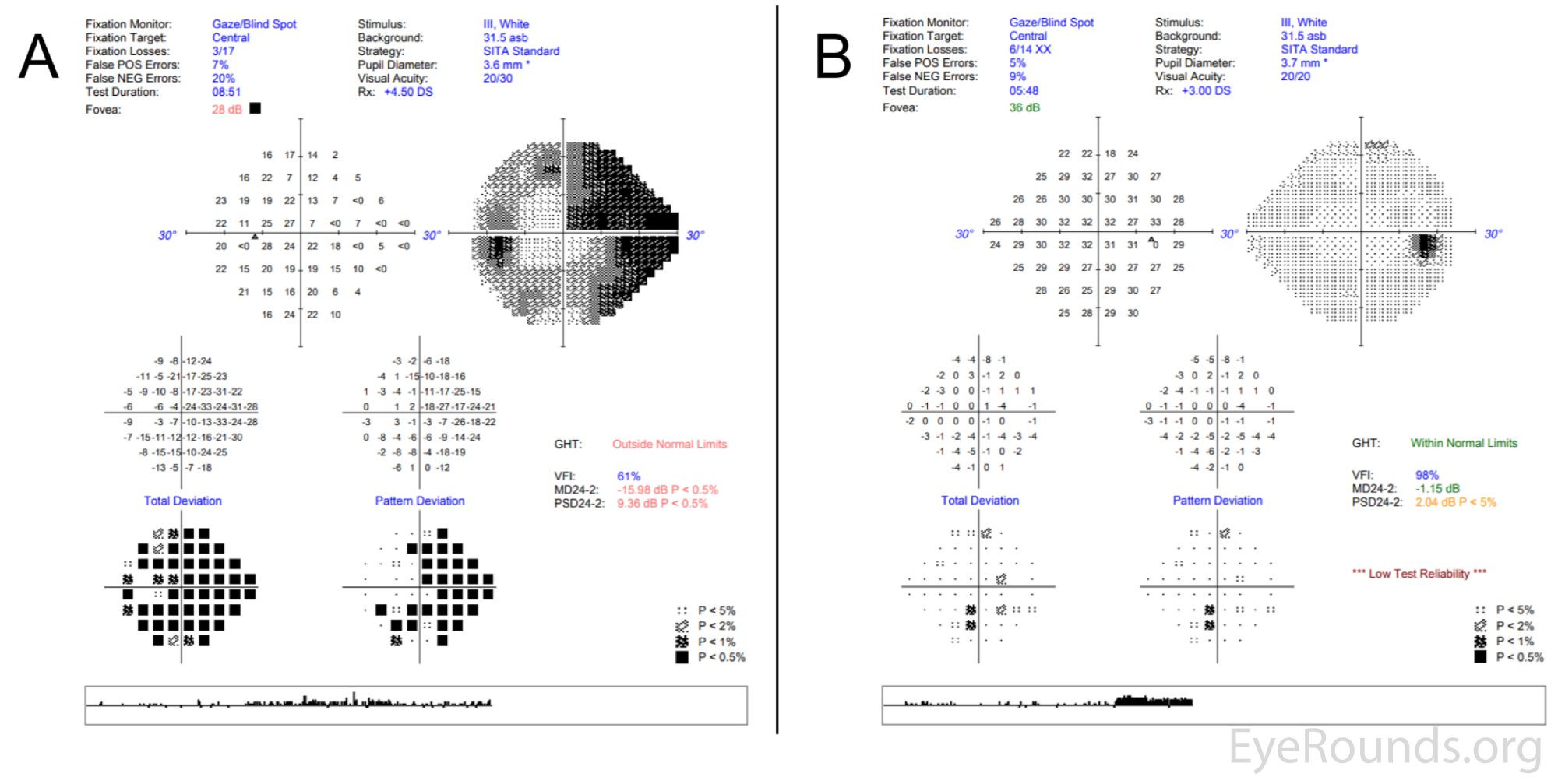
Figure 2: Central 24-2 Humphrey visual field testing of (A) the left eye, which shows diffuse visual field constriction and a pronounced superonasal deficit involving central fixation, and (B) the right eye, which shows mild, nonspecific defects.

Spectral domain optical coherence tomography (SD-OCT) of the macula of (A) the right eye, showing intact retinal laminations, and (B) the left eye, demonstrating patchy perifoveal hyperreflectivity from the inner plexiform to the outer plexiform layers (arrows).

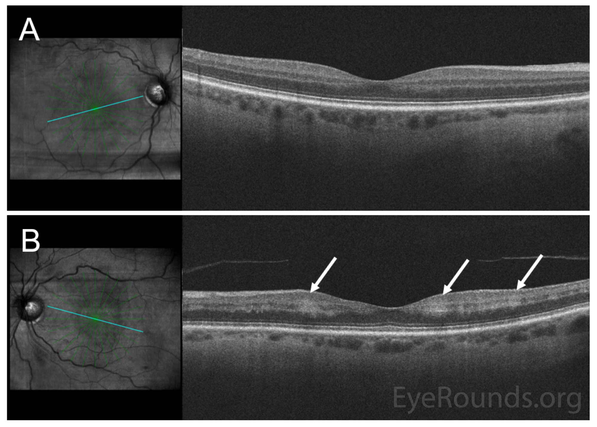
Figure 3: Spectral domain optical coherence tomography (SD-OCT) of the macula of (A) the right eye, showing intact retinal laminations, and (B) the left eye, demonstrating patchy perifoveal hyperreflectivity from the inner plexiform to the outer plexiform layers (arrows).

Optical coherence tomography angiography (3×3 mm) of the superficial capillary plexus. There is mild irregularity of the foveal avascular zone in the right eye (A, angiogram), with homogeneous reflectivity pattern on the corresponding en face structural slab (B). The left eye has an irregular foveal avascular zone with perifoveal vascular flow loss (C), and splotchy irregularity en face (D) corresponding to the hyperreflectivities seen on the OCT macula line scans in Figure 3.

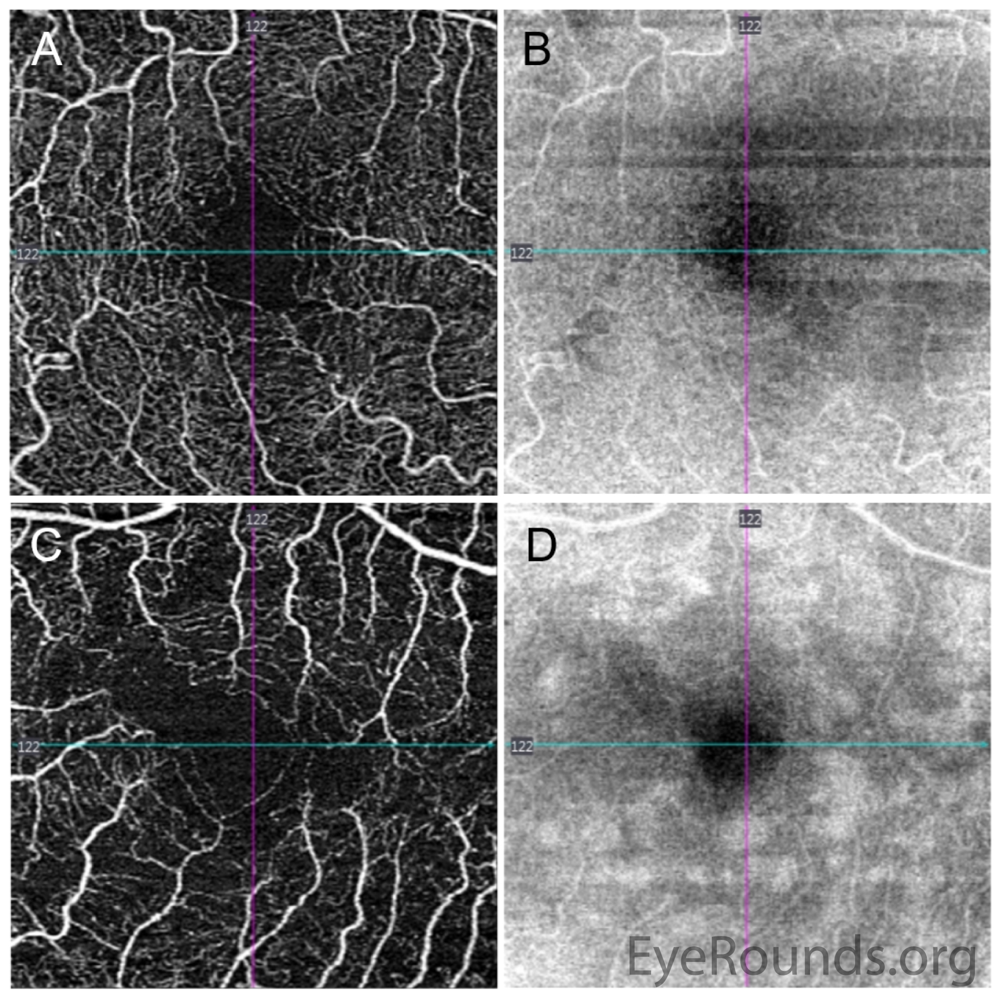
Figure 4: Optical coherence tomography angiography (3×3 mm) of the superficial capillary plexus. There is mild irregularity of the foveal avascular zone in the right eye (A, angiogram), with homogeneous reflectivity pattern on the corresponding en face structural slab (B). The left eye has an irregular foveal avascular zone with perifoveal vascular flow loss (C), and splotchy irregularity en face (D) corresponding to the hyperreflectivities seen on the OCT macula line scans in Figure 3.

Optical coherence tomography angiography (3x3 mm) of the deep capillary plexus. There is relative preservation of the deep capillary plexus vasculature (A) in the right eye, with no abnormalities in the corresponding structural slab (B). In the left eye, there is profound flow loss (C), with corresponding hyperreflectivities on the structural slab (D).

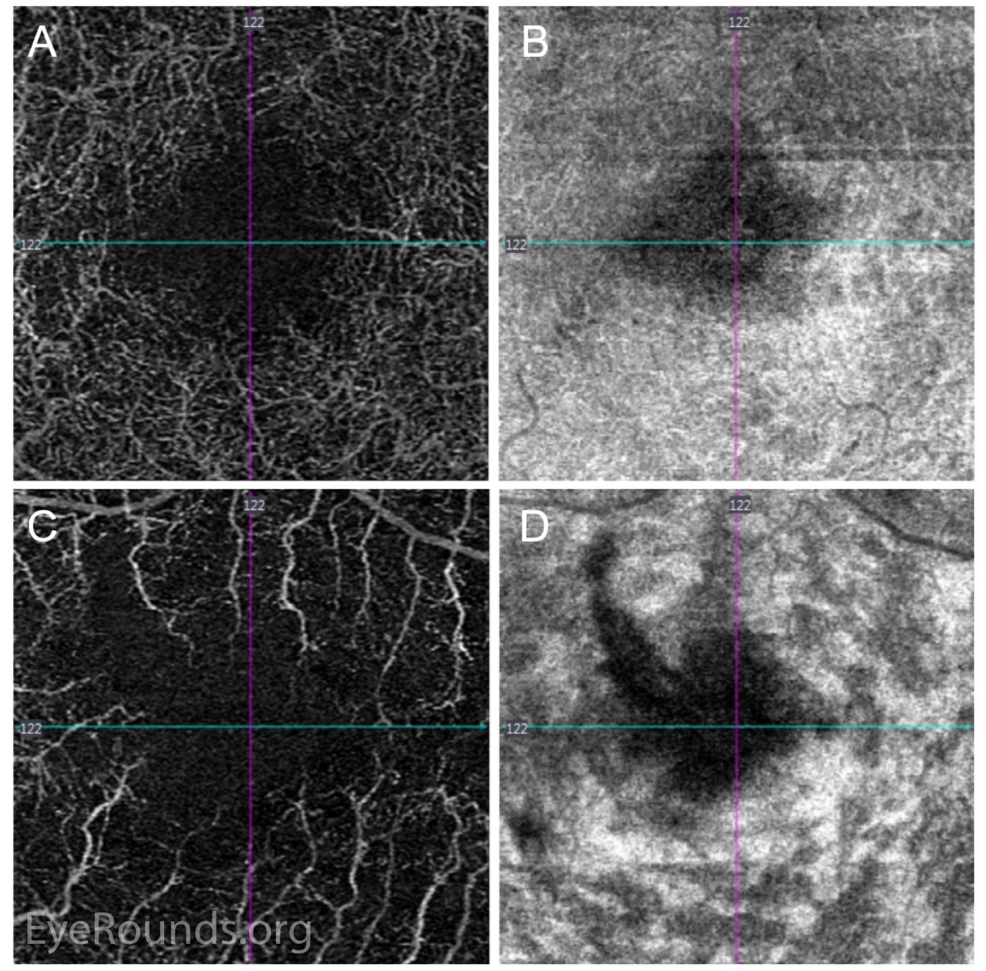
Figure 5: Optical coherence tomography angiography (3x3 mm) of the deep capillary plexus. There is relative preservation of the deep capillary plexus vasculature (A) in the right eye, with no abnormalities in the corresponding structural slab (B). In the left eye, there is profound flow loss (C), with corresponding hyperreflectivities on the structural slab (D).

Differential Diagnosis

  • Paracentral acute middle maculopathy
  • Acute macular neuroretinopathy
  • Giant cell arteritis
  • Reperfused central retinal artery occlusion

CLINICAL COURSE

The patient was seen in the emergency department by the ophthalmology service. Due to the sudden onset of vision changes and the patient's history of cerebrovascular disease, an MRI and MRA of the head and neck were obtained and were negative for acute pathology. Given the initial concern for giant cell arteritis, inflammatory markers were ordered. ESR and CRP were both within normal.

SD-OCT imaging of the macula of the left eye revealed diffuse hyper-reflective bands along the inner nuclear layer (INL), a finding that is typical of paracentral acute middle maculopathy (PAMM). The patient was counseled on optimizing vascular risk factors and instructed to return to the retina clinic in one month for follow up.

DIAGNOSIS

Paracentral acute middle maculopathy (PAMM)

DISCUSSION

Paracentral acute middle maculopathy (PAMM) is primarily a descriptive diagnosis that represents a spectrum of presumed ischemic maculopathies characterized by hyperreflectivity of the inner retinal layers. PAMM was first described by Sarraf et al. in 2013 in a small case series of patients with acute scotomas, parafoveal white-grey lesions, and localization of these lesions to the middle layer of the retina on SD-OCT. These three characteristics continue to be defining hallmarks of the condition [1].

Signs/Symptoms

Patients with PAMM typically present with sudden onset of central or paracentral scotomas that may not resolve [2], and these may be accompanied by mild to moderately decreased visual acuity. There are typically minimal other ocular signs or symptoms, and dilated funduscopic examination may reveal parafoveal areas of grey-white retinal discoloration, though some cases have no visible fundus abnormalities [3]. PAMM may also be seen in cases of retinal vasculature occlusion with emboli along retinal arterial branches [4].

Testing and Imaging Work-up

SD-OCT is the most helpful imaging modality and demonstrates characteristic hyper-reflective thickened bands in the middle retina, which are diagnostic for the condition [5]. Fluorescein angiography (FA) may be unremarkable without delayed filling or evidence of vascular occlusion, and as such, is not needed to diagnose PAMM, though FA may be helpful in excluding other causes of macular ischemia [1,2]. Near-infrared reflectance and fundus autofluorescence are other sensitive imaging techniques that can be used to confirm the diagnosis. Recently, en face OCT and OCT angiography (OCT-A) have provided further insight into disease pathophysiology, as further discussed below [6,7].

Etiology / Pathophysiology

PAMM is thought to be a reflection of intraretinal ischemia predominantly of the intermediate and deep retinal capillary plexuses located in the inner nuclear layer (INL). The ischemic etiology of disease is supported by OCT-A findings of decreased blood flow and abnormal vasculature in the deep capillary plexus (DCP) of PAMM lesions [8,9]. In contrast to PAMM lesions, cotton wool spots are infarcts at the more superficial ganglion cell or nerve fiber layers, and are a brighter white color compared to the deeper, gray-white appearance of the PAMM lesion [3,10].

PAMM has been described in patients with systemic cardiovascular risk factors or in association with other retinal pathology, including retinal vascular occlusions, diabetic retinopathy, retinal vasculitis, and sickle cell retinopathy [4,10-12]. Hypercoaguability and hypertension are also considered a risk factors [13,14]. However, cases without identifiable underlying vascular disease have also been described [15,16].

The hyperreflective lesions in PAMM can be isolated or multiple in number, focal or diffuse in distribution, and globular or fern-like in shape. Moreover, PAMM lesions evolve over time, and some have classified PAMM into acute and chronic phases. In acute lesions, flow within the DCP may appear minimally affected unless there is massive vessel occlusion blocking capillary flow [5,17]. After approximately several weeks, there can be increased flow loss within the DCP and subsequent atrophy corresponding to prior areas of hyperreflectivity [5]. Longitudinally, reports have described decreased DCP flow, capillary dilation and abnormalities, thinning of the INL, thickening of the ONL, and excavation of the inner retinal surface several years after the intial insult [18].

Initially, PAMM was classified as a possible variant of acute macular neuroretinopathy (AMN) since both conditions present similarly with paracentral scotomas and have hyper-reflective bands on OCT [19]. However, debate exists whether PAMM and AMN are part of the same spectrum, or whether these two conditions affect different sections of the retina and choroid. On OCT for example, hyperreflective abnormalities in AMN are deeper in the retina, located at the junction of the outer plexiform layer (OPL) and outer nerve layer (ONL) [2]. In contrast to PAMM lesions, which typically correspond to DCP flow loss, the primary insult in AMN is thought to involve the inner choroid [20,21]. Moreover, there are demographic differences between the patient populations affected by these lesions. PAMM has been associated more frequently with patients who are older (average age >50), male, or have vasculopathic risk factors [8], while AMN tends to affect patients who are younger (average age >30) and female. A shared pathophysiologic mechanism may be sudden changes in blood pressure that overcome the ability of the DCP or choriocapillaris to autoregulate such changes, and in some cases, both choriocapillaris and DCP flow loss can be seen (20). Several studies have demonstrated that areas of inner retinal atrophy resembling PAMM can commonly be seen even in healthy patients, those with hypertension, or with retinal vein occlusions in the fellow eye [14,22].

Treatment / Management

Currently, there is no treatment for PAMM. Symptomatic scotomas may fade but are usually persistent. Because these lesions are often associated with other vasculopathic conditions, it is important to screen patients presenting with PAMM for both local and systemic diseases, including retinal vessel occlusions, carotid artery disease, diabetes, hypertension, and GCA or other vasculitides [11]. Management should be directed at identifying and minimizing vascular risk factors.  Careful surveillance in follow up is also important, as diffuse PAMM lesions can mask occult retinal occlusions that further threaten vision [2].

ETIOLOGY
  • Ischemia of the deep retinal capillary plexus, followed by eventual inner retinal atrophy
  • Likely associated with vascular risk factors
SIGNS
  • Hyper-reflective band-like lesions in the middle retina (inner plexiform, inner nuclear, outer plexiform layers) on SD-OCT
  • Decreased flow in the deep capillary plexus on OCT angiography
SYMPTOMS
  • Acute onset of grey, central or paracentral scotomas
  • Decreased visual acuity
  • May be asymptomatic
TREATMENT/MANAGEMENT
  • No established direct treatment modalities
  • Observation
  • Evaluation for and treatment of underlying systemic cardiovascular risk factors
References
  1. Sarraf D, Rahimy E, Fawzi AA, Sohn E, Barbazetto I, Zacks DN, Mittra RA, Klancnik JM, Jr., Mrejen S, Goldberg NR, Beardsley R, Sorenson JA, Freund KB. Paracentral acute middle maculopathy: a new variant of acute macular neuroretinopathy associated with retinal capillary ischemia. JAMA Ophthalmol 2013;131(10):1275-1287.  https://PubMed.gov/23929382. DOI: 10.1001/jamaophthalmol.2013.4056
  2. Rahimy E, Kuehlewein L, Sadda SR, Sarraf D. Paracentral Acute Middle Maculopathy: What We Knew Then and What We Know Now. Retina 2015;35(10):1921-1930.  https://PubMed.gov/26360227. DOI: 10.1097/iae.0000000000000785
  3. Bailey Freund KSDMWYL. Retinal Vascular Disease. The Retinal Atlas. Philadelphia: Elsevier; 2017; chapter 6; p. 493-650.
  4. Rahimy E, Sarraf D, Dollin ML, Pitcher JD, Ho AC. Paracentral acute middle maculopathy in nonischemic central retinal vein occlusion. Am J Ophthalmol 2014;158(2):372-380.e371.  https://PubMed.gov/24794089. DOI: 10.1016/j.ajo.2014.04.024
  5. Nemiroff J, Phasukkijwatana N, Sarraf D. Optical Coherence Tomography Angiography of Deep Capillary Ischemia. Dev Ophthalmol 2016;56:139-145.  https://PubMed.gov/27022753. DOI: 10.1159/000442806
  6. Sridhar J, Shahlaee A, Rahimy E, Hong BK, Khan MA, Maguire JI, Dunn JP, Mehta S, Ho AC. Optical Coherence Tomography Angiography and En Face Optical Coherence Tomography Features of Paracentral Acute Middle Maculopathy. Am J Ophthalmol 2015;160(6):1259-1268.e1252.  https://PubMed.gov/26386158. DOI: 10.1016/j.ajo.2015.09.016
  7. Pecen PE, Smith AG, Ehlers JP. Optical Coherence Tomography Angiography of Acute Macular Neuroretinopathy and Paracentral Acute Middle Maculopathy. JAMA Ophthalmol 2015;133(12):1478-1480.  https://PubMed.gov/26513596. DOI: 10.1001/jamaophthalmol.2015.4100
  8. Shah A, Rishi P, Chendilnathan C, Kumari S. OCT angiography features of paracentral acute middle maculopathy. Indian J Ophthalmol 2019;67(3):417-419.  https://PubMed.gov/30777975. DOI: 10.4103/ijo.IJO_1249_18
  9. Kulikov AN, Maltsev DS, Leongardt TA. RETINAL MICROVASCULATURE ALTERATION IN PARACENTRAL ACUTE MIDDLE MACULOPATHY AND ACUTE MACULAR NEURORETINOPATHY: A QUANTITATIVE OPTICAL COHERENCE TOMOGRAPHY ANGIOGRAPHY STUDY. Retin Cases Brief Rep 2018;10.1097/icb.0000000000000709.  https://PubMed.gov/29443808. DOI: 10.1097/icb.0000000000000709
  10. Rahimy E, Sarraf D. Paracentral acute middle maculopathy spectral-domain optical coherence tomography feature of deep capillary ischemia. Curr Opin Ophthalmol 2014;25(3):207-212.  https://PubMed.gov/24614148. DOI: 10.1097/icu.0000000000000045
  11. Chen X, Rahimy E, Sergott RC, Nunes RP, Souza EC, Choudhry N, Cutler NE, Houston SK, Munk MR, Fawzi AA, Mehta S, Hubschman JP, Ho AC, Sarraf D. Spectrum of Retinal Vascular Diseases Associated With Paracentral Acute Middle Maculopathy. Am J Ophthalmol 2015;160(1):26-34.e21.  https://PubMed.gov/25849522. DOI: 10.1016/j.ajo.2015.04.004
  12. Christenbury JG, Klufas MA, Sauer TC, Sarraf D. OCT Angiography of Paracentral Acute Middle Maculopathy Associated With Central Retinal Artery Occlusion and Deep Capillary Ischemia. Ophthalmic Surg Lasers Imaging Retina 2015;46(5):579-581.  https://PubMed.gov/26057763. DOI: 10.3928/23258160-20150521-11
  13. Trese MG, Thanos A, Yonekawa Y, Randhawa S. Optical Coherence Tomography Angiography of Paracentral Acute Middle Maculopathy Associated With Primary Antiphospholipid Syndrome. Ophthalmic Surg Lasers Imaging Retina 2017;48(2):175-178.  https://PubMed.gov/28195622. DOI: 10.3928/23258160-20170130-13
  14. Burnasheva MA, Maltsev DS, Kulikov AN, Sherbakova KA, Barsukov AV. Association of Chronic Paracentral Acute Middle Maculopathy Lesions with Hypertension. Ophthalmol Retina 2019;10.1016/j.oret.2019.12.001.  https://PubMed.gov/31948908. DOI: 10.1016/j.oret.2019.12.001
  15. Chen Y, Hu Y. The optical imaging of idiopathic paracentral acute middle maculopathy in a Chinese young man and review of the literature. Photodiagnosis Photodyn Ther 2017;19:383-387.  https://PubMed.gov/28733050. DOI: 10.1016/j.pdpdt.2017.07.004
  16. Haskes C, Santapaola S, Zinn J. An Atypical Case of Paracentral Acute Middle Maculopathy. Optom Vis Sci 2017;94(8):845-850.  https://PubMed.gov/28644209. DOI: 10.1097/opx.0000000000001087
  17. Nemiroff J, Kuehlewein L, Rahimy E, Tsui I, Doshi R, Gaudric A, Gorin MB, Sadda S, Sarraf D. Assessing Deep Retinal Capillary Ischemia in Paracentral Acute Middle Maculopathy by Optical Coherence Tomography Angiography. American Journal of Ophthalmology 2016;162:121-132.e121. DOI: https://doi.org/10.1016/j.ajo.2015.10.026
  18. Nakamura M, Katagiri S, Hayashi T, Aoyagi R, Hasegawa T, Kogure A, Iida T, Nakano T. Longitudinal follow-up of two patients with isolated paracentral acute middle maculopathy. Int Med Case Rep J 2019;12:143-149.  https://PubMed.gov/31191041. DOI: 10.2147/imcrj.S196047
  19. Frisbie RA WA, Risma TBS, Abramoff MD. Acute Macular Neuroretinopathy. EyeRounds.org, 2019. [Last rev.; accessed] https://eyerounds.org/cases/285-Acute-Macular-Neuroretinopathy.htm.
  20. Casalino G, Arrigo A, Romano F, Munk MR, Bandello F, Parodi MB. Acute macular neuroretinopathy: pathogenetic insights from optical coherence tomography angiography. Br J Ophthalmol 2019;103(3):410-414.  https://PubMed.gov/29844084. DOI: 10.1136/bjophthalmol-2018-312197
  21. Lee SY, Cheng JL, Gehrs KM, Folk JC, Sohn EH, Russell SR, Guo Z, Abramoff MD, Han IC. Choroidal Features of Acute Macular Neuroretinopathy via Optical Coherence Tomography Angiography and Correlation With Serial Multimodal Imaging. JAMA Ophthalmol 2017;135(11):1177-1183.  https://PubMed.gov/28973538. DOI: 10.1001/jamaophthalmol.2017.3790
  22. Maltsev DS, Kulikov AN, Burnasheva MA, Chhablani J. Prevalence of resolved paracentral acute middle maculopathy lesions in fellow eyes of patients with unilateral retinal vein occlusion. Acta Ophthalmol 2020;98(1):e22-e28.  https://PubMed.gov/31347293. DOI: 10.1111/aos.14196
Suggested citation format:

Yom KH, Diel RJ, Stiff HA, Johnson AT, Han IC. Paracentral Acute Middle Maculopathy (PAMM). Posted June 10, 2020; Available from: https://eyerounds.org/cases/299-PAMM.htm


last updated: 6/10/2020

Image Permissions:
Creative Commons

Ophthalmic Atlas Images by EyeRounds.org, The University of Iowa are licensed under a Creative Commons Attribution-NonCommercial-NoDerivs 3.0 Unported License.