This site uses tracking information. Visit our privacy policy. Click to agree to this policy and not see this again.

Ophthalmology and Visual Sciences

Pseudoexfoliation syndrome

Pseudoexfoliation syndrome

Category(ies): Cataract, Lens, Glaucoma, Iris, Inherited Eye Disease
Contributor: Lorraine M. Provencher, MD
Photographer: Brice Critser, CRA

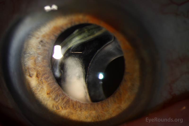
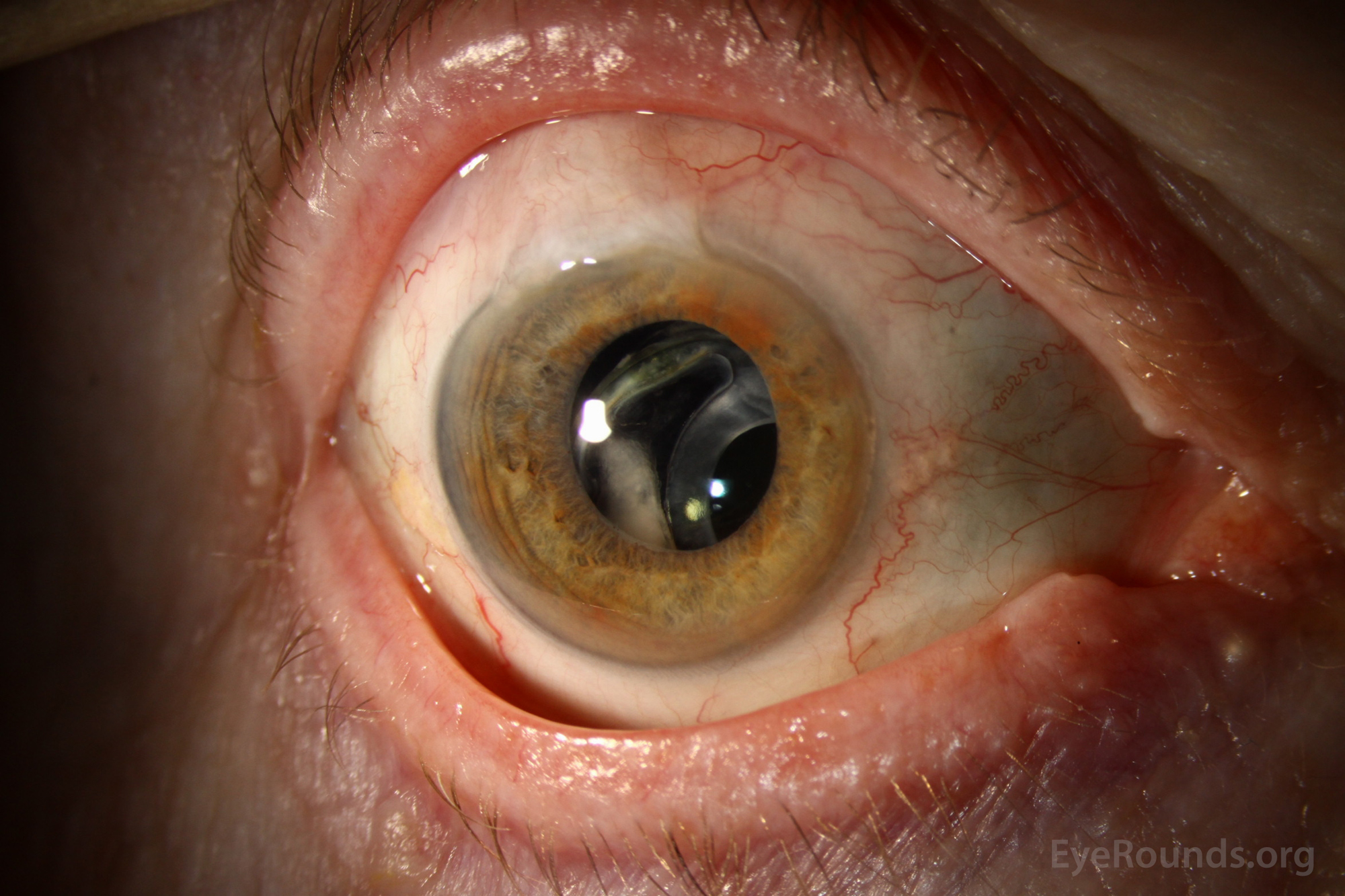
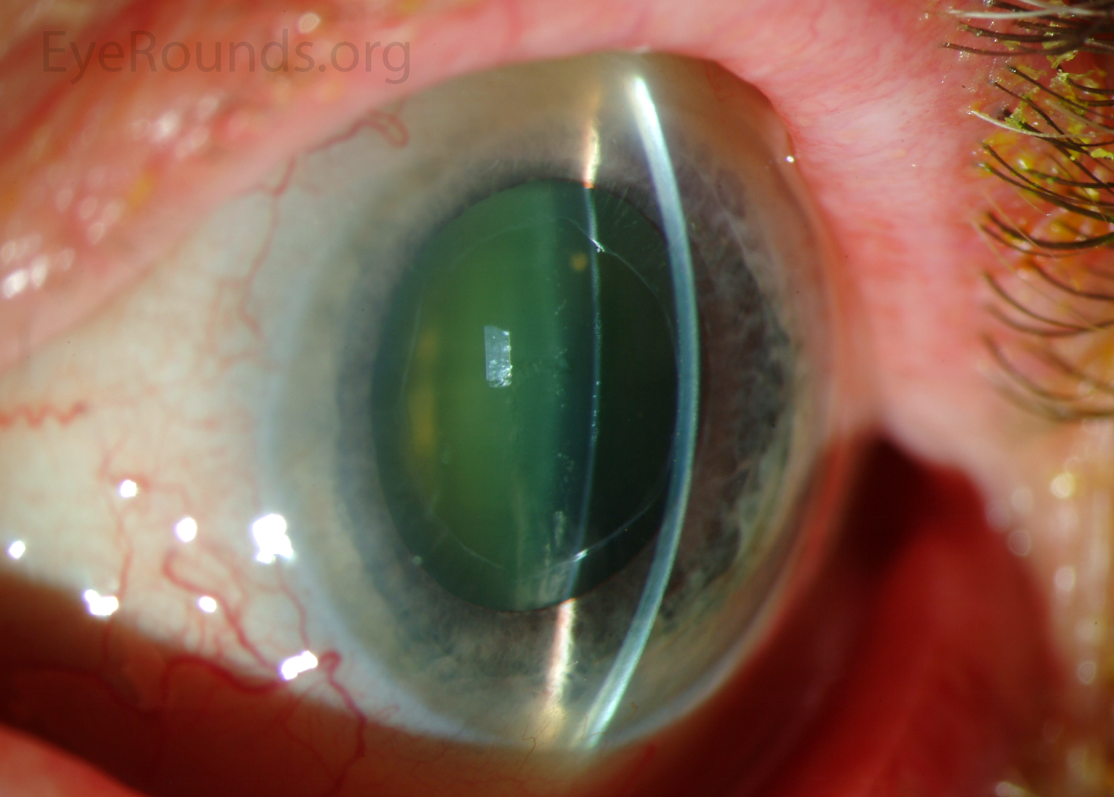
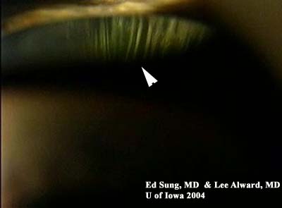
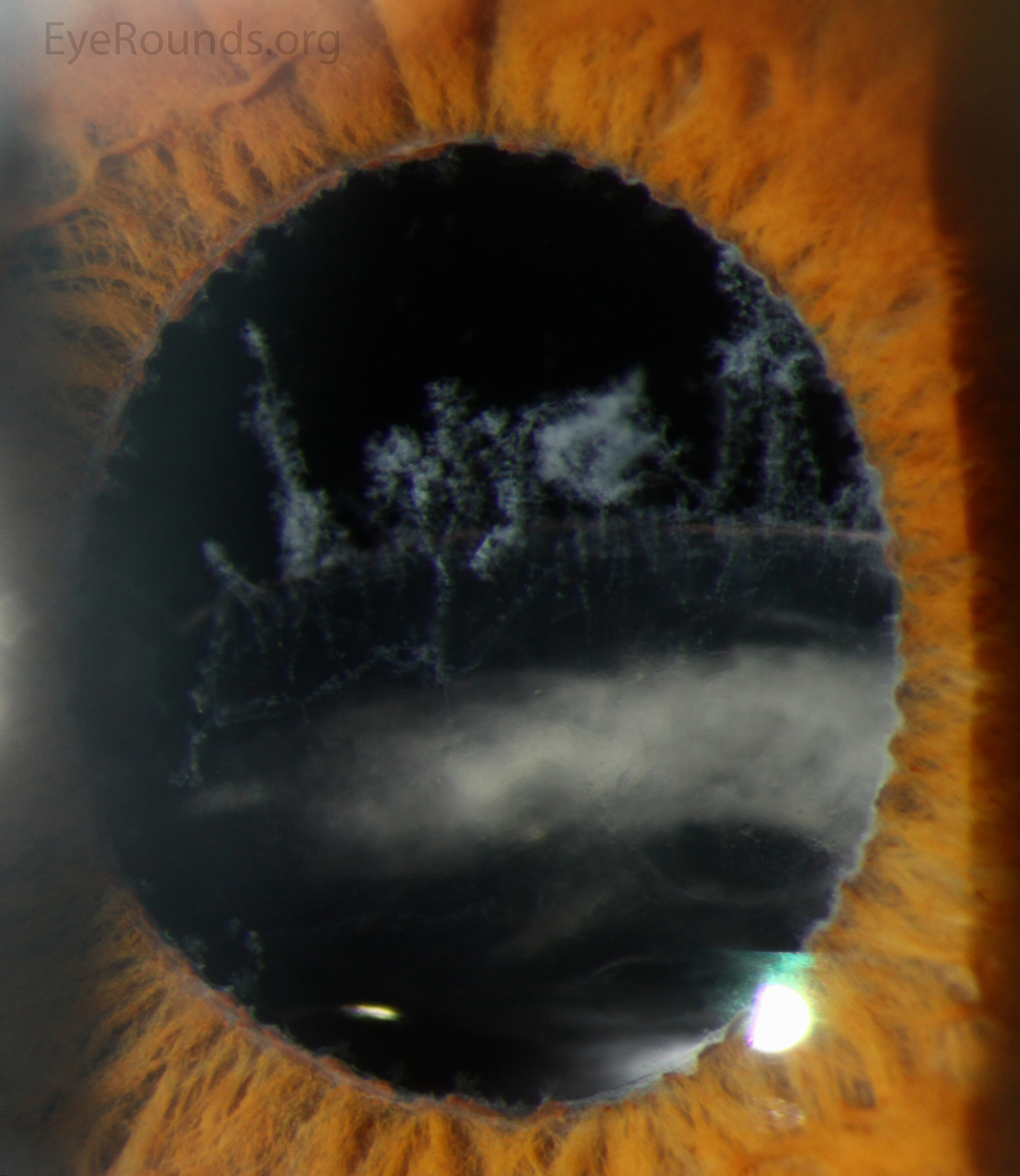
Pseudophakic late in-the-bag lens dislocation in a patient with pseudoexfoliation glaucoma. In this particular case, refixation or lens exchange is more complicated and challenging in the setting of trabeculectomy bleb is shown.


Contributor: Jesse Vislisel, MD; Lucas Lenci, MD
Photographer: Brice Critser, CRA (fig. 1a, 2a,b, 6, 8); Tracy Aly, CRA (fig. 2c,d); Toni Venckus, CRA (fig. 7)

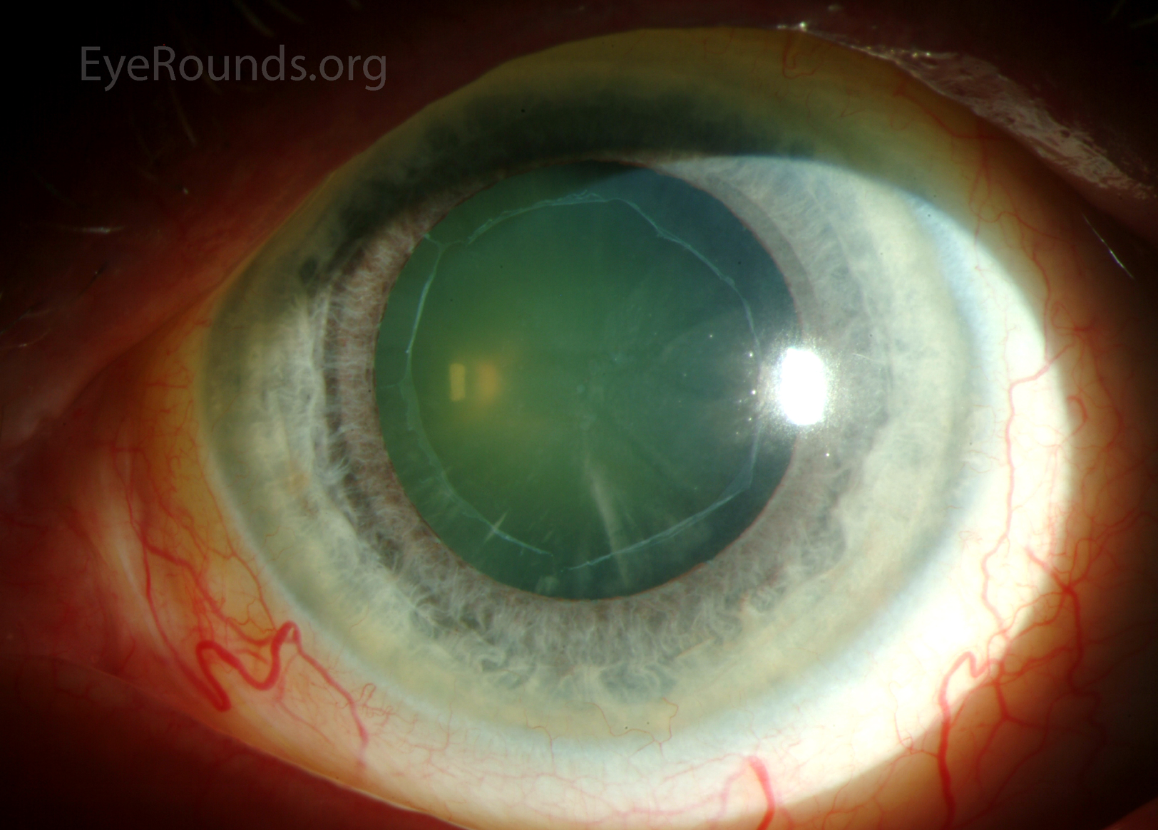
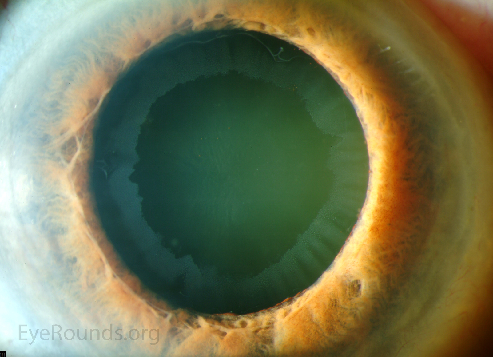
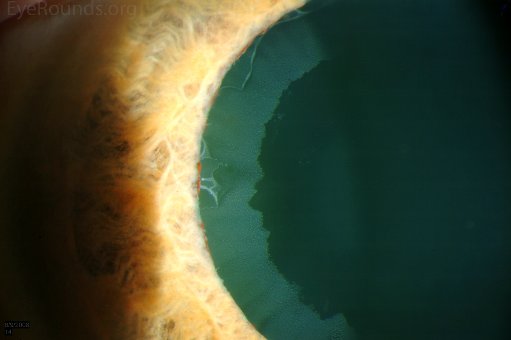
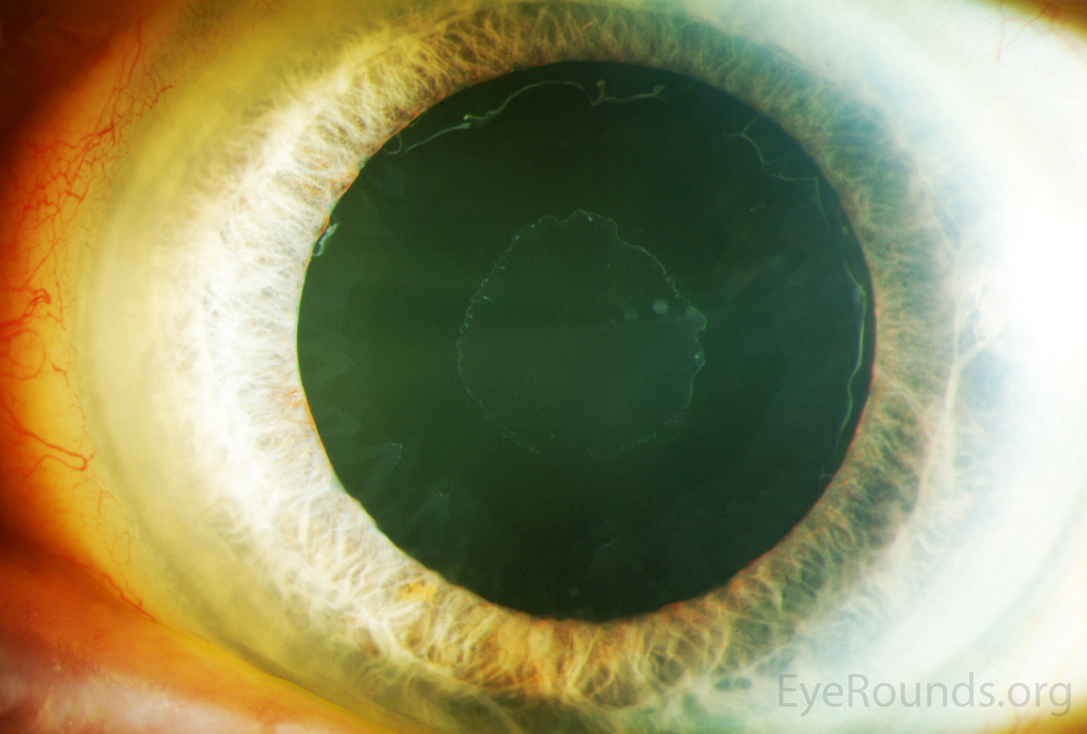
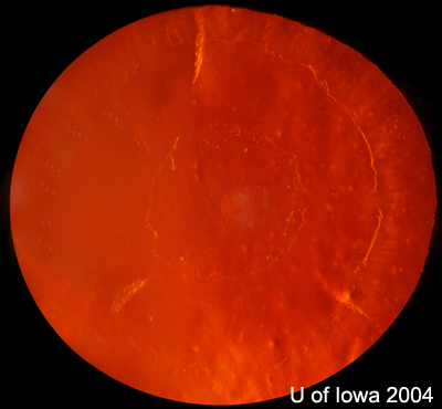
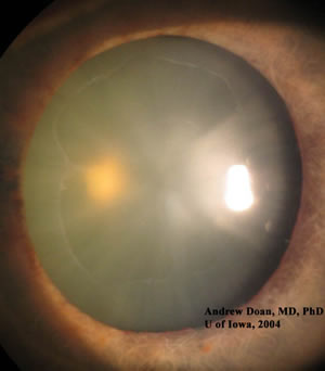
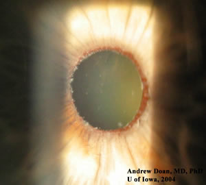
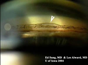
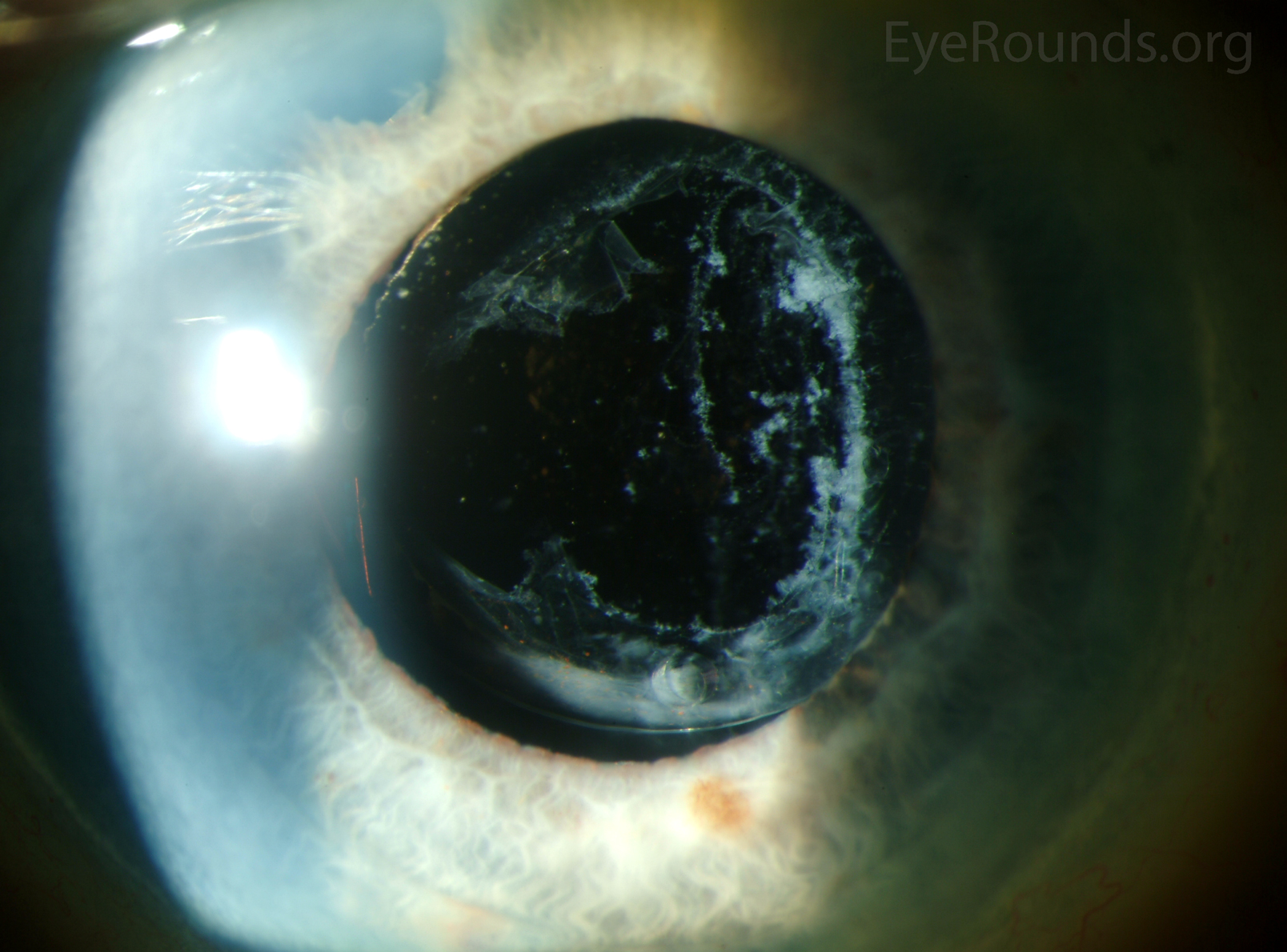
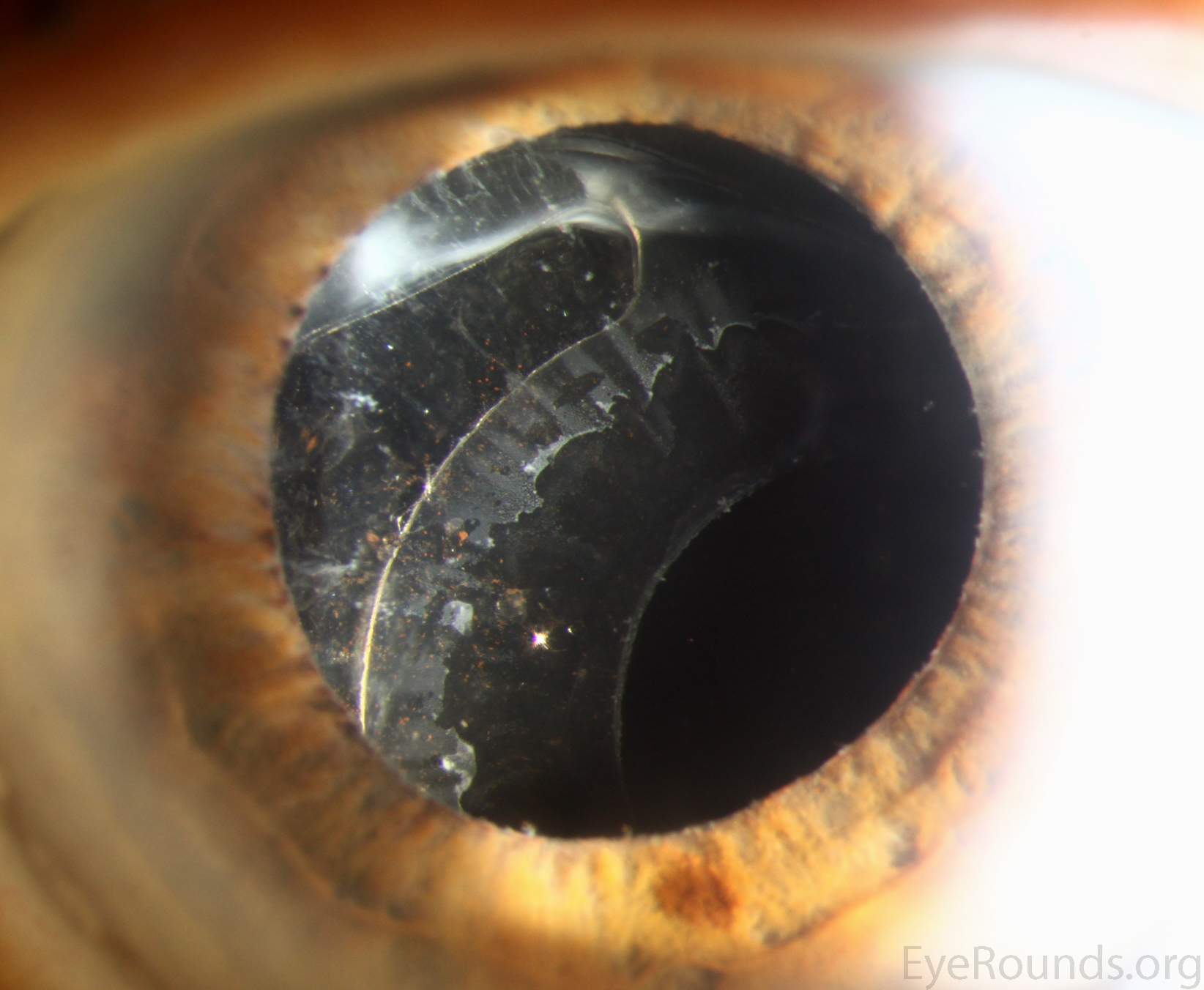
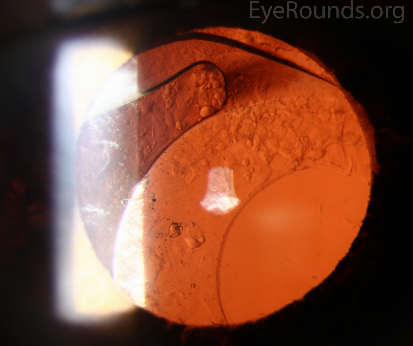
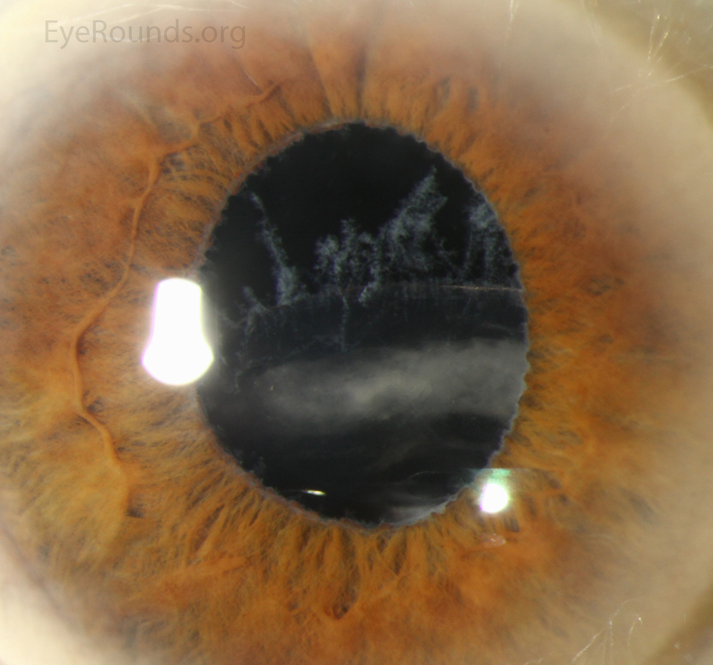
Pseudoexfoliation (PXF) syndrome is a condition in which fibrillar material is deposited in the anterior segment of the eye and other places throughout the body. It has complex inheritance, but is almost always associated with mutations in the LOXL1 gene. Individuals with this condition classically have exfoliative material visible along the pupillary margin and on the anterior lens capsule. The material on the lens capsule is often has a target-like appearance with distribution peripherally and centrally and an intervening clear space in the mid-periphery, presumably because the iris rubs the material off in this location as the pupil changes size. Other findings include "brown sugar" pigment in the trabecular meshwork, "moth-eaten" transillumination defects along the pupillary margin, and potentially zonular laxity which can lead to lens subluxation. It may be associated with open-angle glaucoma if the fibrillar material obstructs the trabecular meshwork and causes elevated intraocular pressure.

Deposition of exfoliative material on the anterior lens capsule
Figure 1a: Deposition of exfoliative material on the anterior lens capsule with intervening clear space. (higher resolution image of 1b not available)
Deposition of exfoliative material on the anterior lens capsule
Figure 1b: Deposition of exfoliative material on the anterior lens capsule with intervening clear space. (higher resolution image of 1b not available)
Exfoliative material on the pupillary margin and anterior lens capsule
Figure 2a: Exfoliative material on the pupillary margin and anterior lens capsule.
Exfoliative material on the pupillary margin and anterior lens capsule
Figure 2b: Exfoliative material on the pupillary margin and anterior lens capsule.
Exfoliative material on the pupillary margin and anterior lens capsule - slit lamp
Figure 2c: Exfoliative material on the pupillary margin and anterior lens capsule.
Exfoliative material on the pupillary margin and anterior lens capsule - high magnification
Figure 2c: Exfoliative material on the pupillary margin and anterior lens capsule.
 Exfoliative material on an undilated pupillary margin
Figure 3a: Exfoliative material on an undilated pupillary margin. (higher resolution image not available)
 Exfoliative material on an undilated pupillary margin
Figure 3b: Exfoliative material on an undilated pupillary margin. (higher resolution image not available)
Pigment deposition within the trabecular meshwork
Figure 4: Pigment deposition within the trabecular meshwork and Sampaolesi line on gonioscopy. (higher resolution image not available)
Exfoliative material visible on the lens zonules
Figure 5: Exfoliative material visible on the lens zonules. (higher resolution image not available)
 Exfoliative material accumulating on the posterior capsular remnants
Figure 6: Exfoliative material accumulating on the posterior capsular remnants after YAG laser posterior capsulotomy.
Subluxated intraocular lens implant occurring spontaneously many years after cataract surgery
Figure 7a: Subluxated intraocular lens implant occurring spontaneously many years after cataract surgery. Fibrillar material is visible on the anterior capsule and along the pupillary margin.
Subluxated intraocular lens implant occurring spontaneously many years after cataract surgery
Figure 7b: Subluxated intraocular lens implant occurring spontaneously many years after cataract surgery. Fibrillar material is visible on the anterior capsule and along the pupillary margin.
Inferiorly displaced posterior chamber intraocular lens located in the capsular bag
Figure 8a: Inferiorly displaced posterior chamber intraocular lens located in the capsular bag. There is pseudoexfolation material on the visible superior zonules and pupillary margin.
Inferiorly displaced posterior chamber intraocular lens located in the capsular bag
Figure 8b: Inferiorly displaced posterior chamber intraocular lens located in the capsular bag. There is pseudoexfolation material on the visible superior zonules and pupillary margin.