This site uses tracking information. Visit our privacy policy. Click to agree to this policy and not see this again.

Ophthalmology and Visual Sciences

Grouped congenital albinotic spots of the retinal pigmented epithelium (polar bear tracks)

Grouped congenital albinotic spots of the retinal pigmented epithelium (polar bear tracks)

Category(ies): Retina, Vitreous
Contributor: Jesse Vislisel, MD
Photographer: Brice Critser, CRA

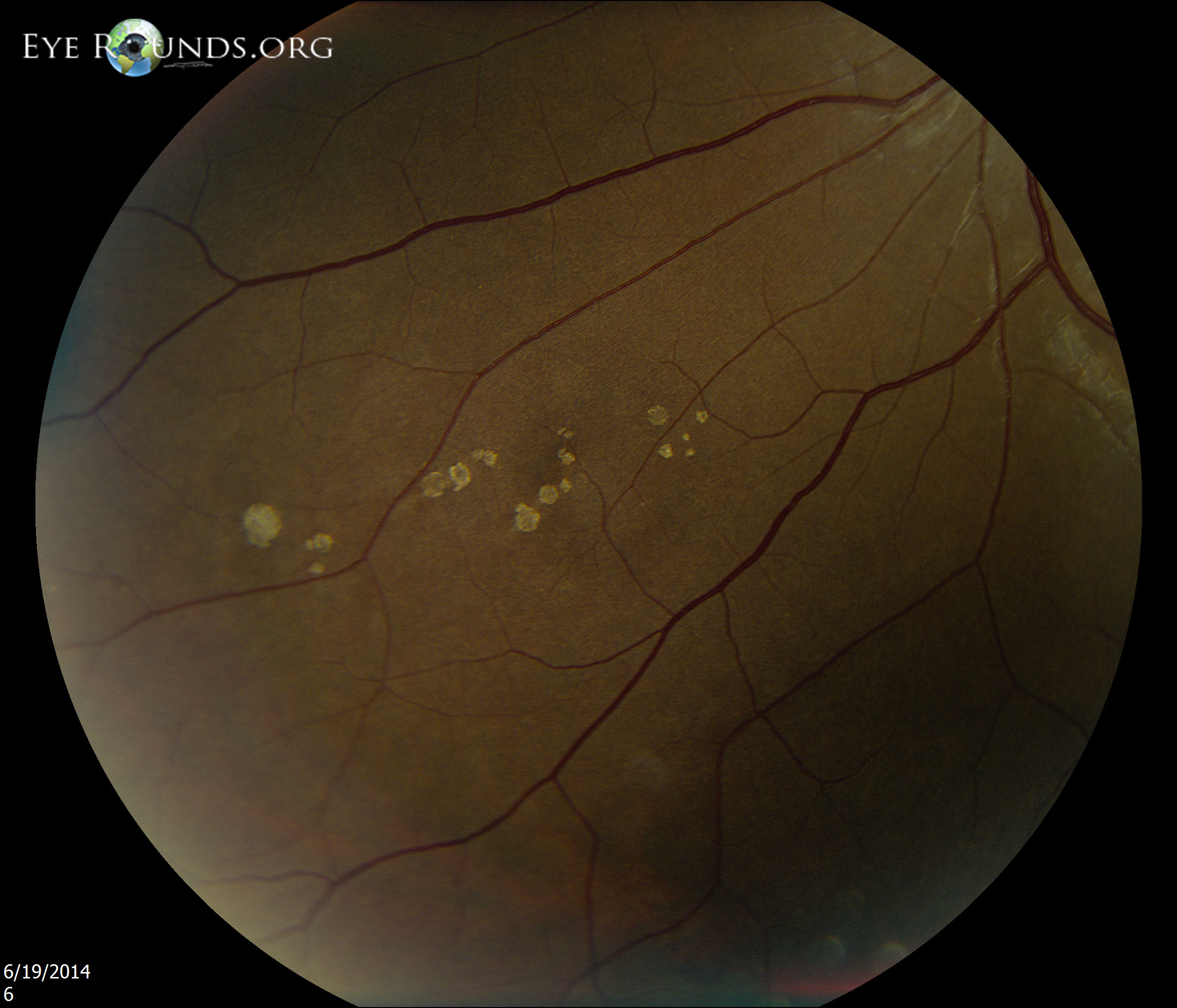
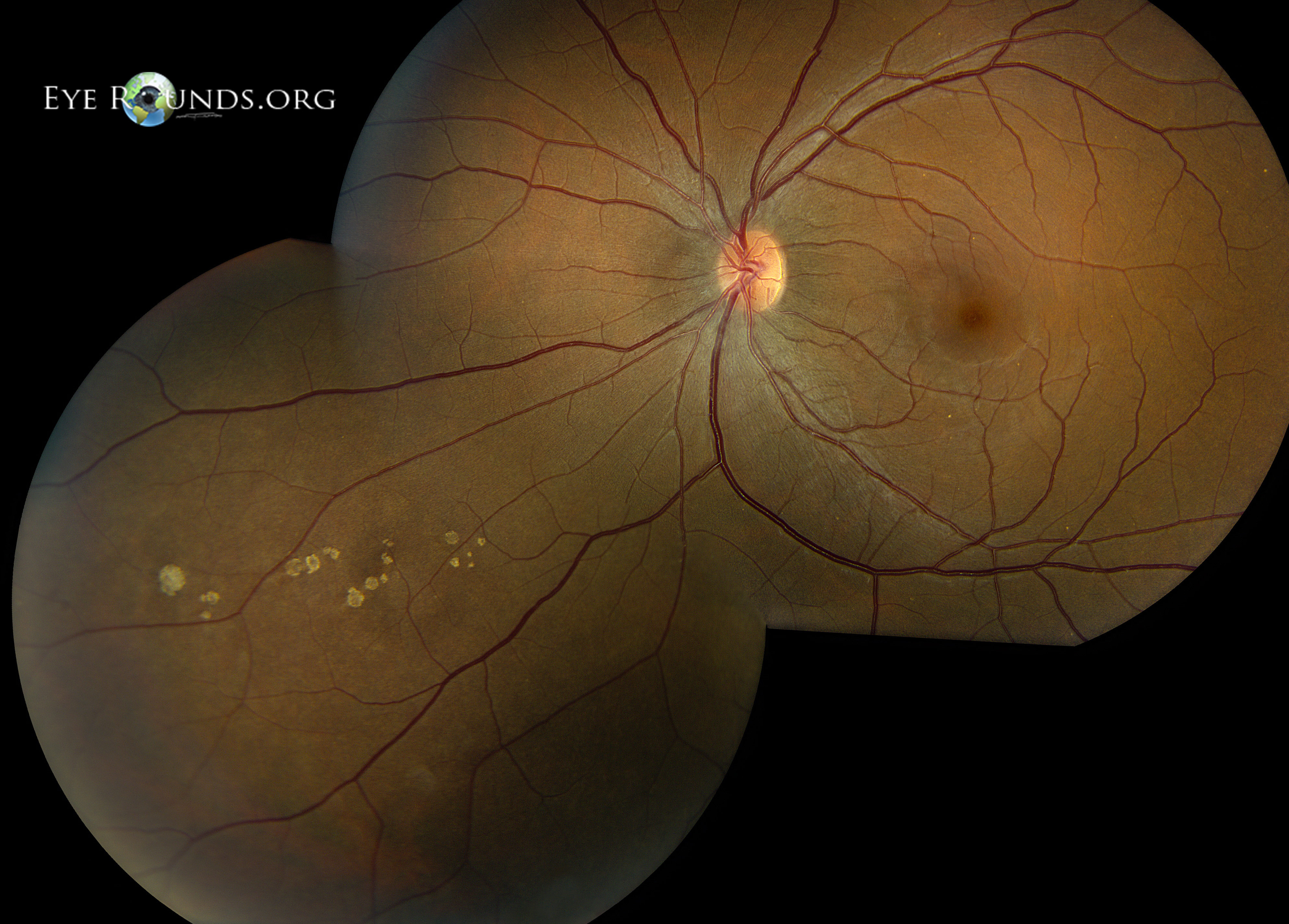
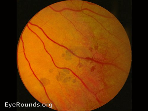
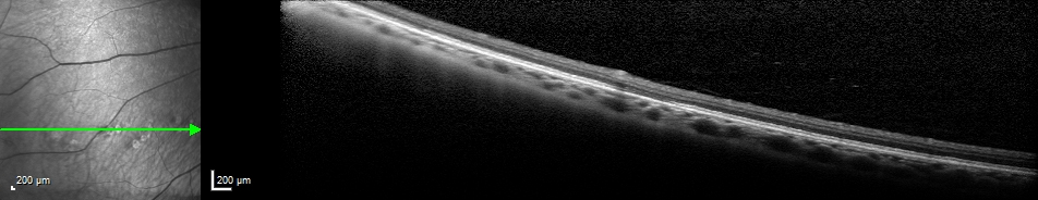
Congenital albinotic spots of the retinal pigmented epithelium (CASRPE) can be configured in solitary or grouped configurations. Groups of the spots, as seen here, are commonly referred to as "polar bear tracks" due to their color and resemblance of animal footprints. It is believed that these spots may develop secondary to deposition of an abnormal white material in retinal pigmented epithelial (RPE) cells rather than the usual darkly pigmented melanin. The lesions are flat, sharply circumscribed, placoid, chalky white, and lie at the level of the RPE. Optical coherence tomography (OCT) demonstrates a disruption of the highly-reflective signal from the ellipsoid zone in the region of the lesions, as indicated by the orange line on the scan.

oct of polar bear tracks, congenital albinotic spots of the retinal pigmented epithelium (CASRPE)
Optical coherence tomography (OCT) demonstrates a disruption of the highly-reflective signal from the ellipsoid zone in the region of the lesions, as indicated by the orange line on the scan.
Reference:
  • Gass JD. Focal congenital anomalies of the retinal pigment epithelium. Eye (1989) 3;1-18.
  • Kim DY, Hwang JC, Moore AT, Bird AC, Tsang SH. Fundus Autofluorescence and Optical Coherence Tomography of Congenital Grouped Albinotic Spots. Retina. Sep 2010; 30(8): 1217–1222.

Image Permissions:
Creative Commons

Ophthalmic Atlas Images by EyeRounds.org, The University of Iowa are licensed under a Creative Commons Attribution-NonCommercial-NoDerivs 3.0 Unported License.