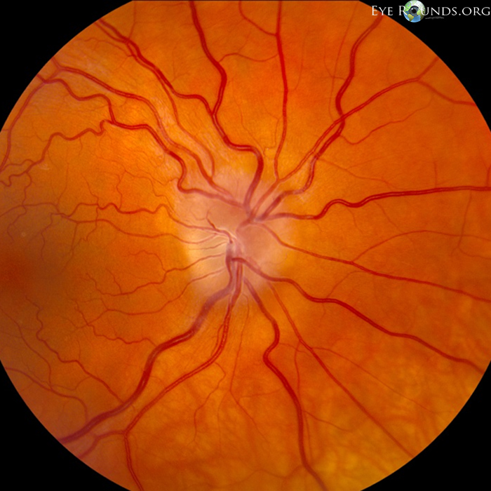
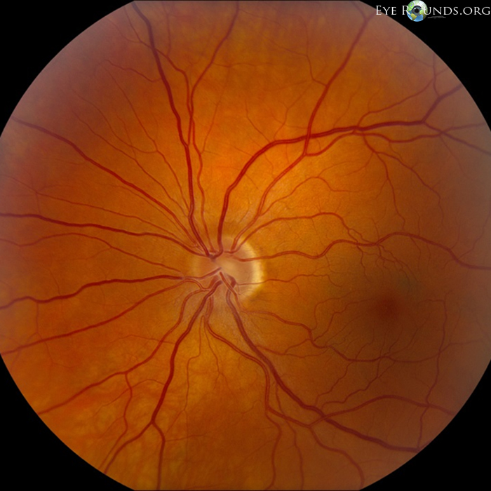
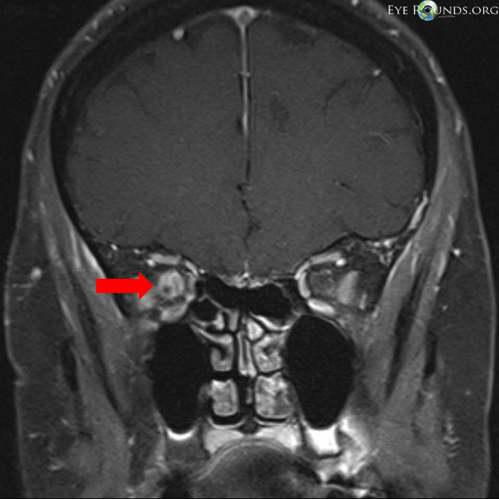
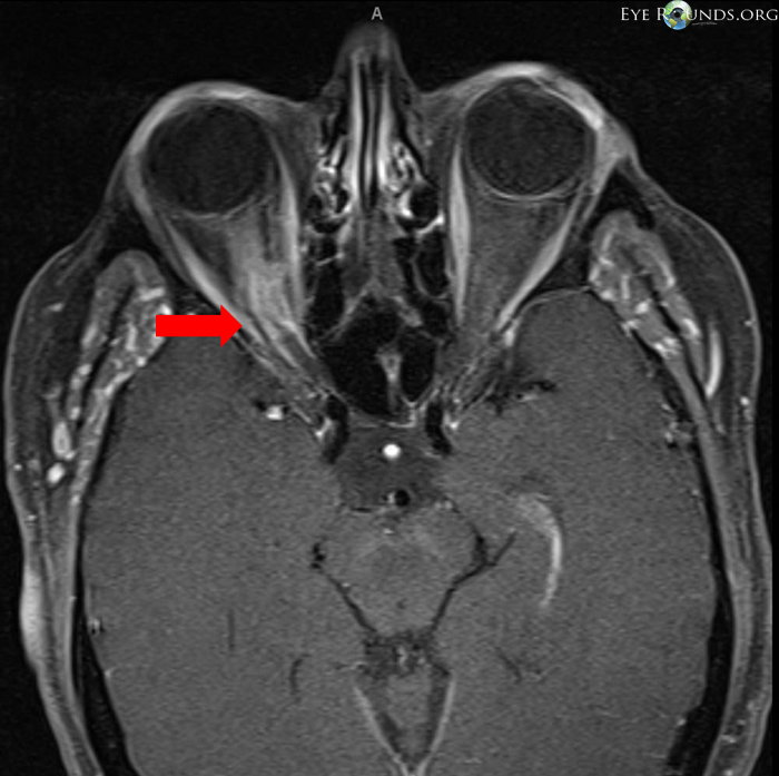
This patient presented with slowly progressive painless vision loss in the right eye. Dilated fundus examination demonstrates mild chronic appearing disc edema and dilated veins in the right eye, while the left eye is unaffected. MRI T1-weighted images with contrast show enlargement and enhancement of the optic nerve sheath consistent with an optic nerve sheath meningioma. The attenuated optic nerve is surrounded by the enhancing mass causing a "bull's eye" appearance on coronal and "tram track" appearance on axial images (red arrows).

Ophthalmic Atlas Images by EyeRounds.org, The University of Iowa are licensed under a Creative Commons Attribution-NonCommercial-NoDerivs 3.0 Unported License.


Address
University of IowaLegal
Related Links