This site uses tracking information. Visit our privacy policy. Click to agree to this policy and not see this again.

Ophthalmology and Visual Sciences

Ocular Toxoplasmosis

Ocular Toxoplasmosis

Category(ies): Retina, Vitreous, Uveitis
Contributor: Eric Chin, MD and Christi Carter, MD
Photographer: Toni Venckus, CRA

A 20-year-old male was referred by an optometrist because of a pigmented scar in the right eye. The patient complained of black spots in his vision for the past week. He had no pain or irritation. He was born in Peru, but came to the United States at the age of 14 and lived in Tennessee on a farm and had been exposed to many cats in the past. He had no other complaints or medical problems.

  • Vision with correction: OD 20/25-1; OS 20/20
  • Intraocular pressure: normal OU
  • Slit lamp examination OU: unremarkable; no AC cells; no posterior synechiae; clear lens
Dilated fundus examination of the right eye showed a well-circumscribed small old peripheral pigmented scar just beyond the inferotemporal arcades
Fig 1: Dilated fundus examination of the right eye showed a well-circumscribed small old peripheral pigmented scar just beyond the inferotemporal arcades, with a patchy area of active chorioretinitis inferiorly. There was no hemorrhage or vitritis.
magnification inferotemporally showed an old pigmented scar with an adjacent area of active chorioretinitis. The yellow arrows point to faint Kyrieleis' vascular plaques.
Fig 2a-b: Higher magnification inferotemporally showed an old pigmented scar with an adjacent area of active chorioretinitis. The yellow arrows point to faint Kyrieleis' vascular plaques.
magnification inferotemporally showed an old pigmented scar with an adjacent area of active chorioretinitis
Fig 2a-b: Higher magnification inferotemporally showed an old pigmented scar with an adjacent area of active chorioretinitis. The yellow arrows point to faint Kyrieleis' vascular plaques.
Dilated fundus examination of the left eye was unremarkable without any pigmented scars or active areas of chorioretinitis.
Fig 3: Dilated fundus examination of the left eye was unremarkable without any pigmented scars or active areas of chorioretinitis.

Ocular Toxoplasmosis

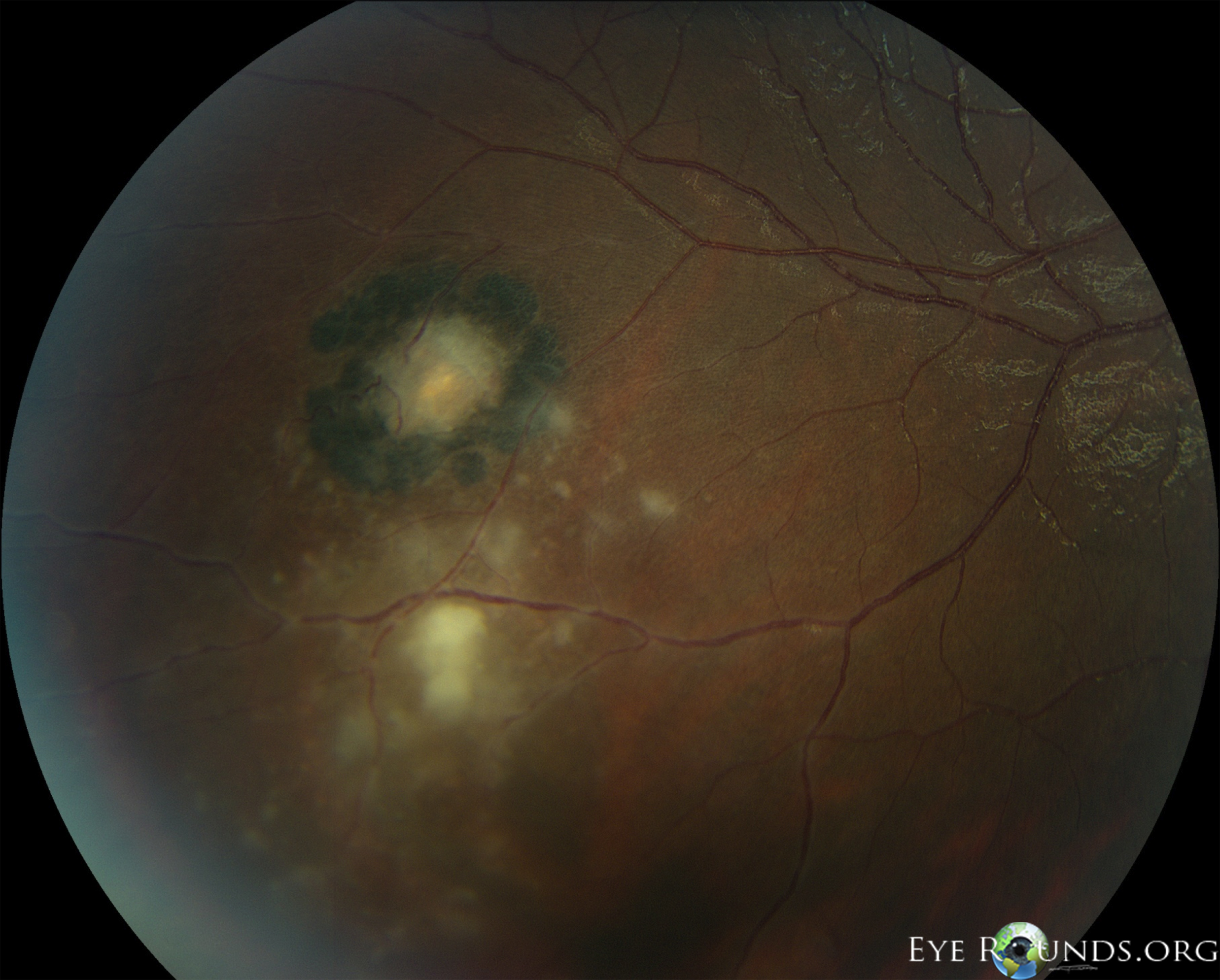
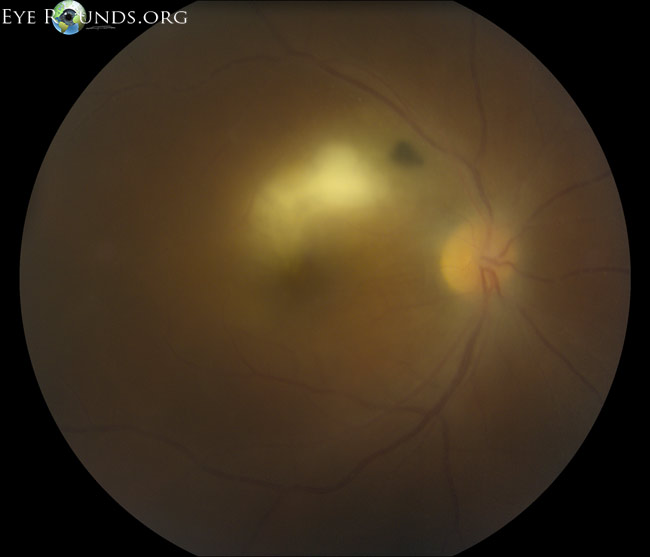
Toxoplasmic retinochoroiditis lesions have similar fundus characteristics, whether they result from congenital or acquired infections. Acute and new lesions are usually intensely white, focal lesions with overlying vitreous inflammatory haze. Active lesions with a severe vitreous inflammatory reaction will have the classic "headlight in the fog" appearance.[2]

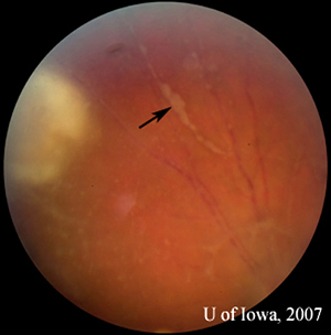
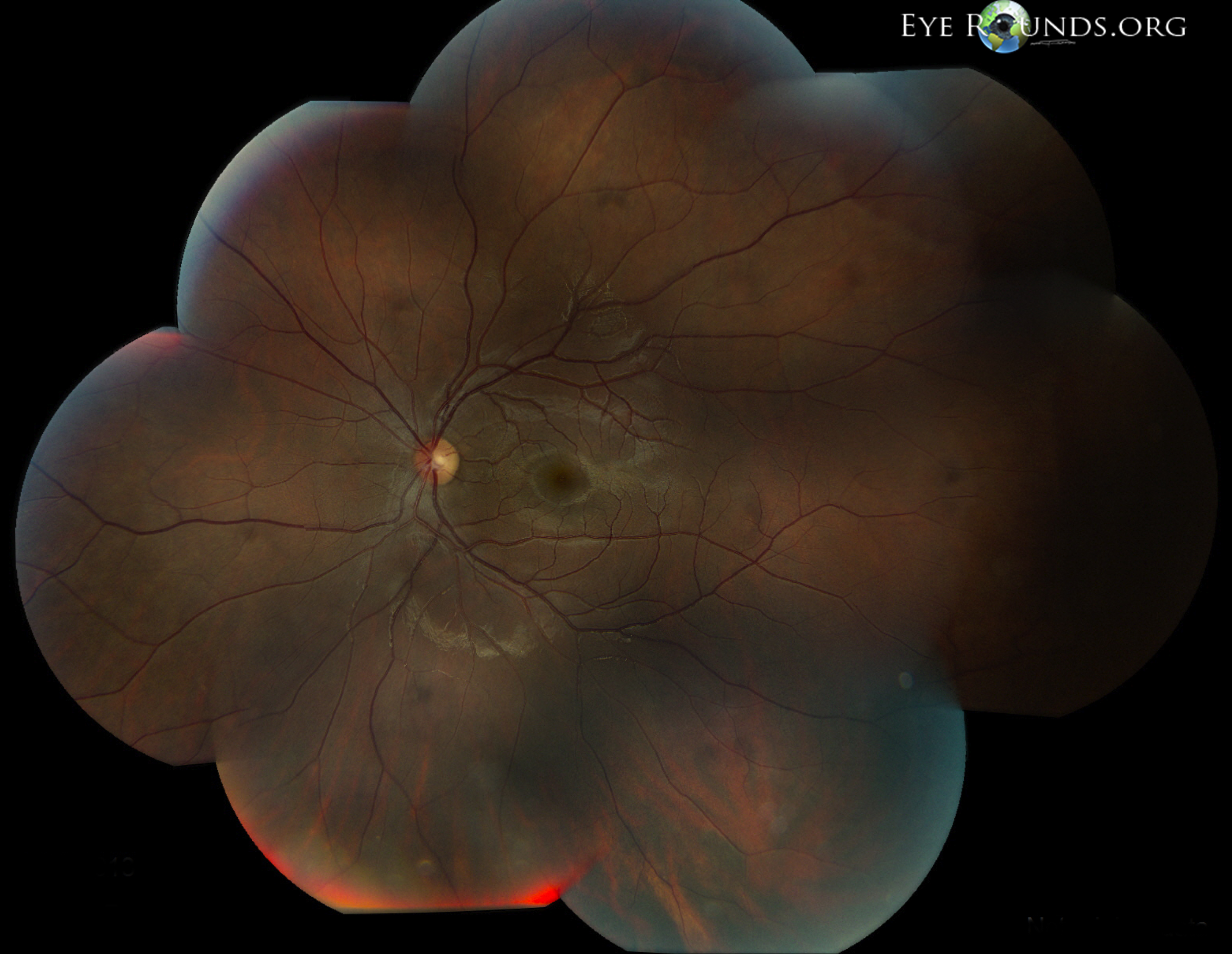
Patients with active toxoplasmic retinochoroiditis may develop retinal vasculitis with vascular sheathing and hemorrhages in response to reactions between circulating antibodies and local Toxoplasma gondii antigens.[3]

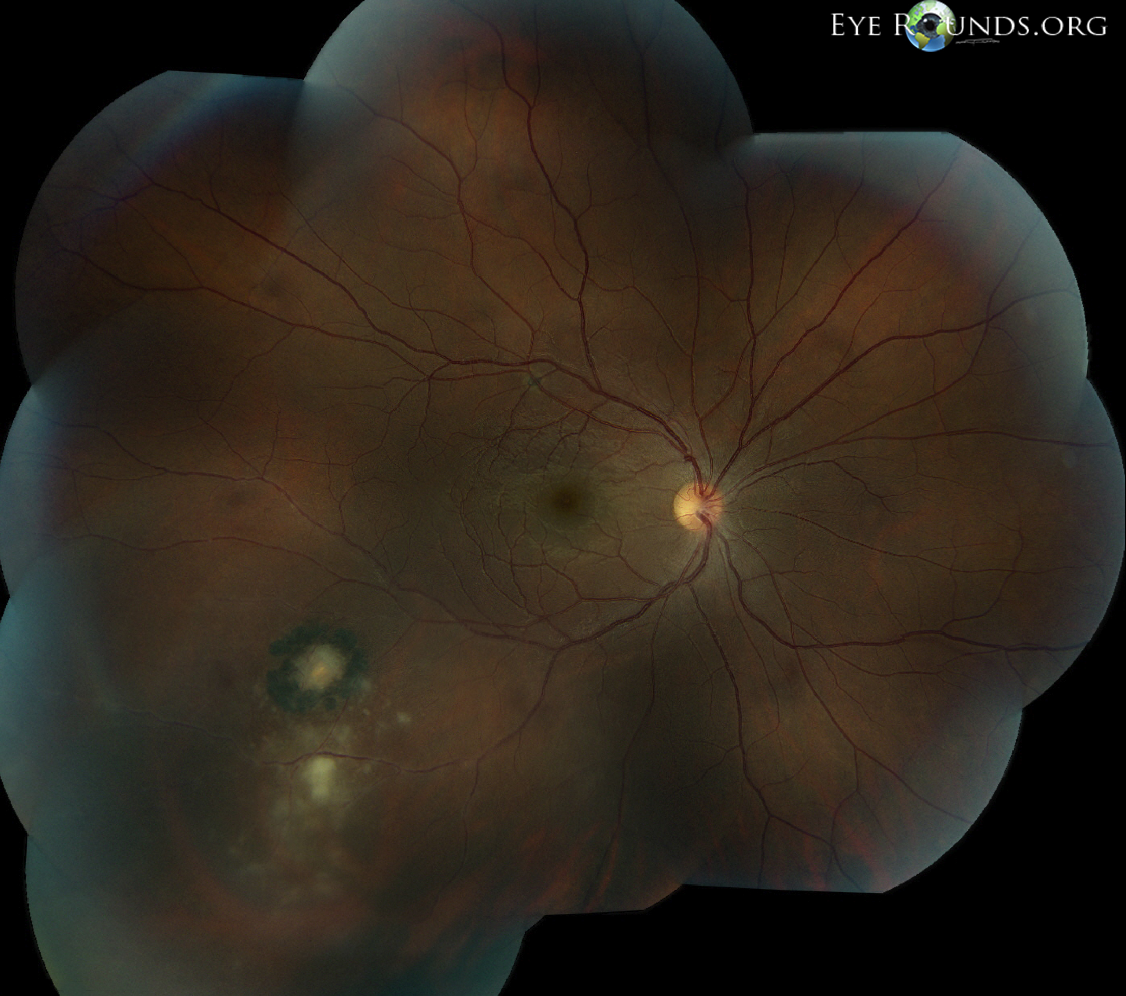
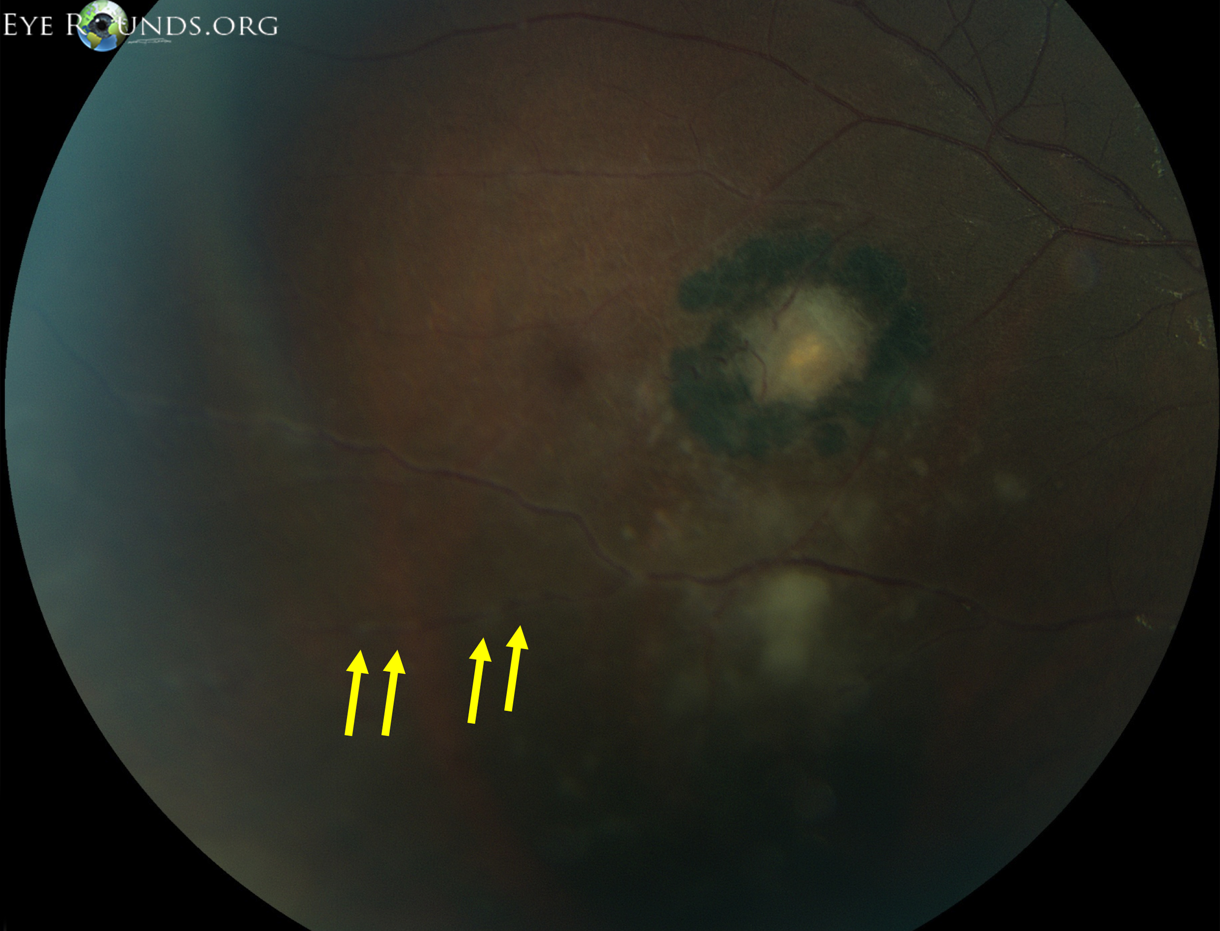
Typically, hyperpigmented scars of old and inactive lesions are present with recurrent "satellite lesions" at the border of healed scar. As a lesion heals, its borders become more defined and hyperpigmented. Large scars will have an atrophic center devoid of all retinal and choroidal elements. The underlying sclera gives the lesion its white center.[4]

"Kyrieleis plaques" describe nodular peri-arteritis or a beaded pattern of vascular inflammation. These plaques are predominately associated with toxoplasmosis infections but were originally reported in association with ocular tuberculosis in 1933.[5] It may be less commonly seen with syphilis, tuberculosis, cytomegalovirus, herpes zoster, and others.

Reference:
  1. Rothova A. Ocular manifestations of toxoplasmosis (Review). Curr Opin Ophthalmol 2003;14(6):384-388.
  2. Smith JR, Cunningham Jr ET. Atypical presentations of ocular toxoplasmosis (Review). Curr Opin Ophthalmol 2002;13:387-392.
  3. Hovakimyan A, Cunningham Jr ET: Ocular toxoplasmosis (Review). Ophthalmol Clin North Am 2002;15(3):327-332.
  4. Kyrieleis W. Uber atypische gerfaesstuberkulose der netzhaut (periarteritis "nodosa" tuberculosa). Arch Augenheilkd 1933;107:182-90.
  5. Holland GN, Crespi CM, ten Dam-van Loon N, et al. Analysis of recurrence patterns associated with toxoplasmic retinochoroiditis. Am J Ophthalmol 2008;145(6):1007-1013.

Image Permissions:
Creative Commons

Ophthalmic Atlas Images by EyeRounds.org, The University of Iowa are licensed under a Creative Commons Attribution-NonCommercial-NoDerivs 3.0 Unported License.