see case: Weed M, Johns AT, Thurtell M. Ocular Syphilis Presenting with Posterior Subcapsular Cataract and Optic Disc Edema: 30-year-old female with progressive bilateral vision loss EyeRounds.org. Posted Oct 10, 2012. Available from http://www.EyeRounds.org/cases/157-ocular-syphilis.htm.
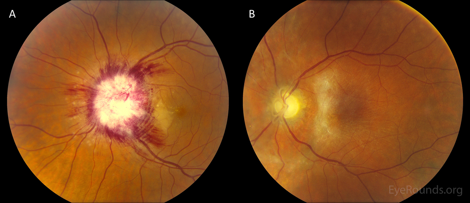
A 50-year-old male presented with 10 days of progressive vision loss in the left eye (OS) as well as fevers, fatigue, and unintentional weight loss. Visual acuity was hand motion OS. Slitlamp examination showed 1+ anterior vitreous cells. On funduscopy, he had severe optic disc edema with 360 degrees of peripapillary cotton wool spots and nerve fiber layer hemorrhages, along with a serous retinal detachment with peripapillary yellowish inflammation in the outer retina around the disc (A). He was found to have active syphilis with AIDS (Acquired Immunodeficiency Syndrome). He was treated with penicillin at that time and anti-retroviral therapy was ultimately initiated by the infectious diseases service. At 10 months follow up, his vision was light perception. He had severe optic atrophy, retinal thinning around the optic nerve, and an epiretinal membrane (B).
Patients with ocular syphilis may present with findings most pronounced in the area near the optic disc. The macular chorioretinal lesion just temporal to the optic disc is consistent with the typical placoid lesions seen with syphilitic uveitis.

Ophthalmic Atlas Images by EyeRounds.org, The University of Iowa are licensed under a Creative Commons Attribution-NonCommercial-NoDerivs 3.0 Unported License.

Address
University of IowaLegal
Related Links