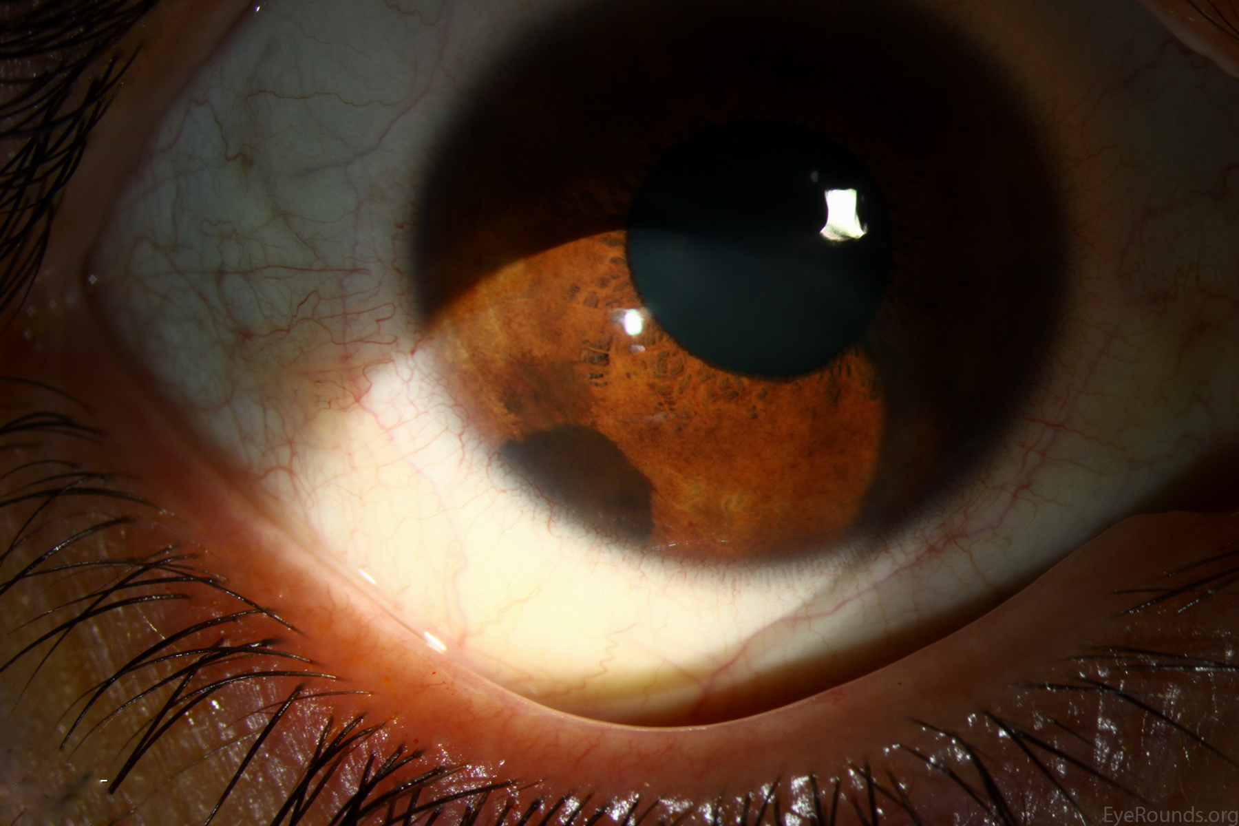
Iris nevi typically appear as hyperpigmented regions of the iris with minimal disruption of the normal iris architecture. However, they may occasionally be associated with ectropion uveae or sectoral cataract. There are two types of iris nevi, Circumscribed or Diffuse. Circumscribed iris nevi are discrete and often nodular. Diffuse iris nevi, as shown in the first photograph, involve an entire sector or rarely the entire iris. Iris nevi have low risk for malignant transformation. In a large case series, only 8% of iris nevi referred for evaluation at an ocular oncology center transformed into melanoma.
An ABCDEF mnemonic was proposed by Shields to help remember the risk factors for growth:
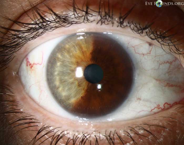
This is an example of a circumscribed iris nevus. On slit lamp examination, a well-circumscribed, brown mass is situated on the inferior iris without distortion of the surrounding iris architecture or pupil. There is no prominent vascularity to the lesion or "sentinel vessels" seen on the adjacent conjunctiva./figcaption>
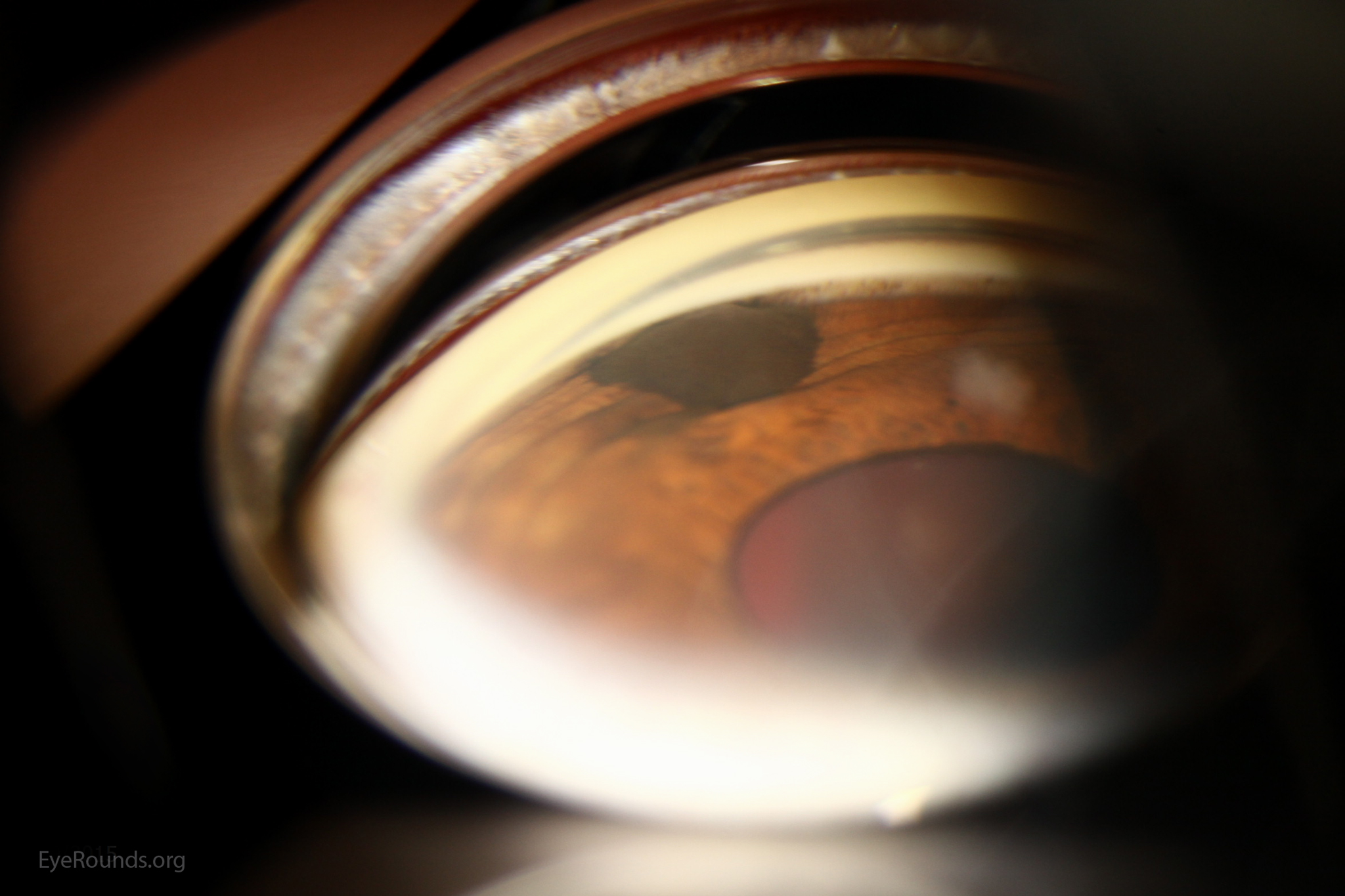
On gonioscopy, the lesion appears to sit on top of the iris without distorting or disrupting the normal iris architecture. There is no extension into the surrounding trabecular meshwork/figcaption>
University of Iowa
Roy J. and Lucille A. Carver College of Medicine
Department of Ophthalmology and Visual Sciences
200 Hawkins Drive
Iowa City, IA 52242
University of Iowa
Roy J. and Lucille A. Carver College of Medicine
Department of Ophthalmology and Visual Sciences
200 Hawkins Drive
Iowa City, IA 52242