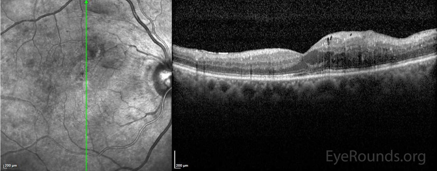
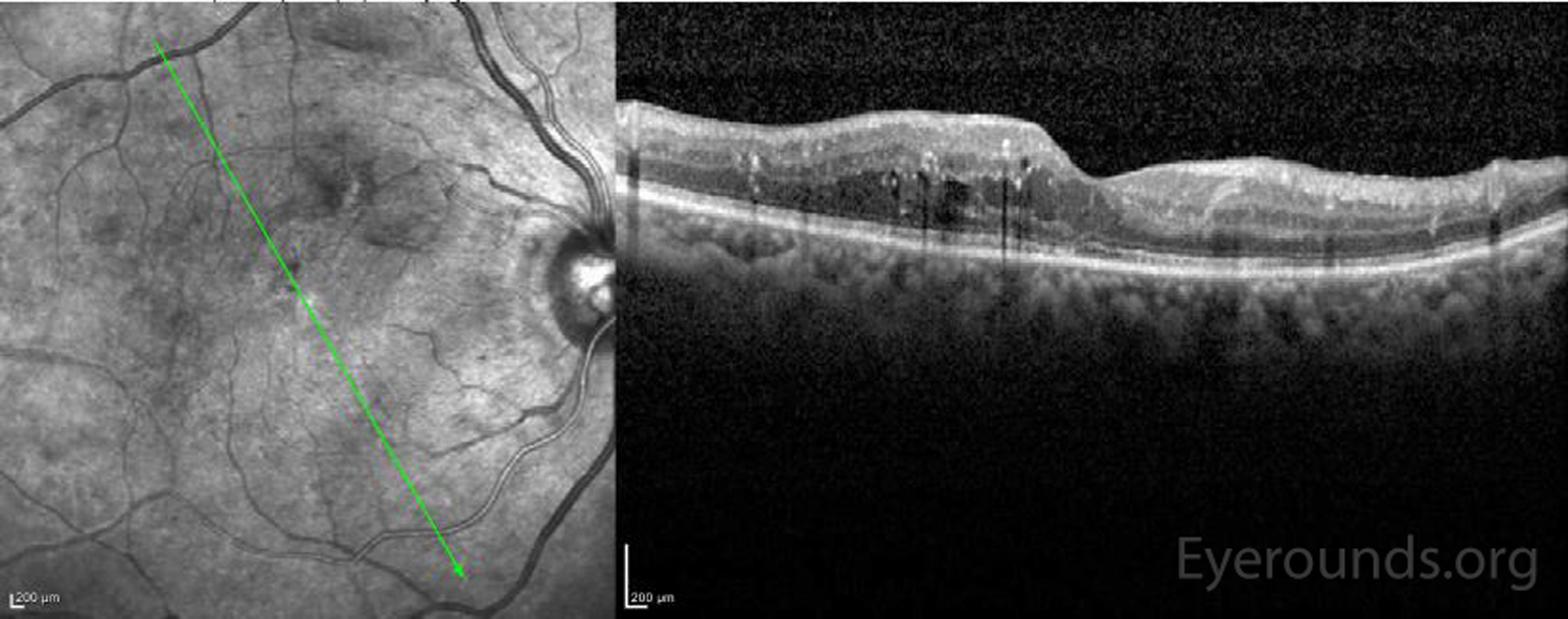
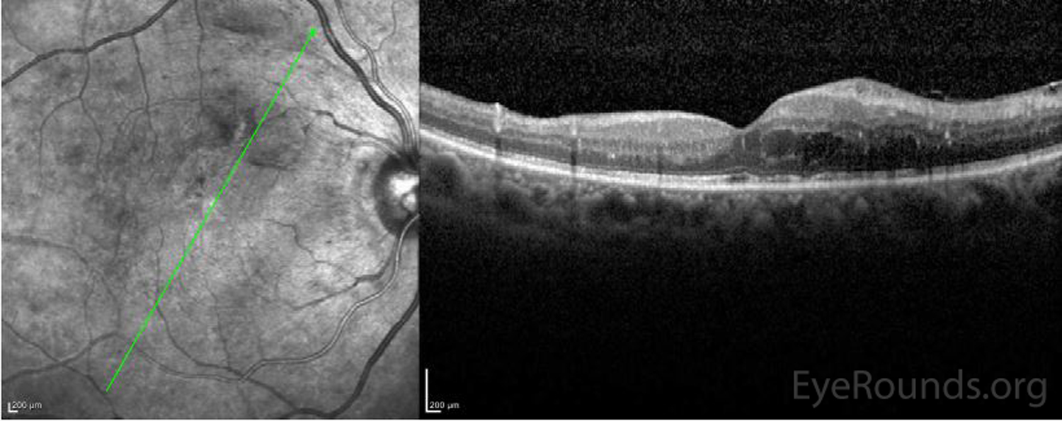
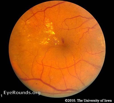
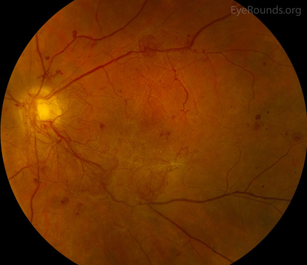
This patient has a history of severe non-proliferative retinopathy and recurrent diabetic macular edema in both eyes due to type II diabetes mellitus. On the optical coherence tomogram below, there are parafoveal intraretinal cysts with exudates.
Diabetic macular edema can result in diffuse or focal areas of swelling and retinal thickening and may be associated with hard exudates. Diabetic macular edema is often classified based on the Early Treatment Diabetic Retinopathy Study (ETDRS), which defined clinically significant diabetic macular edema. However, these findings are based on the funduscopic examination and in classifying this edema based on optical coherence tomography (OCT), the term center-involving macular edema is often used. Treatment includes the use of anti-VEGF drugs but may also include the use of focal or grid pattern photocoagulation or pars plana vitrectomy.
Diabetic macular edema. In: Basic and clinical science course (BCSC) Section 12: Retina and vitreous. Chapter 5 Retinal vascular disease: diabetic retinopathy. San Francisco, CA: American Academy of Ophthalmology. 2016-2017 p92-100

Ophthalmic Atlas Images by EyeRounds.org, The University of Iowa are licensed under a Creative Commons Attribution-NonCommercial-NoDerivs 3.0 Unported License.

Address
University of IowaLegal
Related Links