Cavernous sinus syndrome is characterized by lesions that affect multiple cranial nerves within the cavernous sinus. The clinical presentation typically consists of various combinations of ocular motor nerve impairments (Oculomotor Nerve - CN III, Trochlear Nerve - CN IV, and Abducens Nerve - CN VI), Horner syndrome, and sensory loss of the first (V1) or second (V2) division of the trigeminal nerve (CN V). There are a variety of etiologies that can cause this.
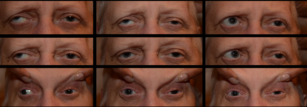
This patient presented with a left-sided ptosis, anisocoria and near complete ophthalmoplegia of the left eye. She demonstrated severe supraduction, infraduction, adduction and abduction deficits on the left and had 10 degrees of excyclotorison on double Maddox rod testing. She also had V1 and V2 distribution numbness. There was no evidence of visual field loss, a relative afferent pupillary defect, or decrease in vision. She is demonstrating palsies of CN III, CN IV, CN VI and the first two divisions of CN V on the left that localizes her lesion to the cavernous sinus. Imaging showed a large sphenoid wing meningioma that was infiltrating the cavernous sinus.

Ophthalmic Atlas Images by EyeRounds.org, The University of Iowa are licensed under a Creative Commons Attribution-NonCommercial-NoDerivs 3.0 Unported License.

Address
University of IowaLegal
Related Links