Calcific band keratopathy (BK) results from calcium hydroxyapetite deposition in the superficial cornea, mainly Bowman layer. It can be caused by chronic ocular inflammation, hypercalcemia, hyperphosphatemia, chronic exposure to mercurial vapors or preservatives, silicone oil, primary hereditary transmission. It is often idiopathic. BK usually begins as fine, yellow-white deposits in the periphery that may coalesce over time to form a horizontal band across the cornea.
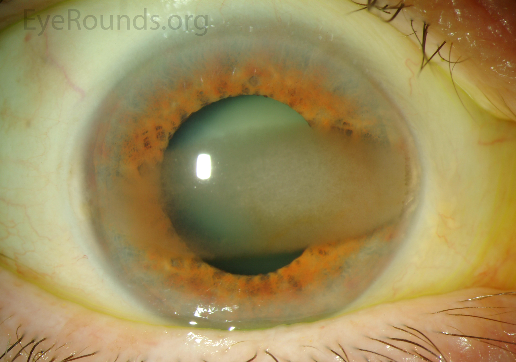
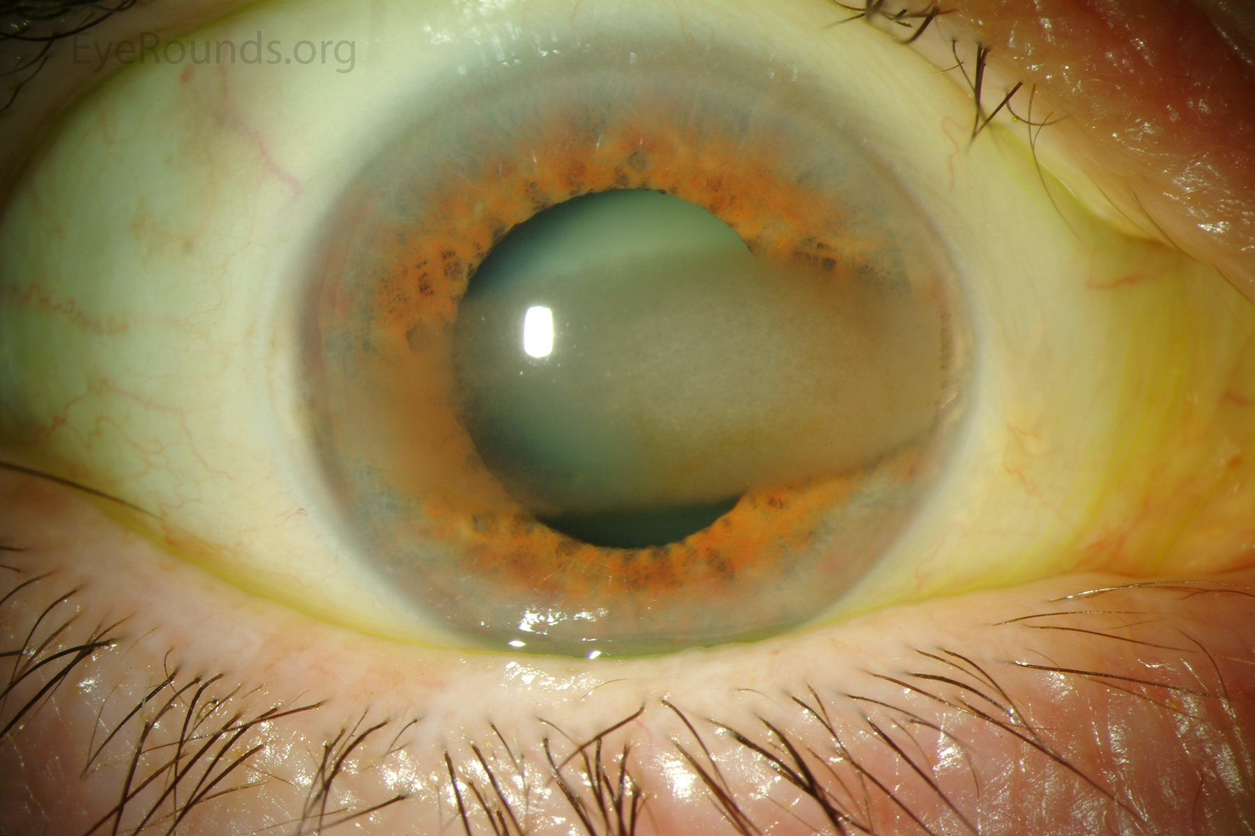
Calcific band keratopathy secondary to hyperparathyroidism
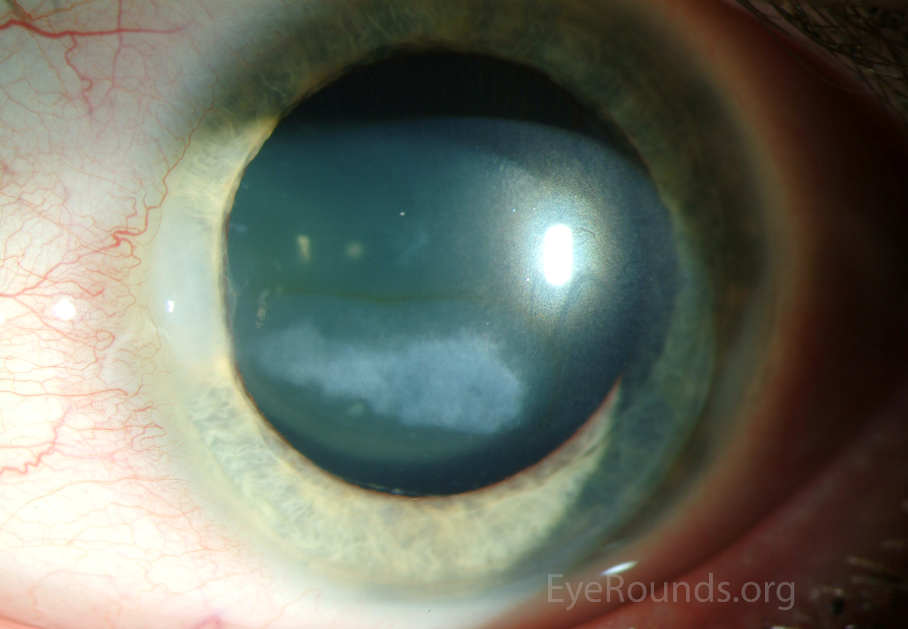
Early band keratopathy in a patient with uveitis associated with juvenile idiopathic arthritis. The accompanying iron line denotes the chronicity of these corneal changes.
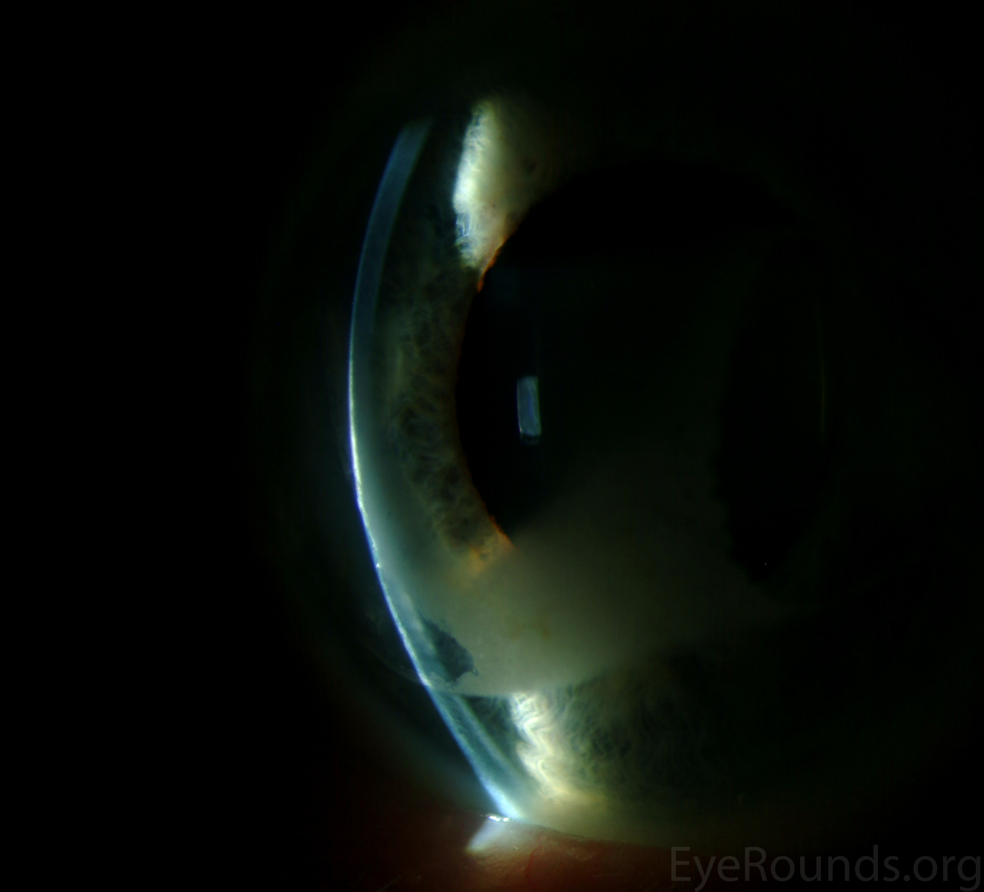
Late band keratopathy with a chalky, white consistency.
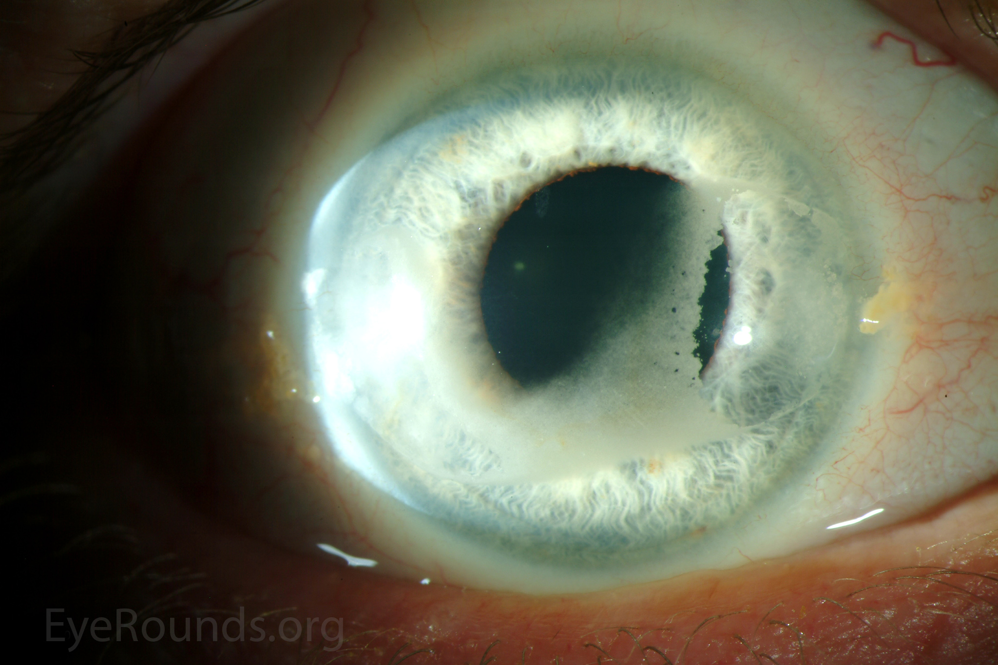
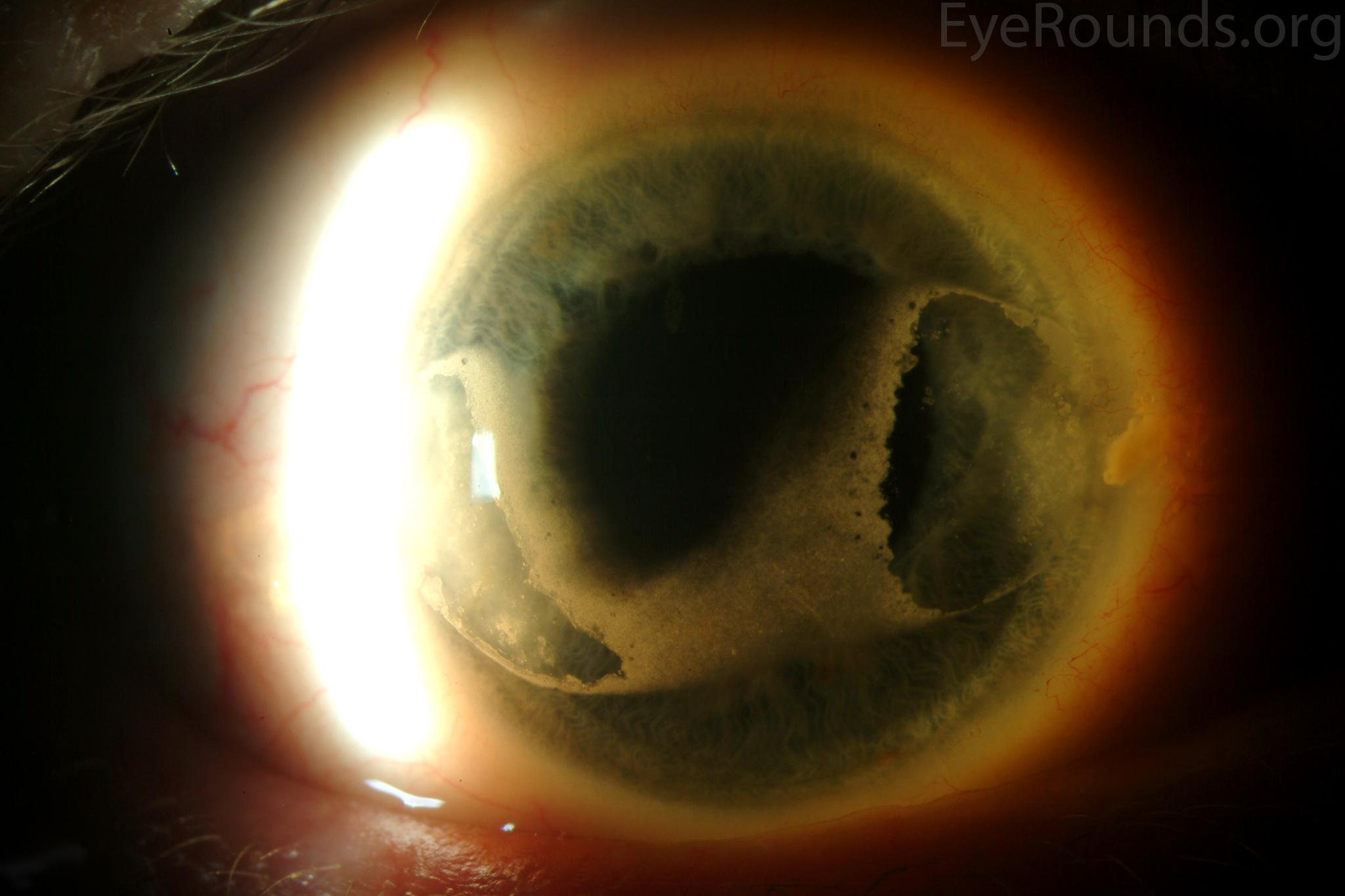
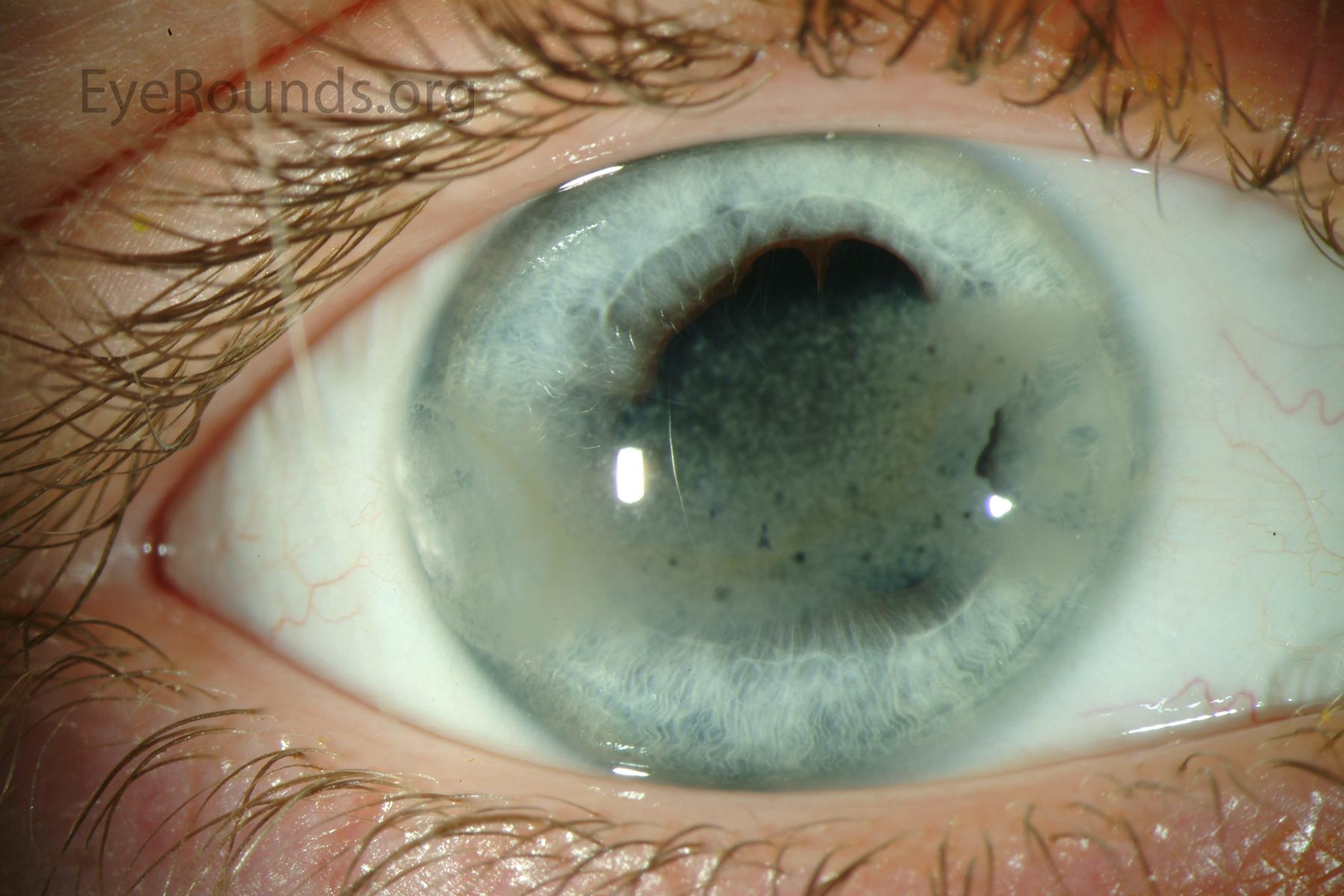
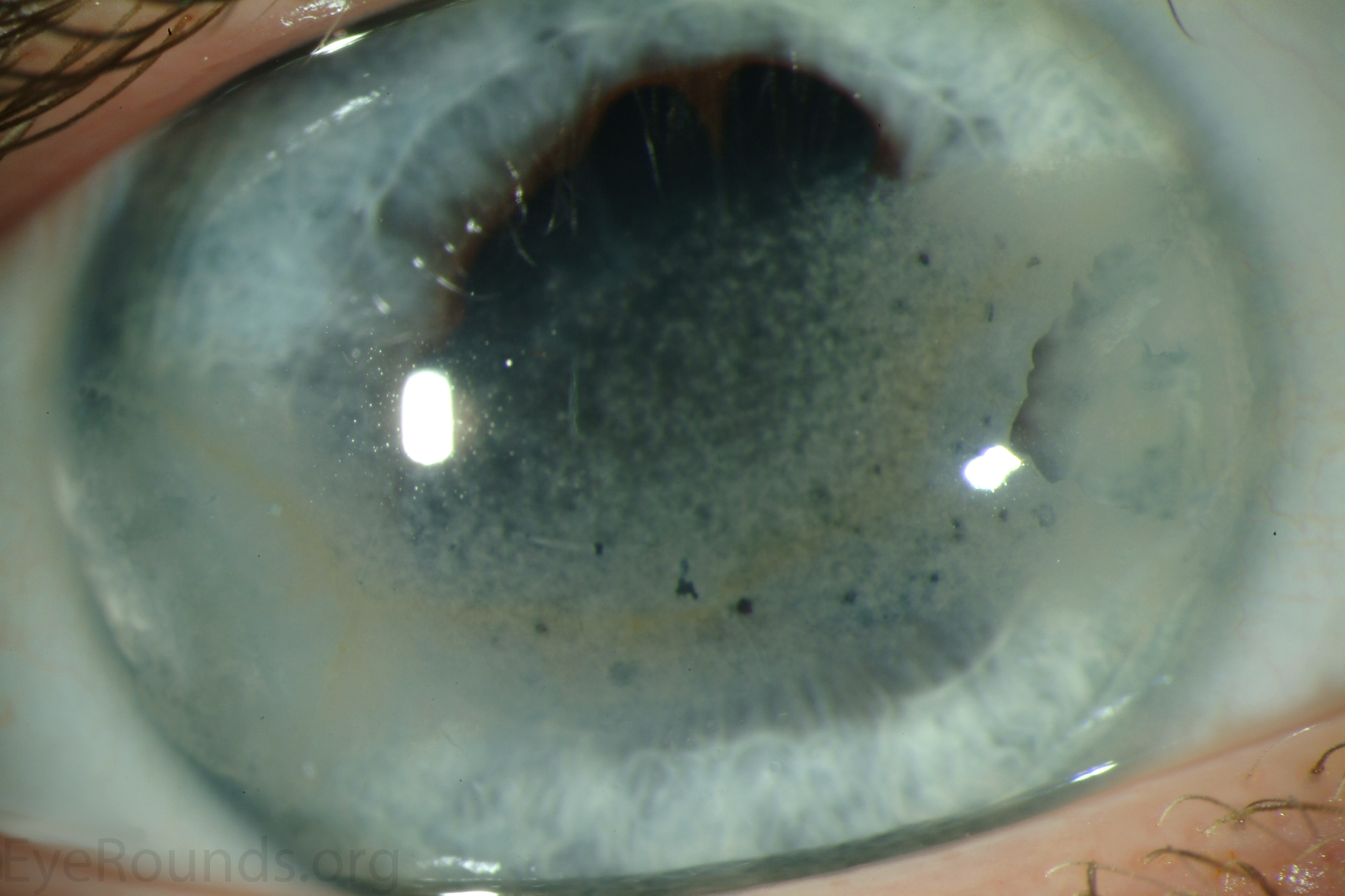
Band keratopathy in a patient with HLA-B27-associated anterior uveitis. The "Swiss cheese" appearance derives from lucent "holes" in the calcific pattern due to corneal nerve penetration through Bowman's layer.
Calcific band keratopathy

Ophthalmic Atlas Images by EyeRounds.org, The University of Iowa are licensed under a Creative Commons Attribution-NonCommercial-NoDerivs 3.0 Unported License.

Address
University of IowaLegal
Related Links