This site uses tracking information. Visit our privacy policy. Click to agree to this policy and not see this again.

Ophthalmology and Visual Sciences

Stargardt disease

Stargardt disease

Category(ies): Genetics, Inherited Eye Disease, Retina
Contributor: Jeffrey D. Welder, MD
Photographer: D. Brice Critser, CRAt

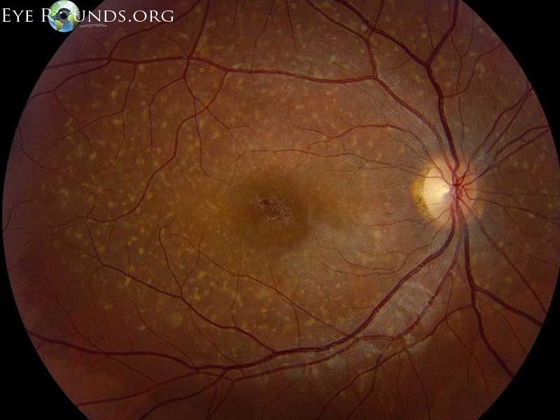
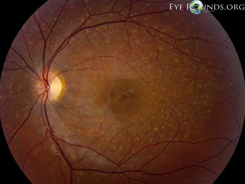
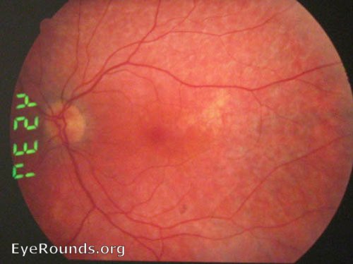
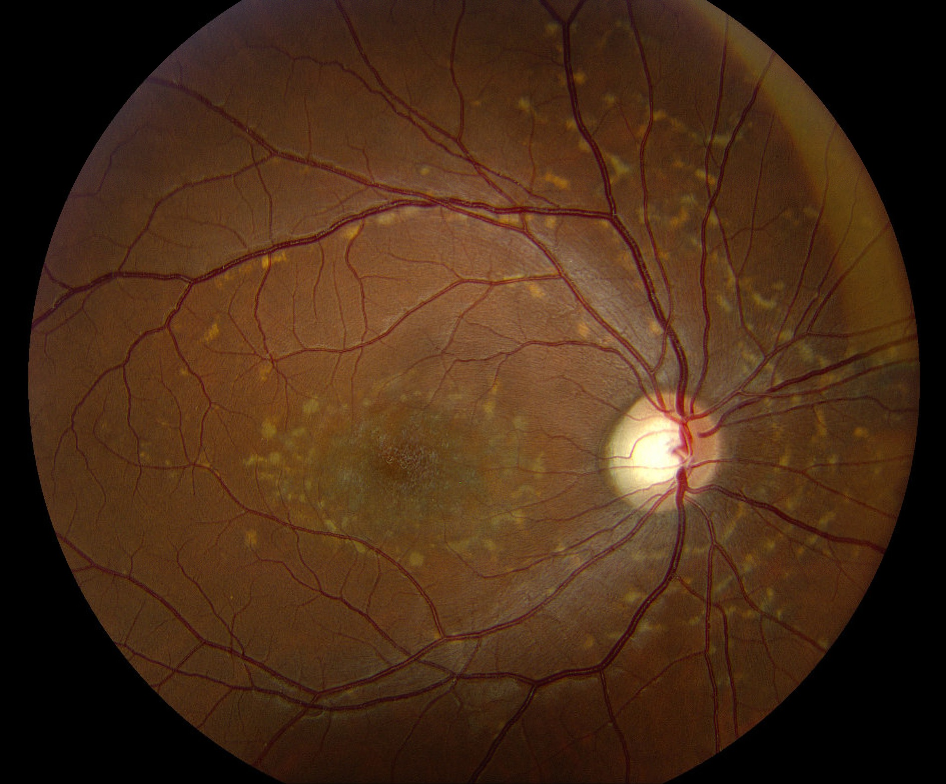
20-year-old male with molecularly confirmed Stargardt disease (ABCA4 mutation). Note the diffuse yellow pisciform flecks within the arcades and central macular mottling in both eyes.

OMIM #248200, #600110, #603786


Stargardt's Disease (macular dystrophy)

Stargardt's Disease (macular dystrophy)

Contributor: Andrew Doan, MD, PhD
Fundus Bullseye maculopathy
1) Bullseye maculopathy.
Fundus Pisciform flecks in the retina
2) Pisciform flecks in the retina.

Ed Stone, MD, PhD teaches the following:

A photoreceptor cell-specific ATP-binding transporter gene (ABCR) is mutated in recessive Stargardt macular dystrophy.

Most common mutation is Gly1961Glu

Clinical features:

  • • bullseye maculopathy
  • • vermillion (orange) fundus and masked choroid
  • • pisciform flecks (due to lipofuscin A2E deposits) → If flecks are seen, then patients will usually decline to 20/200 vision within 1 year
  • • patients with no flecks do much better, but may develop flecks later-- less than 20/200 vision in 5 yrs after becoming 20/40.

If patients are 20/40, then they'll be 20/200 in 5 years on average.

Patients have exuberant response to incidental ocular trauma- keloid scars in macular. Avoid contact sports.

FFA demonstrates masked choroid.

Heidelberg autofluoresence is present.

OMIM #248200, #600110, #603786


Stargardt Disease and an Optic Disc Pit

Stargardt Disease and an Optic Disc Pit

Category(ies): Inherited Eye Diseases,Retina / Vitreous
Contributor: Lauren A. Tomlinson MD; Tina Hendricks MS MD
Photographer: Alexandra Copple
Posted: 06/02/2025

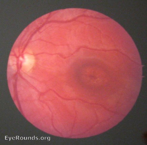
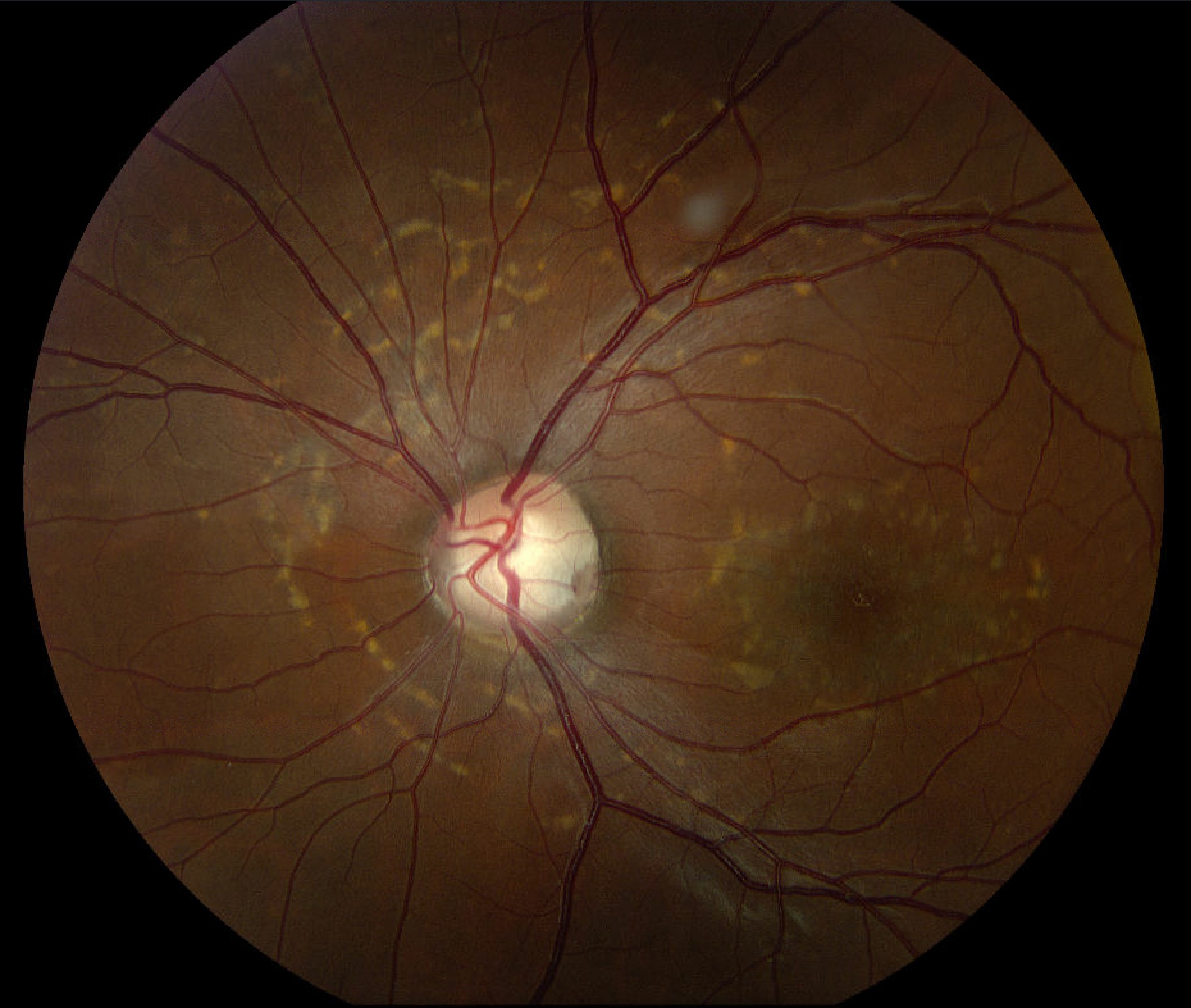
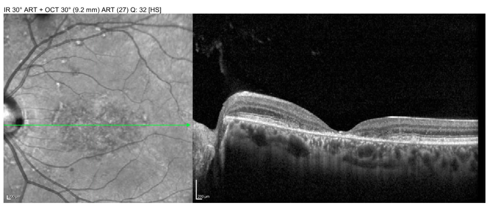
25 year old female with autosomal recessive ABCA4 Stargardt Disease. BCVA with correction 20/160+2 OD and 20/125-1 OS. Fundus exam notable for central macular atrophy and pisciform flecks in both eyes with an inferonasal optic disc pit in the left eye. There was no evidence of maculopathy on OCT imaging.

Fundus photography of the right eye showing pisciform yellow deposits characteristic of Stargardt disease.
Fundus photography of the left eye again showing pisciform yellow deposits but also an inferonasal optic disc pit.