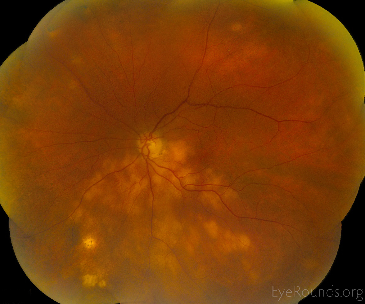
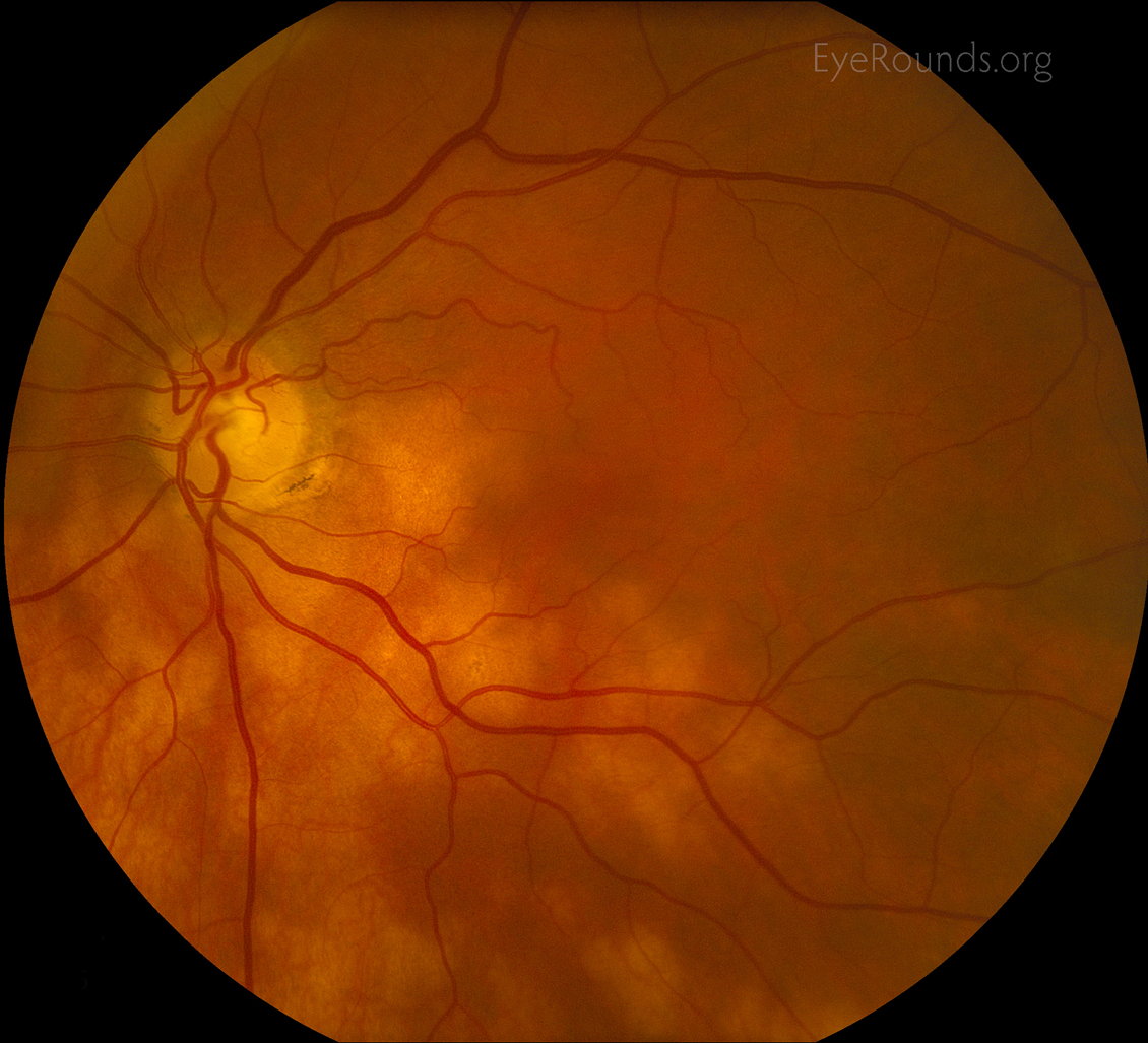
76-year-old female with biopsy proven systemic sarcoidosis presented for routine eye exam and the numerous flat yellowish lesion were noted in the fundus of the left eye more so than the right. She was started on methotrexate which controlled her infiltrates. The methotrexate was stopped and she developed photopsias and the lesions appeared more active. Thus the methotrexate was restarted with resolution of symptoms and control of lesion growth was obtained after several months. The three slightly brighter more superfical lesion in the inferior nasal periphery where felt to be inactive presumed ocular histoplasmosis scars. Choroidal granulomas manifesting as the sole lesion in ocular sarcoidosis has been previously described [1].
The differential includes lymphoma infiltrate or syphilitic lesions but review of systems, previous diagnosis of sarcoidosis, and the improvement on immunosuppresion make these extremely unlikely.

Ophthalmic Atlas Images by EyeRounds.org, The University of Iowa are licensed under a Creative Commons Attribution-NonCommercial-NoDerivs 3.0 Unported License.

Address
University of IowaLegal
Related Links