This site uses tracking information. Visit our privacy policy. Click to agree to this policy and not see this again.

Ophthalmology and Visual Sciences

Pellucid marginal degeneration

Pellucid marginal degeneration

Category(ies): Cornea and External Disease
Contributor: Jesse Vislisel, MD
Photographer: Carol Chan, CRA

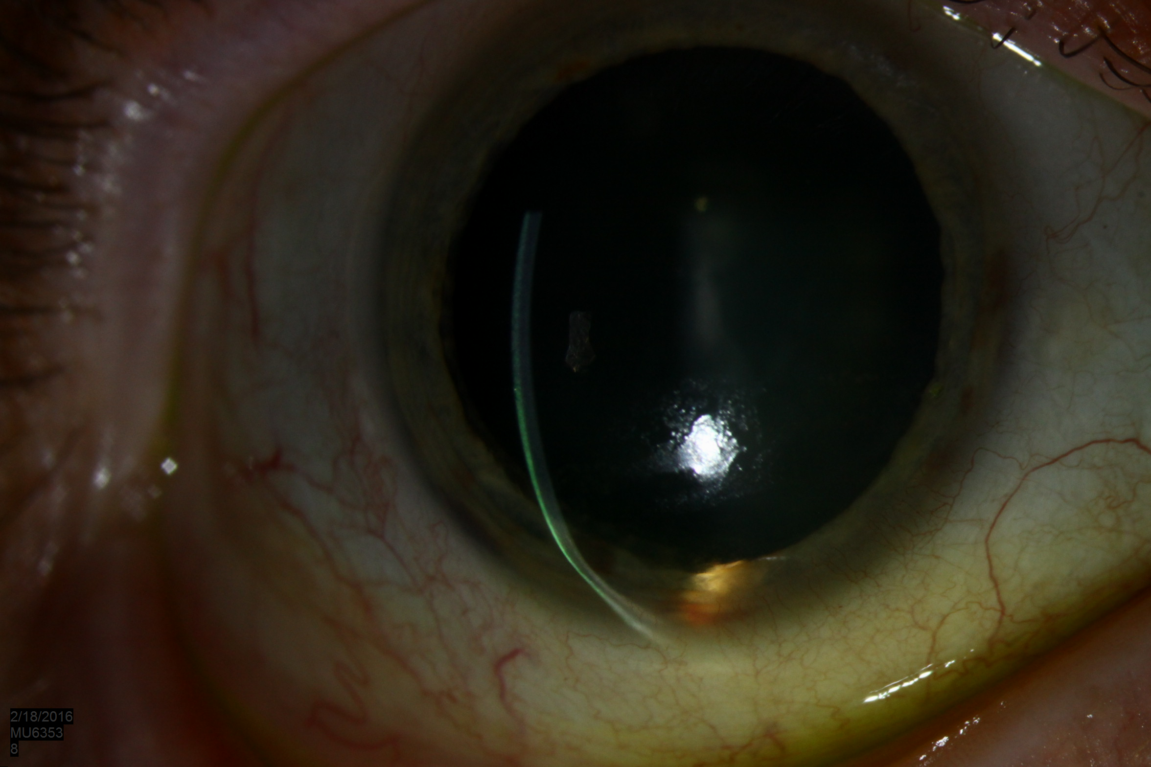
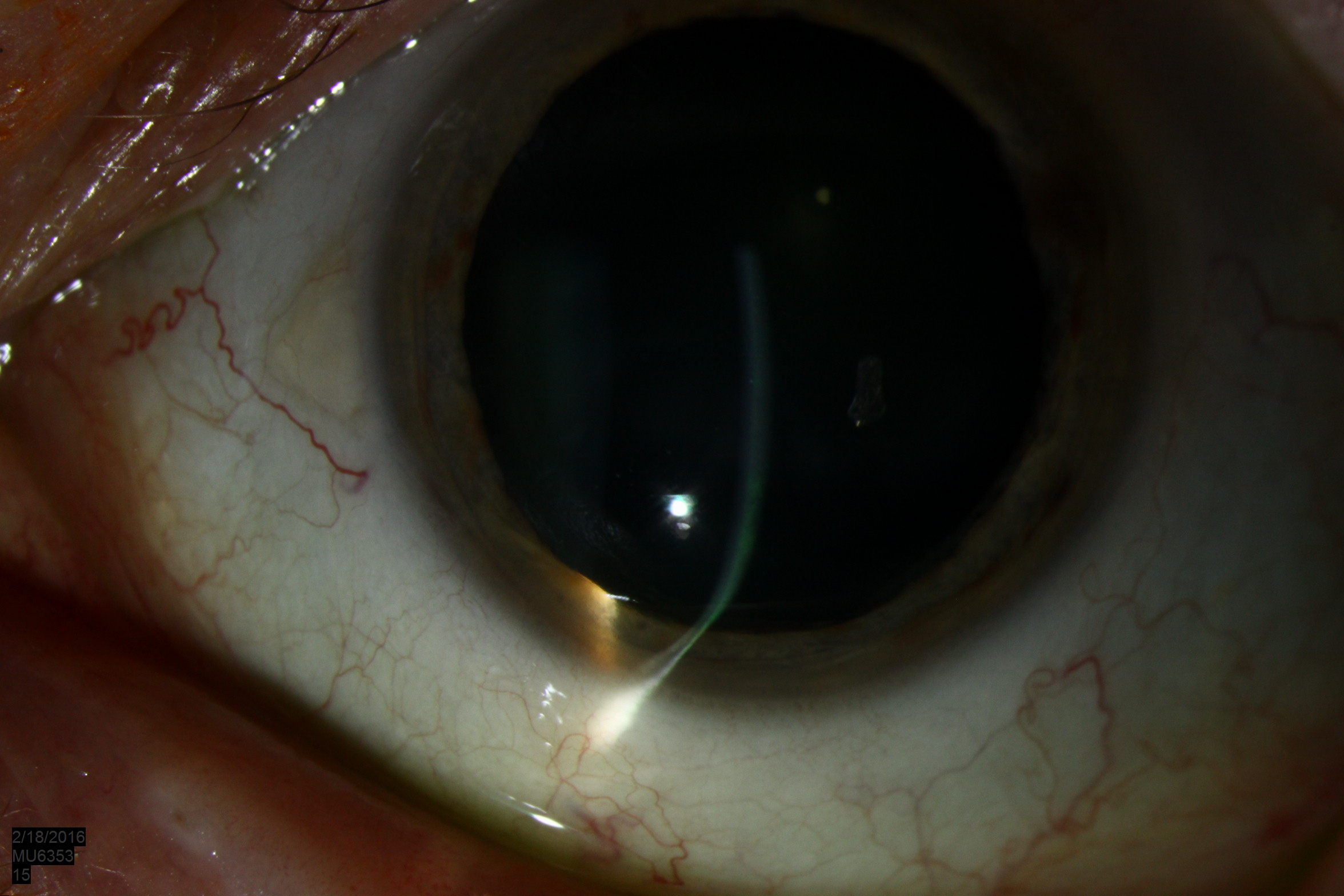
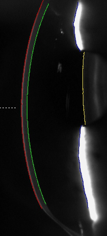
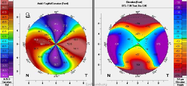
Pellucid marginal degeneration is a bilateral corneal ectasia that results in inferior corneal thinning with a corneal protrusion above the thinnest area. This is in contrast to keratoconus where maximal thinning occurs at the apex of the protrusion.

slit lamp corenal protrusion OD
Figure 1a: OD These slitlamp photographs demonstrate the corneal protrusion above the area of inferior thinning in a patient with pellucid marginal degeneration.
slit lamp corenal protrusion OS
Figure 1b: OS These slitlamp photographs demonstrate the corneal protrusion above the area of inferior thinning in a patient with pellucid marginal degeneration.
Pentacam Scheimpflug cross-sectional image
Figure 2: The Pentacam Scheimpflug cross-sectional images from the patient in Figures 1 show the contour changes associated with the inferior stromal thinning
Pentacam Scheimpflug cross-sectional image
Figure 3: The axial curvature and anterior float Pentacam topography maps from the patient in Figure 1 reveal the characteristic high against-the-rule astigmatism pattern seen in patients with pellucid marginal degeneration. Inferior steepening is also present, as seen grossly in the images in Figures 1 and 2, but is too peripheral in the case to be captured on the topography scans, thus the absence of the classic "crab claw" topographic appearance.