This site uses tracking information. Visit our privacy policy. Click to agree to this policy and not see this again.

Ophthalmology and Visual Sciences

Posterior Polymorphous Corneal Dystrophy (PPMD)

Posterior Polymorphous Corneal Dystrophy (PPMD)

Category(ies): Cornea
Contributor: Jesse Vislisel, MD

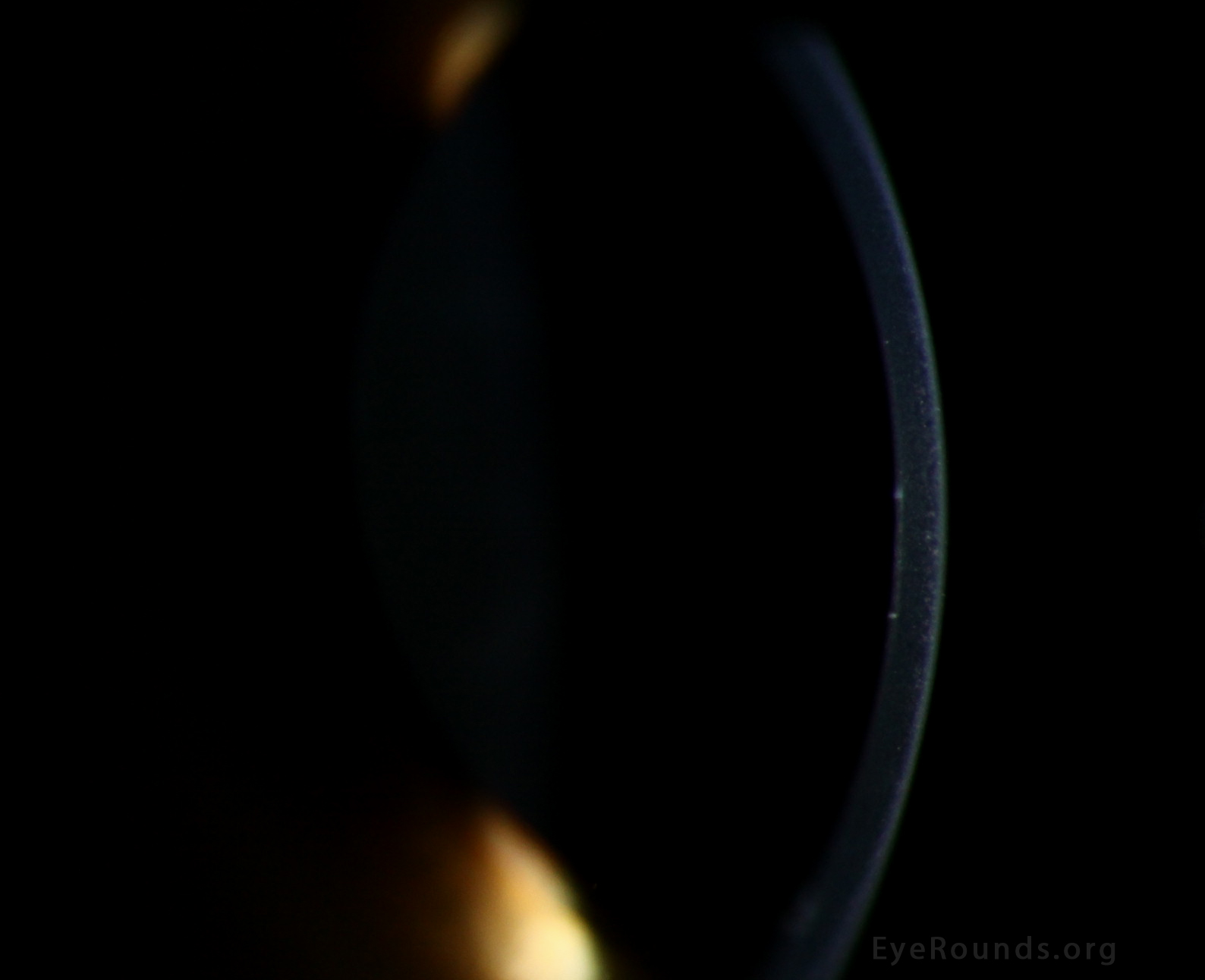
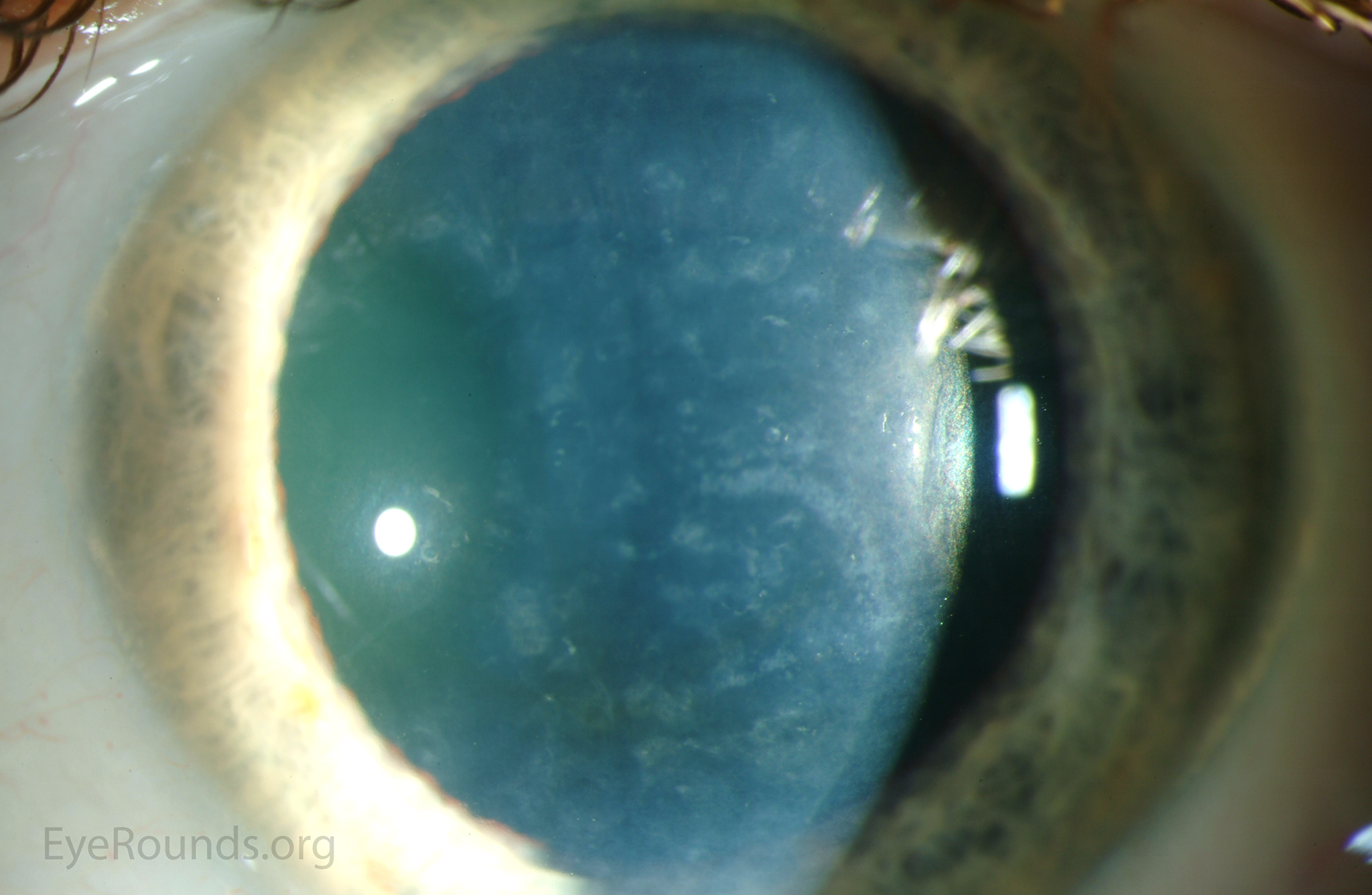
Posterior polymorphous corneal dystrophy (PPMD, PPCD) is a rare, bilateral, autosomal dominant inherited corneal dystrophy. The corneal abnormality in PPMD occurs at the level of Descemet's membrane and endothelium, and rarely will result in corneal edema or elevated intraocular pressure. The three main patterns in which PPMD may present include endothelial vesicle-like lesions, band lesions, and diffuse deep stromal opacities.


Figure 1: Vesicle-like lesions in PPMD

Figure 1: Vesicle-like lesions in PPMD

Contributor: Jesse Vislisel, MD and Kenneth M. Goins, MD
Photographer: Brice Critser, CRA

March 2,2015

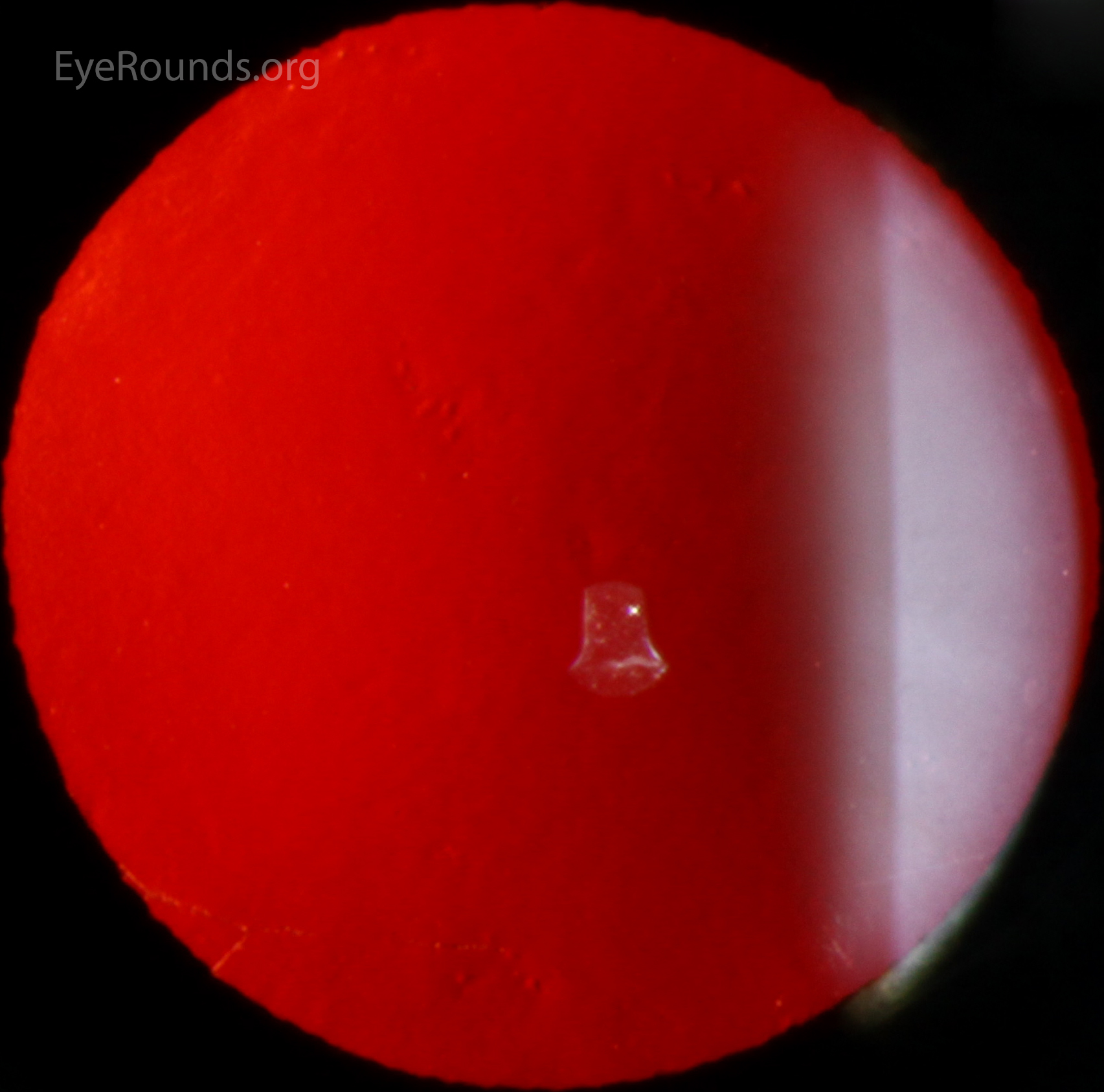
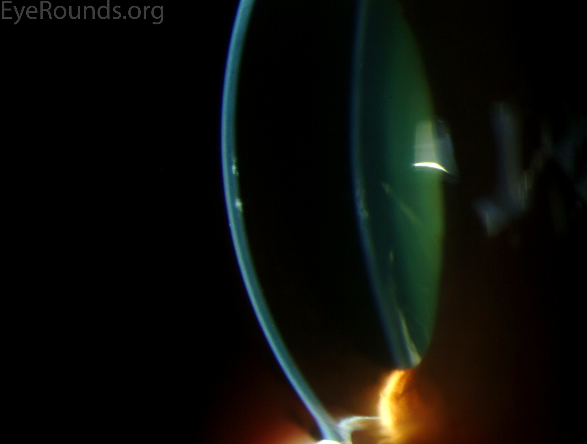
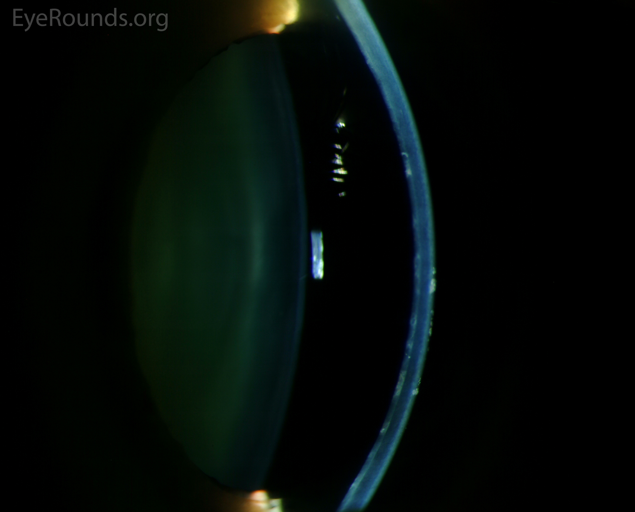
Vesicle-like lesions at the level of Descemet's membrane and endothelium are the hallmark lesions of PPMD. They appear as transparent cystic lesions surrounded by gray halos and commonly occur in lines or clusters. The figures below demonstrate the classic appearance of these lesions with direct illumination, retroillumination, and on specular microscopy.

classic appearance of these lesions with direct illumination
Classic appearance of these lesions with direct illumination
classic appearance of these lesions with retroillumination
Classic appearance of these lesions with retroillumination
classic appearance of these lesions with specular microscopy
Classic appearance of these lesions with specular microscopy

Figure 2: Band lesions in PPMD

Figure 2: Band lesions in PPMD

Contributor: Jesse Vislisel, MD and Kenneth M. Goins, MD
Photographer: Brice Critser, CRA

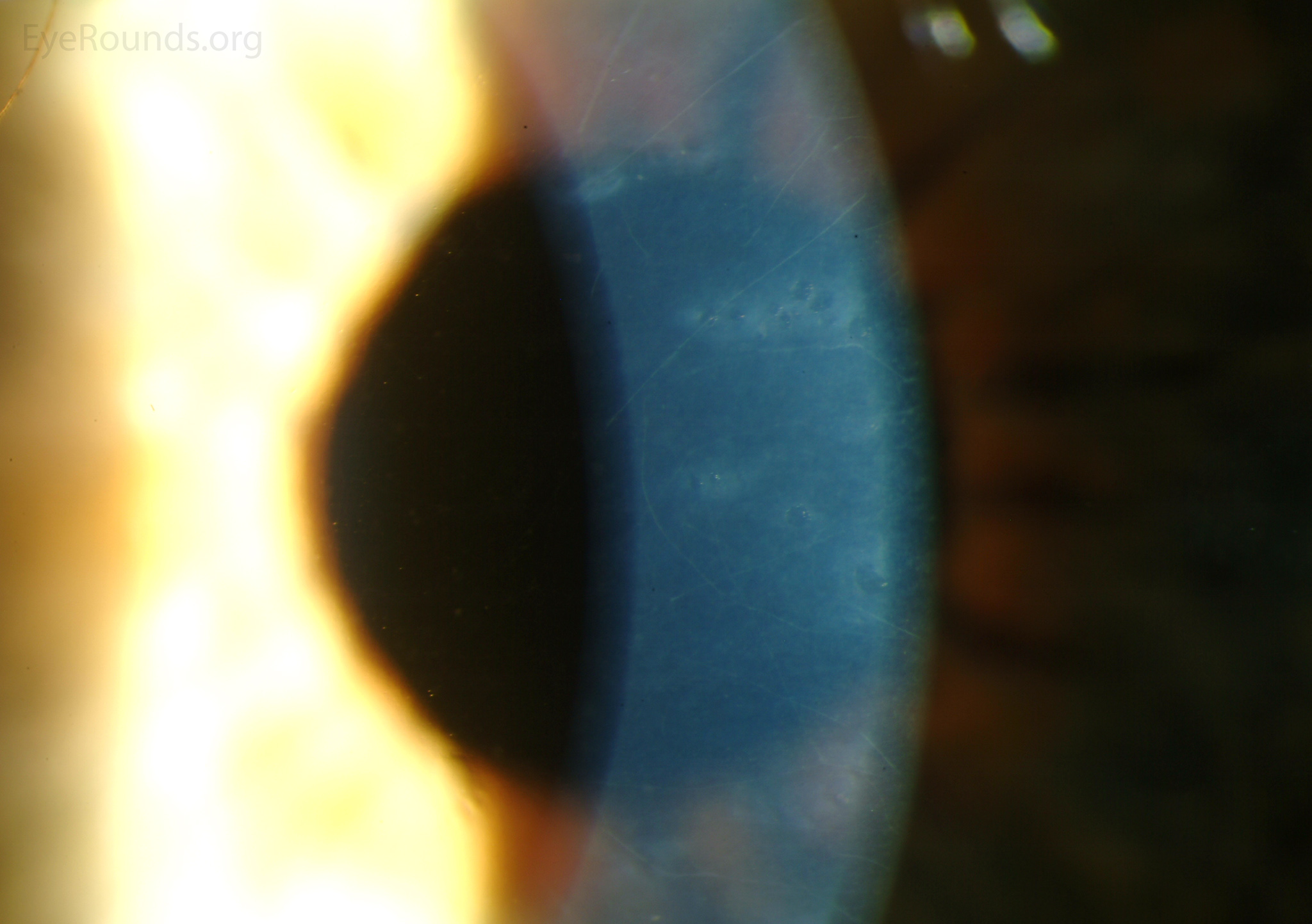
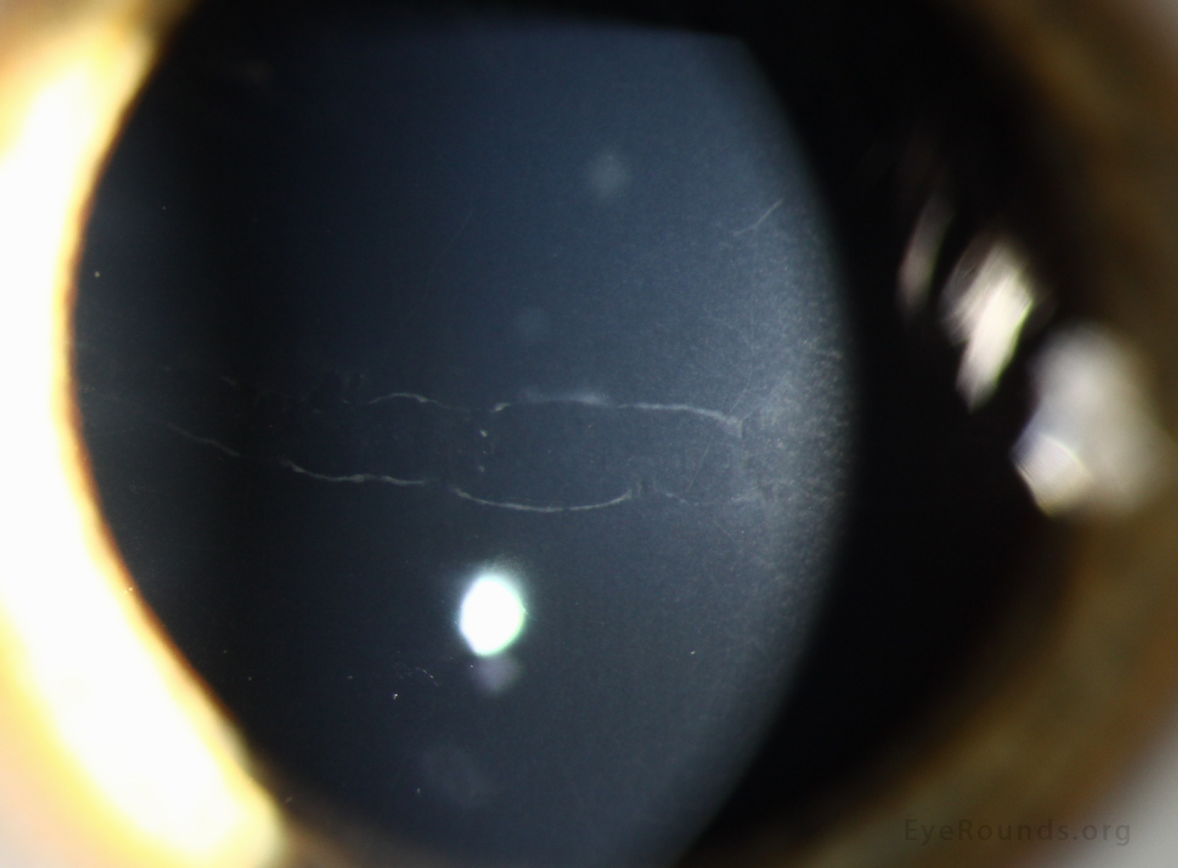
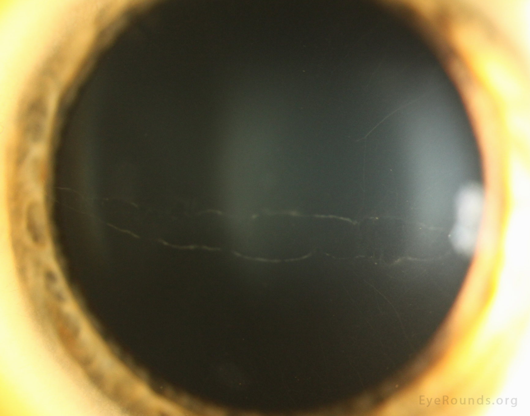
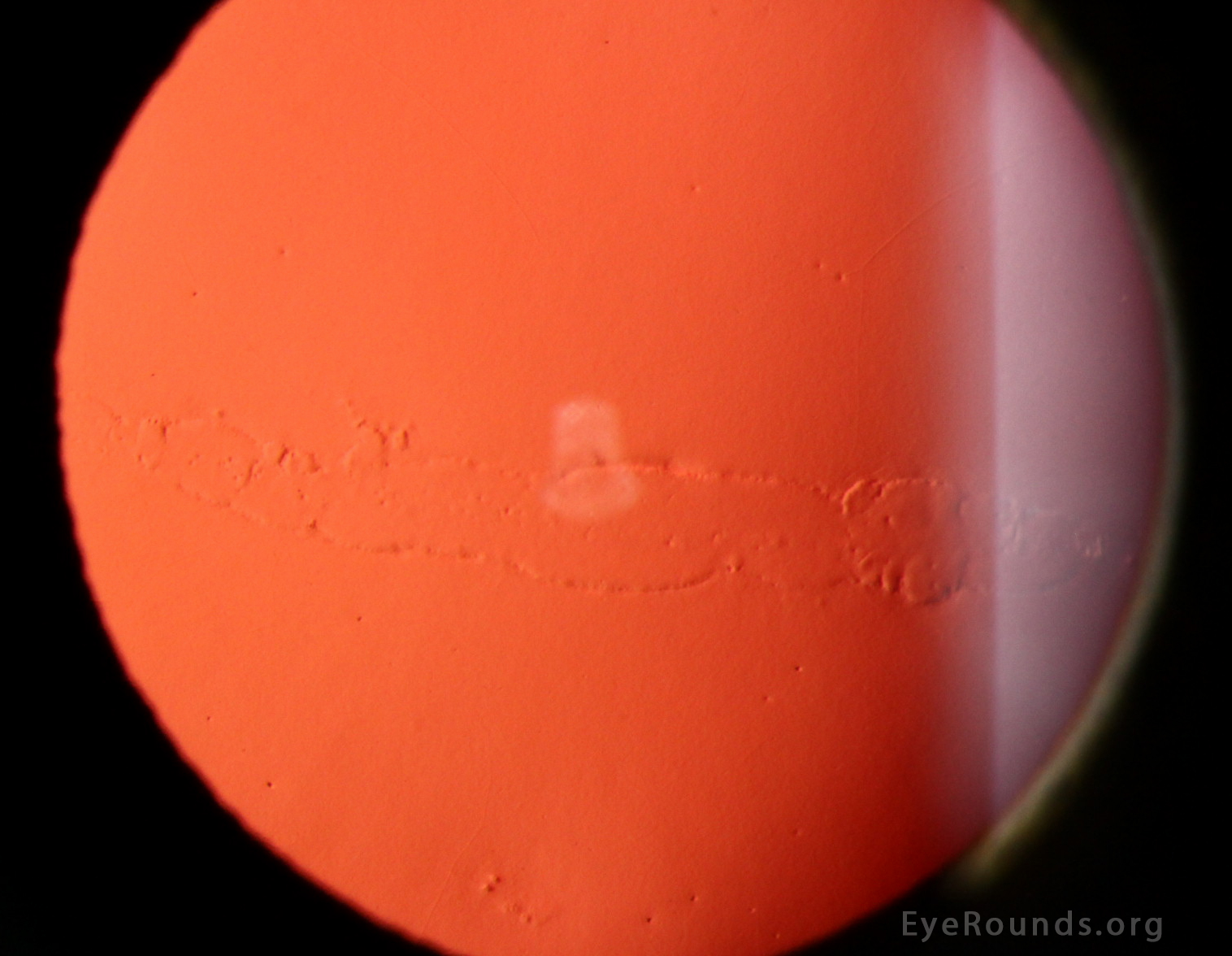
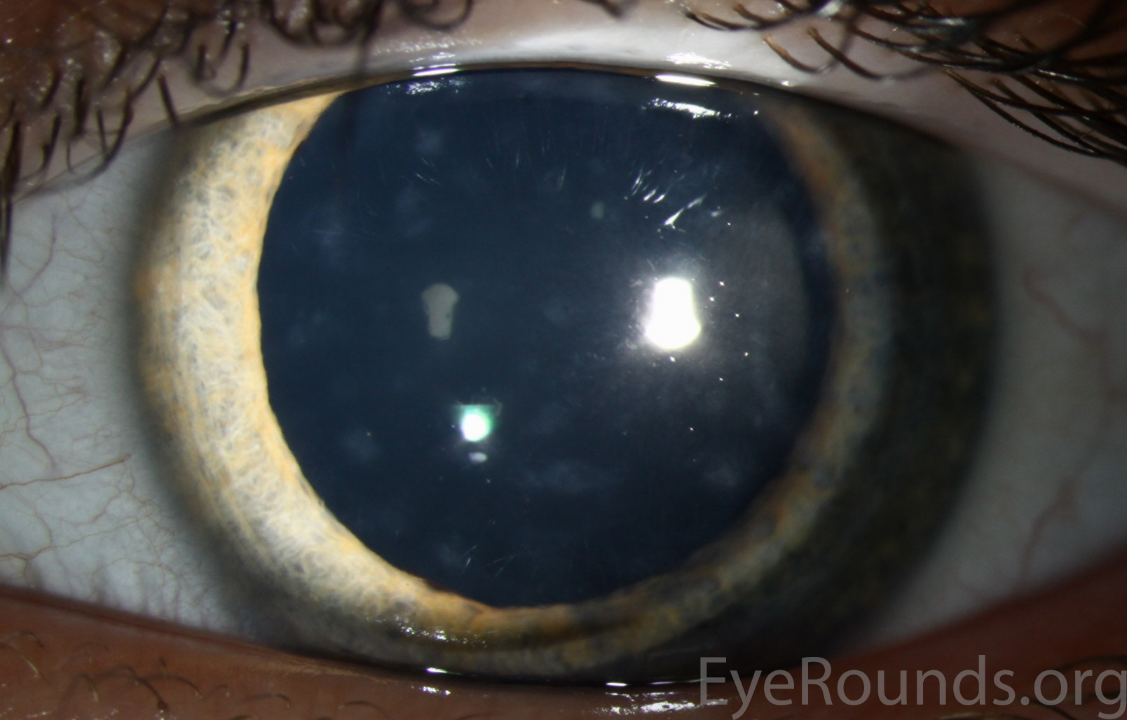
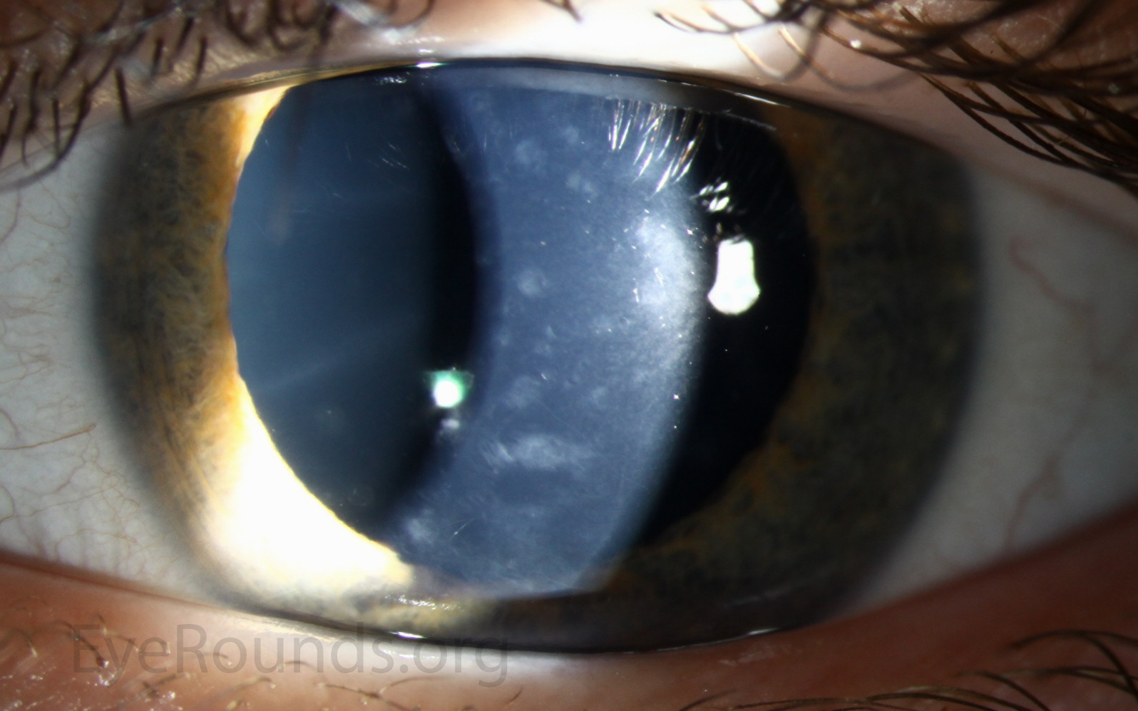
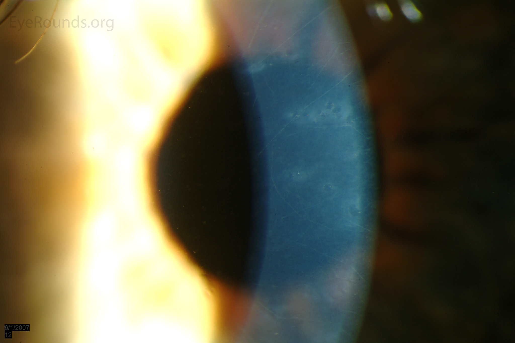
Band lesions, sometimes called "snail tracks," are classically horizontal lesions with parallel, scalloped, non-tapering edges at the level of the posterior cornea. Below are examples of these lesions in 2 separate patients.

Band lesions, sometimes called 'snail tracks,' are classically horizontal lesions with parallel, scalloped, non-tapering edges at the level of the posterior cornea
Patient 2a
patient 2a Band lesions, sometimes called 'snail tracks,' are classically horizontal lesions with parallel, scalloped, non-tapering edges at the level of the posterior cornea
Patient 2a
patient 2a Band lesions retroillumination
Patient 2a
patient 2a Band lesions slit lamp
Patient 2a
patient 2b Band lesions
Patient 2b
patient 2b Band lesions slit lamp
Patient 2b

Figure 3: Diffuse posterior stromal opacities in PPMD

Figure 3: Diffuse posterior stromal opacities in PPMD

Contributor: Jesse Vislisel, MD and Kenneth M. Goins, MD
Photographer: Brice Critser, CRA

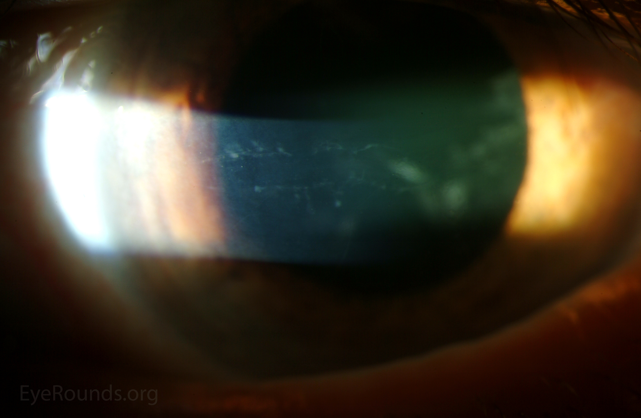
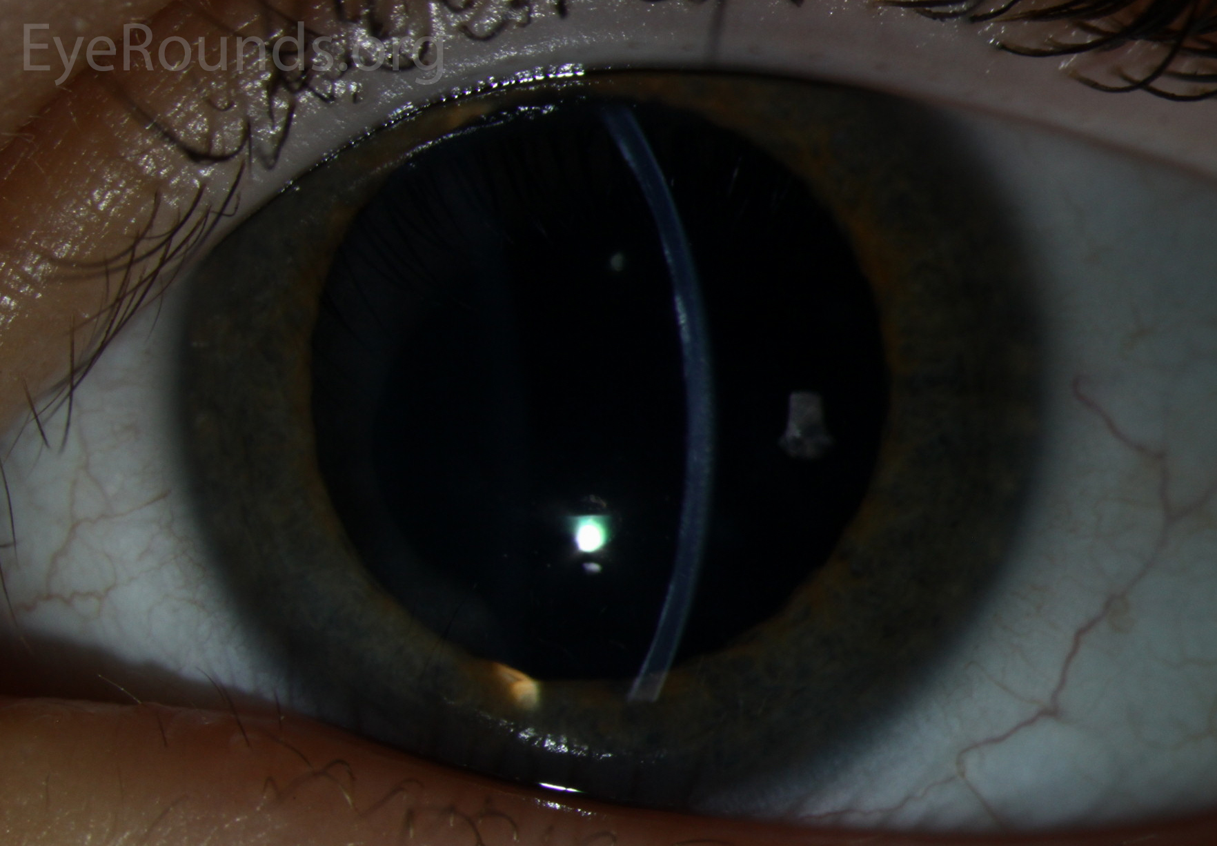
PPMD may present with diffuse, gray-white opacities at the level of Descemet's membrane. There may be deep stromal haze adjacent to the lesions. Below are slit lamp photographs of two patients with such lesions.

patient 3a PPMD may present with diffuse, gray-white opacities
Patient 3a
patient 3a PPMD may present with diffuse, gray-white opacities, slit lamp
Patient 3a
patient 3b PPMD may present with diffuse, gray-white opacities, slit lamp
Patient 3b
patient 3b PPMD may present with diffuse, gray-white opacities
Patient 3b
patient 3b PPMD may present with diffuse, gray-white opacities
Patient 3b

Figure 4: Confocal microscopy of affected endothelium in PPMD

Figure 4: Confocal microscopy of affected endothelium in PPMD

Contributor: Andrew Doan, MD, PhD, University of Iowa

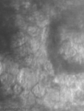
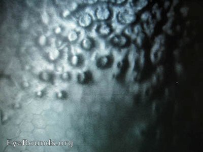
Confocal microscopy of the endothelium in PPMD shows epithelial-like cells. Normal endothelial cells are hexagonal and flat.