Lipid keratopathy is characterized by the stromal deposition of yellow or cream-colored lipids in areas of corneal neovascularization, often secondary to herpes simplex virus, varicella zoster virus, or trachoma. Argon laser and topical and subconjunctival bevacizumab have been reported to reduce corneal neovascularization and lipid deposition.
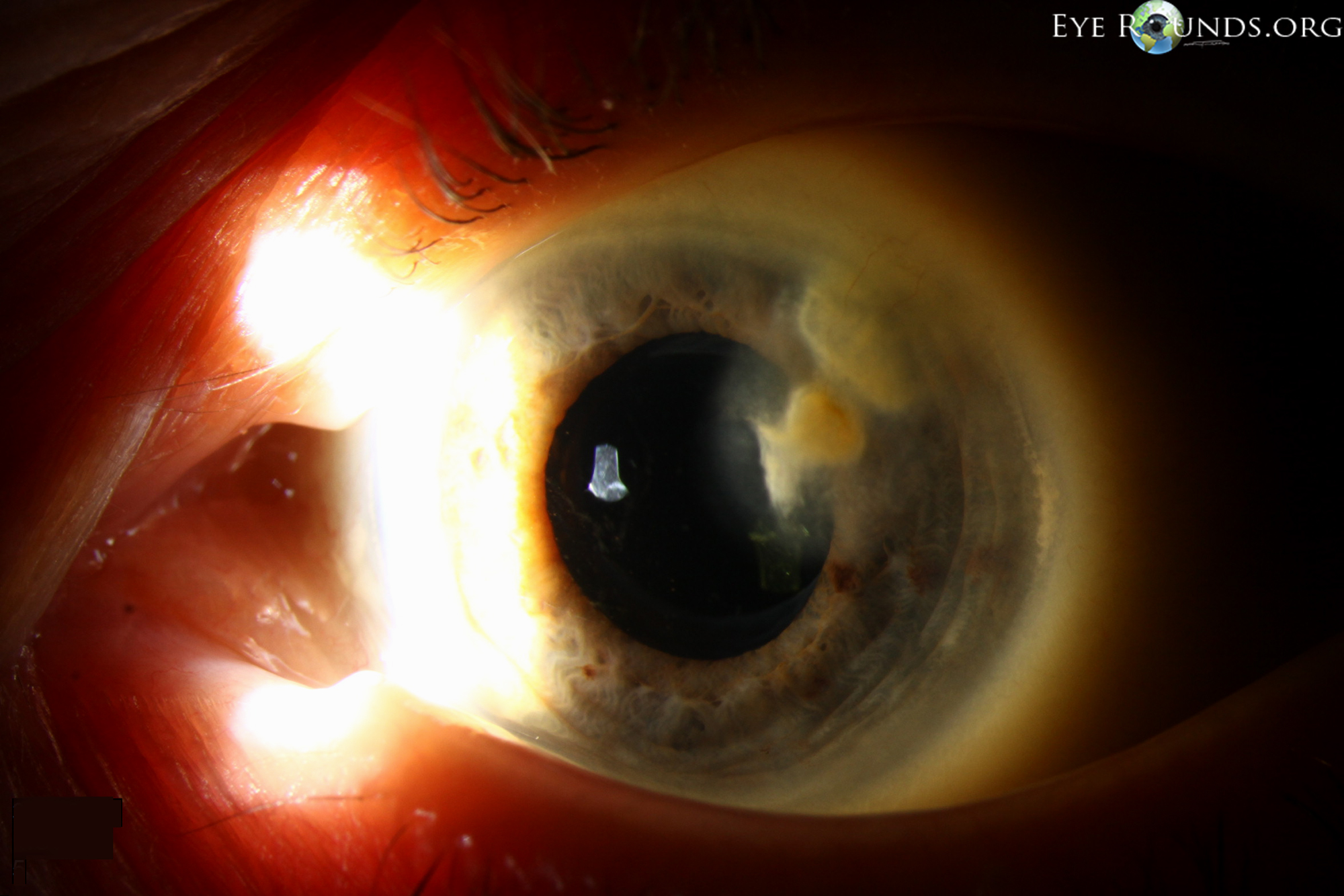
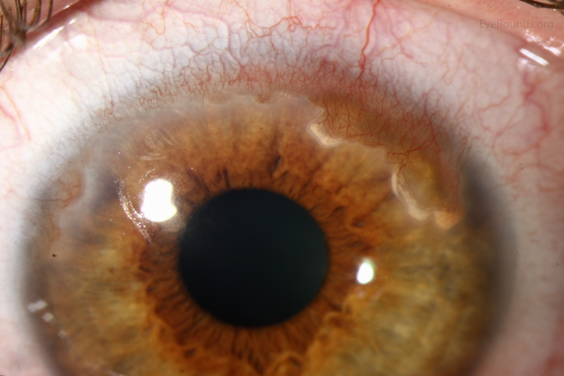
Stromal neovascularization and lipid keratopathy secondary to chronic herpes zoster ophthalmicus. Note the dense cream-colored opacifications with surrounding haze which represent inflammation and extravasation of cholesterol and fatty acids.
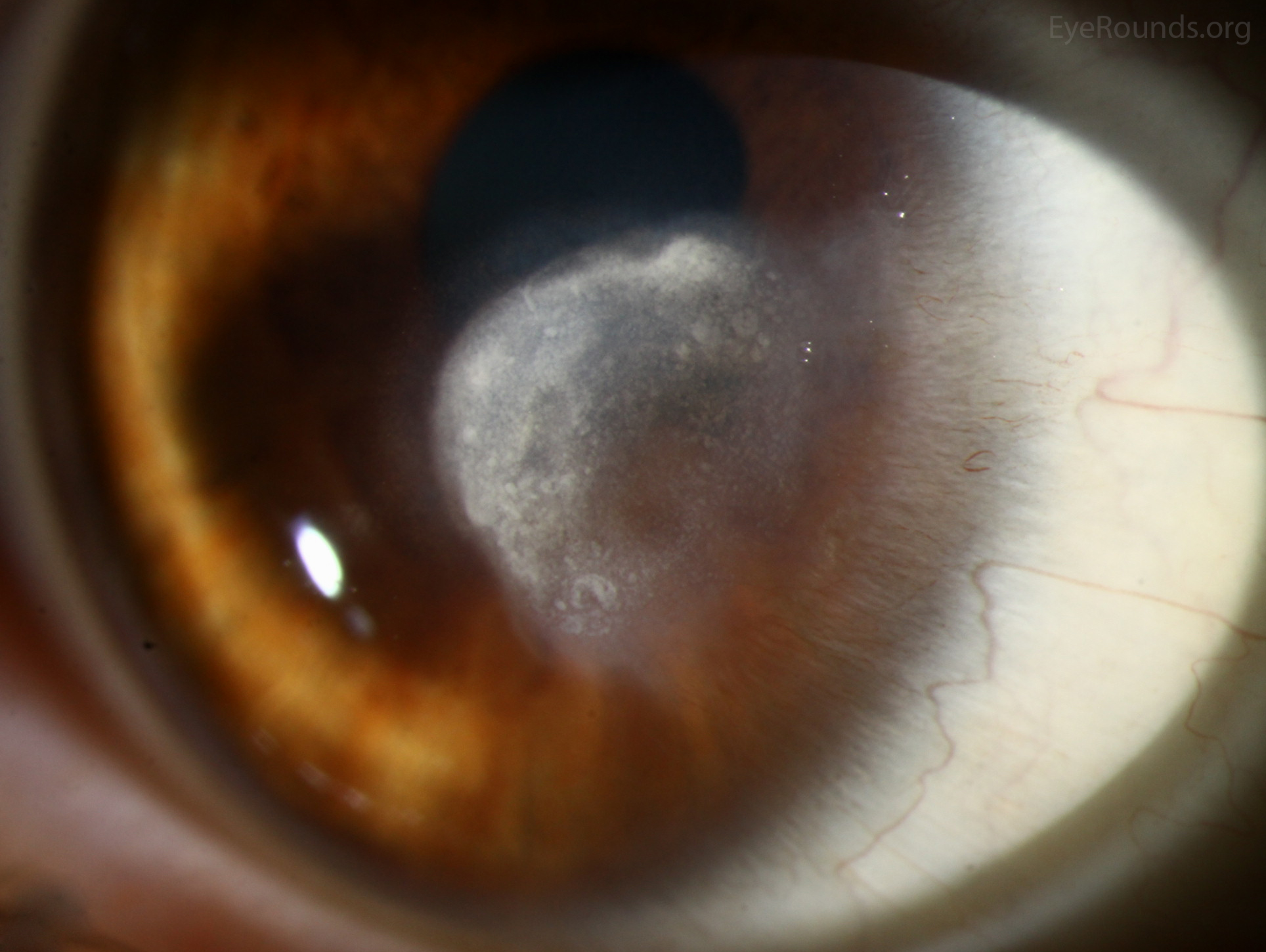
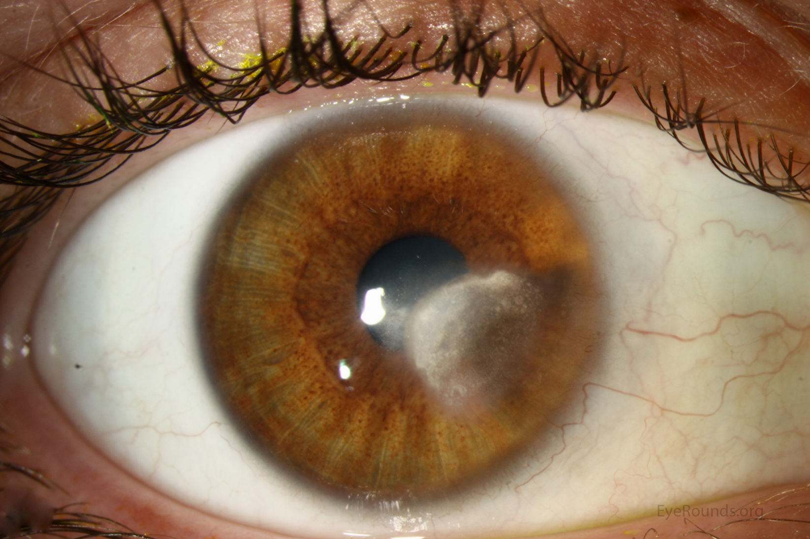
Lipid keratopathy in a region of corneal scarring and neovascularization resulting from previous Pseudomonas keratitis.
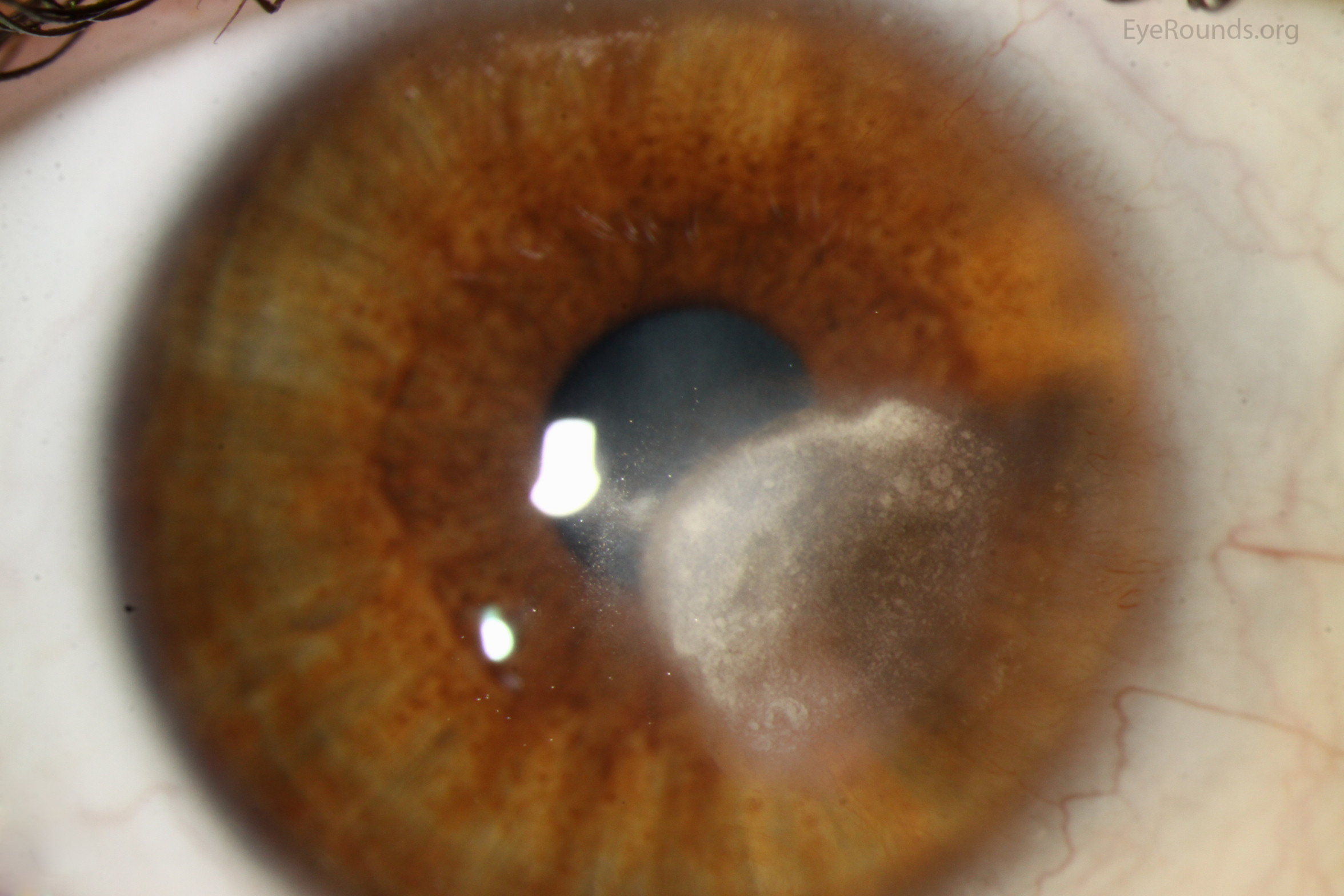
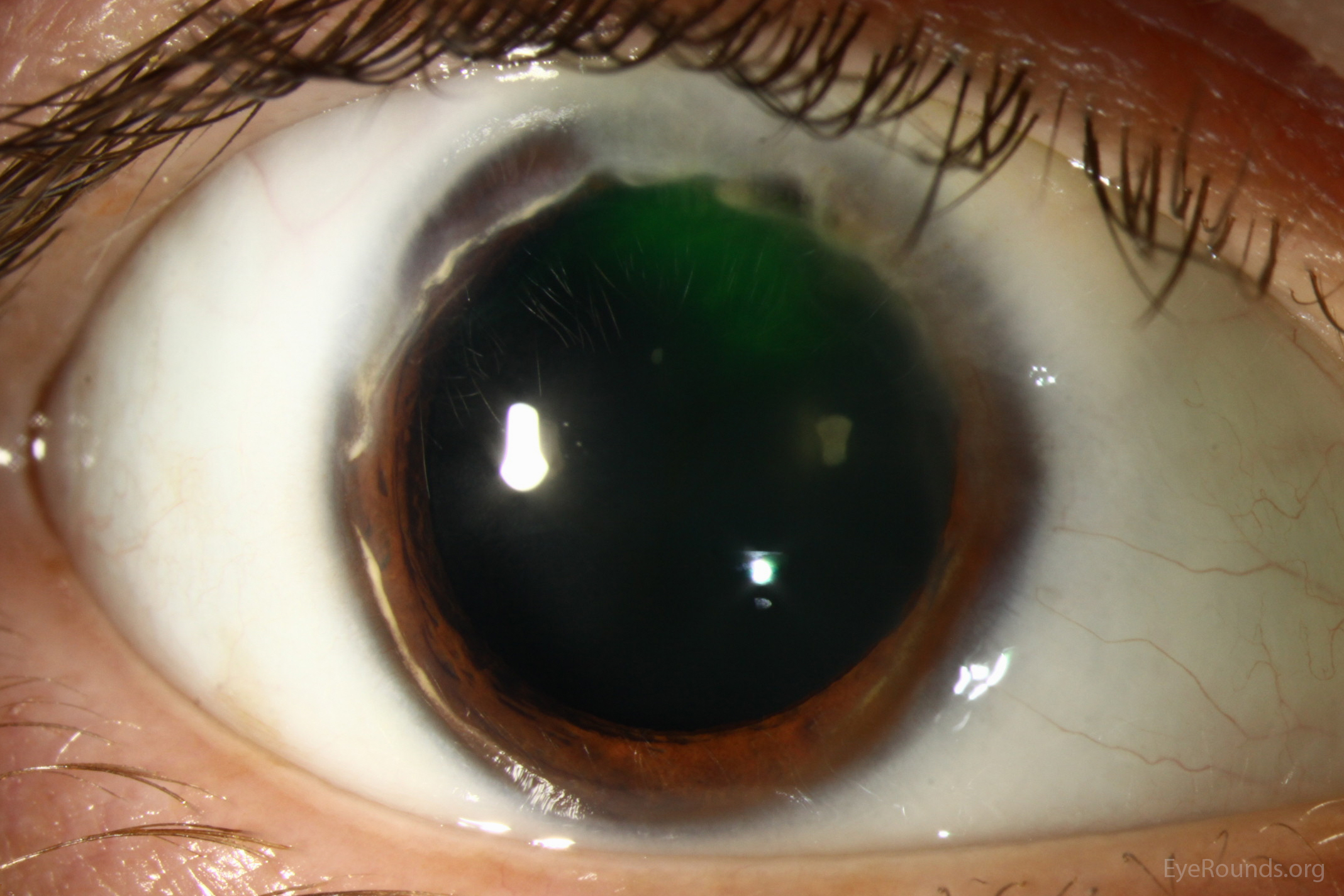
Lipid deposition at the leading edge of superior pannus associated with Terrien marginal degeneration
Lipid keratopathy. In: External Disease and Cornea, Section 8. Basic and Clinical Science Course (BCSC). San Francisco: American Academy of Ophthalmology, 2011-2012, p. 342.

Ophthalmic Atlas Images by EyeRounds.org, The University of Iowa are licensed under a Creative Commons Attribution-NonCommercial-NoDerivs 3.0 Unported License.

Address
University of IowaLegal
Related Links