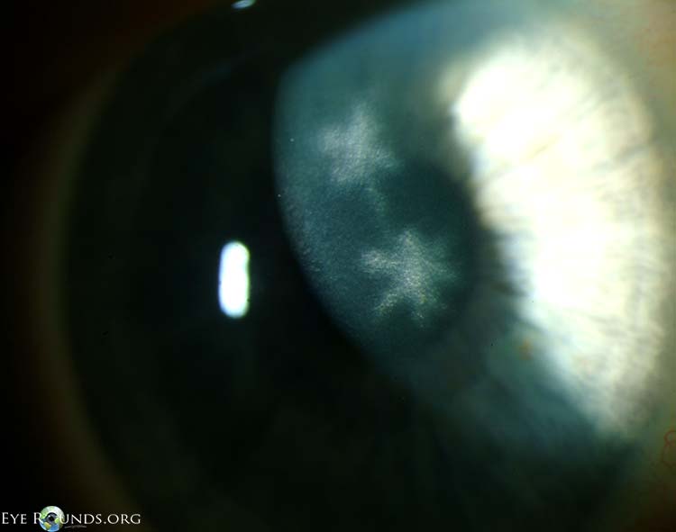
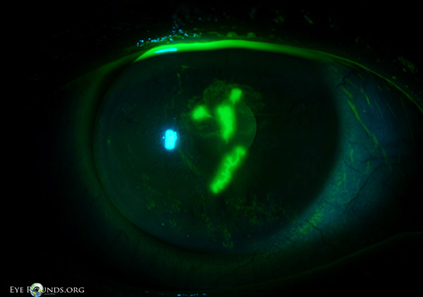
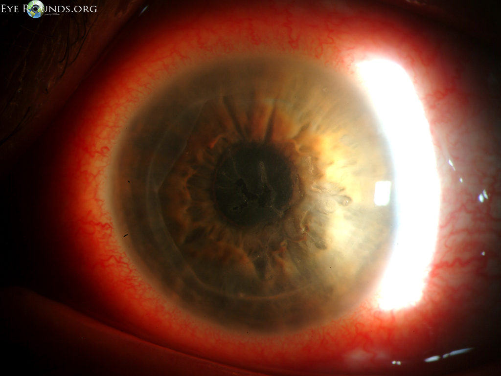
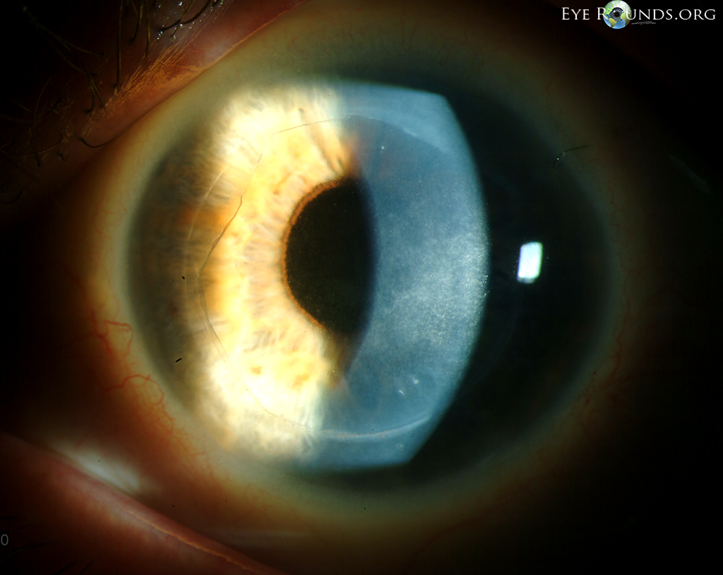
This patient was on chronic topical steroids after undergoing a deep lamellar endothelial keratoplasty (DLEK) when she developed Herpes simplex virus (HSV) epithelial keratitis. The dendritic ulcer is visible using the sclerotic scatter technique (top) and it becomes readily apparent with fluorescein staining (middle image). HSV dendritic ulcers have terminal bulbs and their base stains positively with fluorescein. They can coalesce to form larger geographic ulcers, as seen centrally here, especially in the presence of topical steroids. After resolution of the keratitis, residual subepithelial infiltration and scarring can be seen just deep to the area of the previous ulcer, resulting in "ghost dendrites", as seen in the bottom photo.
Krachmer, Jay H., Mark J. Mannis, Edward J. Holland. Cornea. St. Louis: Mosby, 2011.

Ophthalmic Atlas Images by EyeRounds.org, The University of Iowa are licensed under a Creative Commons Attribution-NonCommercial-NoDerivs 3.0 Unported License.

Address
University of IowaLegal
Related Links


 Caption -->