This site uses tracking information. Visit our privacy policy. Click to agree to this policy and not see this again.

Ophthalmology and Visual Sciences

Exophthalmos secondary to a carotid cavernous fistula

Exophthalmos secondary to a carotid cavernous fistula

Contributor: Brittni A. Scruggs, MD, PhD
Photographer: Cynthia Montague
Category(ies): Neuro-ophthalmology, Glaucoma
Posted: September 26, 2017

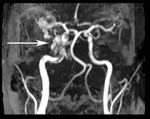
A carotid-cavernous fistula (CCF) is a pathologic shunt between the carotid artery and the venous cavernous sinus; such hemodynamic lesions often lead to the triad of exophthalmos, epibulbar arterialized loops, and glaucoma. Diagnosis is by clinical examination, computed tomography or magnetic resonance angiography, and/or catheter angiography. A direct CCF (type a) connects the intracavernous portion of the internal carotid artery (ICA) to the sinus, whereas indirect, or dural, fistulas (type b) are low-flow fistulas that connect ICA branches to the cavernous sinus (1). If vision-threatening signs develop, then embolization must be considered.

External photography and slit lamp examination of both eyes.
External photography and slit lamp examination of both eyes. Upper image: External photo demonstrating the difference in appearance between the right (OD) and left eye (OS). Left image: The right eye had a completely normal exam. Right image: Dilated corkscrew vessels extended to the limbus OS.The patient also had periorbital edema on the left with mild abduction deficit and proptosis.
Slit lamp examination OS
Slit lamp examination OS. There was evidence of numerous dilated corkscrew vessels extending to the limbus prior to definitive treatment of an indirect carotid-cavernous fistula.
Pre-operative CT imaging of the orbits.
Pre-operative CT imaging of the orbits. Left image: There was prominent proptosis OS (red lines) with a 3 mm difference in Hertel measurements. Right image: Congestion of the left extraocular muscles due to venous stasis in the left orbit (red arrows).
Pre-operative CT imaging
Pre-operative CT imaging (left images) and post-operative MR angiography (right image).There was early filling of the left cavernous sinus on the arterial phase images (circled in red). This represented leakage of contrast from the internal carotid artery to the left cavernous sinus..
Color fundus photography of both eyes
Color fundus photography of both eyes. Left image: The right eye had a normal optic disc, macula, and vessels. Right image: The left eye had dilated and tortuous veins, a normal macula, and scattered dot blot hemorrhages. Central retinal vein occlusion, or venous stasis retinopathy, occurred as a result of CCF.
References:
  1. R.J.W. Keizer (2003) Carotid-cavernous and orbital arteriovenous fistulas: ocular features, diagnostic and hemodynamic considerations in relation to visual impairment and morbidity, Orbit, 22:2, 121-142

Image Permissions:
Creative Commons

Ophthalmic Atlas Images by EyeRounds.org, The University of Iowa are licensed under a Creative Commons Attribution-NonCommercial-NoDerivs 3.0 Unported License.