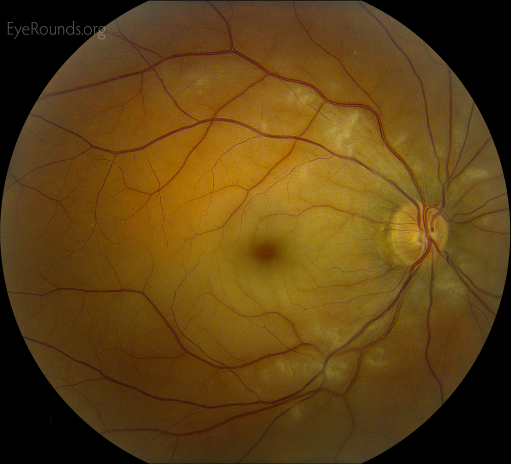
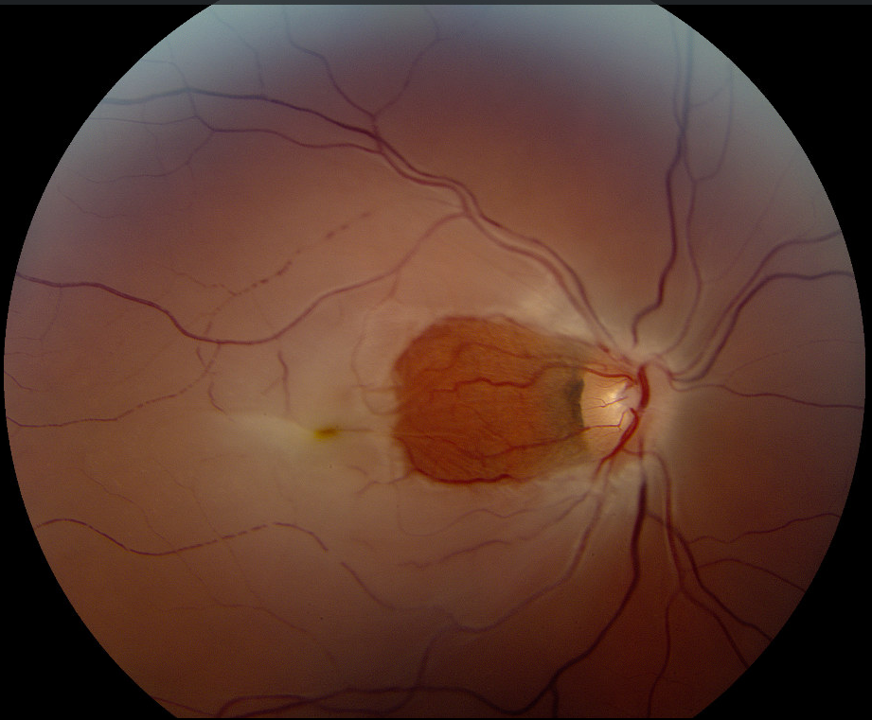
Central retinal artery occlusion (CRAO) occurs when occlusion of the central retinal artery, a branch of the ophthalmic artery, results in infarction of the inner retina with subsequent, typically severe, vision loss. The picture above exhibits a CRAO with sparing of the foveal retina due to the presence of a cilioretinal artery. A cilioretinal artery has been found to be present in 49.5% of patients. The cilioretinal artery branches off the ophthalmic artery and supplies the inner retina in these individuals. This is in contradistinction to the posterior ciliary arteries, which branch off the ophthalmic artery and supply the choroid and outer retina. Patients with a cilioretinal artery may have preserved central vision in the unfortunate event of a CRAO.
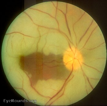
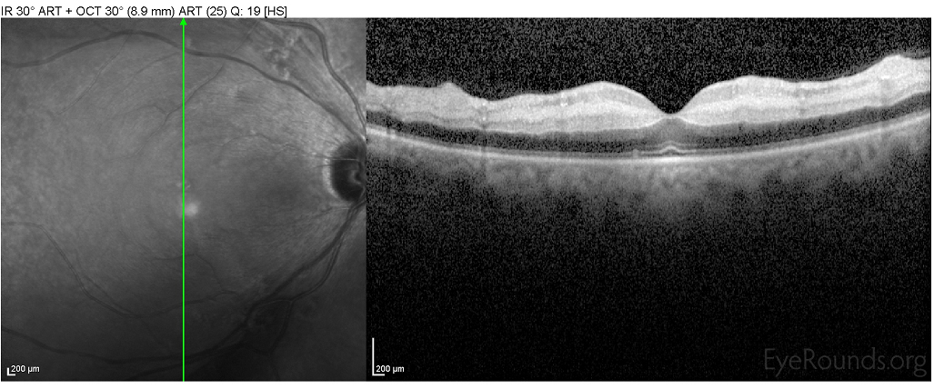
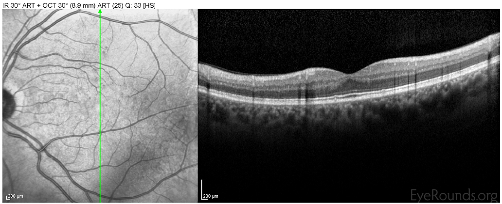
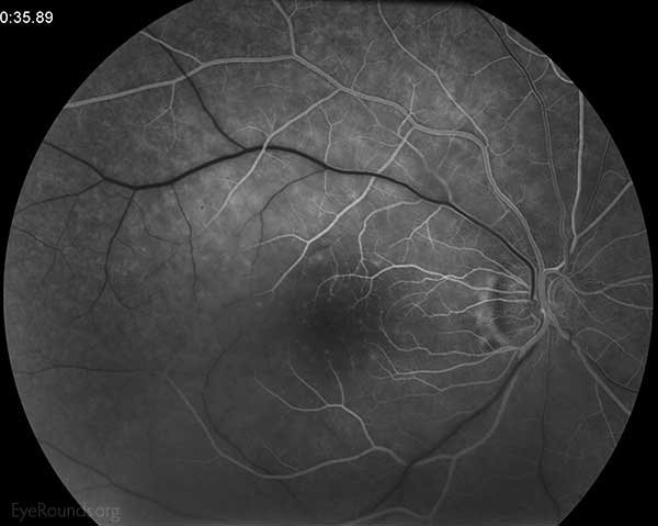
This middle-aged male presented with an abrupt decrease in vision in the right eye 4 days prior. Note the retinal whitening, classic cherry red spot, and scatted cotton wool spots. The OCT shows inner retinal edema in the right eye (OD) compared to the normal retinal OCT in the left (OS). Also note the delayed filling of the macular arteries on the fluorescein angiogram.
A 34-year-old female with history of recent methamphetamine use presented with several hours of painless vision loss in the right eye. Exam was significant for retinal whitening with sparing of the nasal macula due to vascular preservation via the cilioretinal artery.

Ophthalmic Atlas Images by EyeRounds.org, The University of Iowa are licensed under a Creative Commons Attribution-NonCommercial-NoDerivs 3.0 Unported License.

Address
University of IowaLegal
Related Links