This site uses tracking information. Visit our privacy policy. Click to agree to this policy and not see this again.

Ophthalmology and Visual Sciences

Cytomegalovirus (CMV) Retinitis

Cytomegalovirus (CMV) Retinitis

Category(ies): Retina, Vitreous
Contributor: Eric Chin, MD
Photographers: Toni Venckus, CRA (images 1 & 2); Brice Critser, CRA (images 3 & 4)

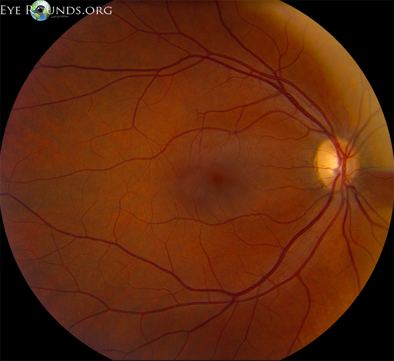
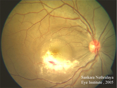
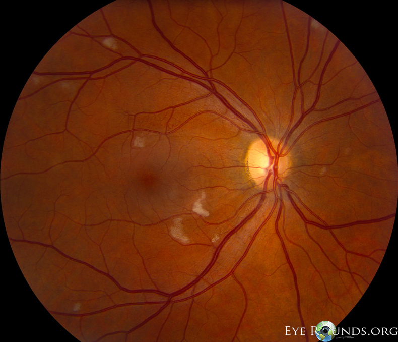
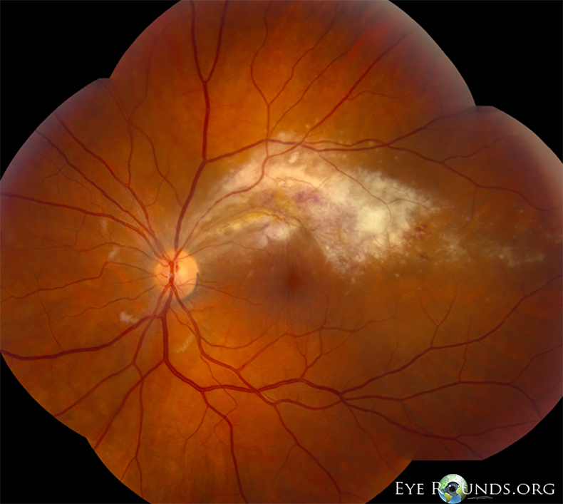
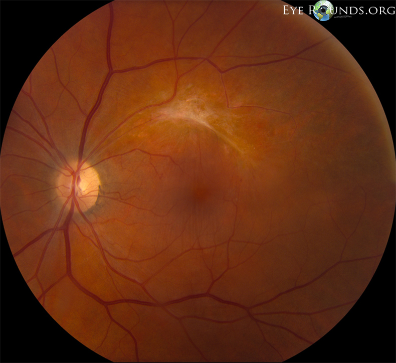
41-year-old male with HIV, presented with blurry vision in the left eye. His CD4 count at presentation was 2 cells/mm3 (normal 500-1,000

  • Visual acuity
    • OD sc: 20/40, PH to 20/30
    • OS sc: 20/100, PH to 20/50
  • OP: 15 and 14
  • SLE: OD normal; OS: AC quiet; 2+ vitreous cell

June

OD fundus with cotton wool spots
Right eye – multiple cotton wool spots adjacent to the nerve and throughout the macula, consistent with HIV retinopathy
OS fundus with intraretinal hemorrhages along the superior arcades
Left eye - broad retinal whitening and intraretinal hemorrhages along the superior arcades, with cotton wool spots adjacent to the nerve consistent with CMV retinitis and HIV retinopathy
  • The patient was treated with intravitreal foscarnet in the left eye, and five days of intravenous ganciclovir while as an inpatient for other systemic manifestations. He was discharged on oral valganciclovir (induction dose 900 mg BID for two weeks) then 900 mg daily maintenance until CD4 count was above 50.
  • At future follow-up in October 2011, the patient had resolution of his CMV retinitis and HIV retinopathy but with persistently low CD4 count of 1. It was thought that valganciclovir contributed to his low CD4 count, and was therefore discontinued.

October

OD fundus resolved cotton wool spots
Right Eye: resolution of cotton wool spots
Vision sc: 20/50
OD fundus resolved cotton wool spots
Left Eye: glial scars along superior arcade; resolution of cotton wool spots adjacent to the nerve
Vision sc: 20/63
Summary: CMV Retinitis
  • CMV retinitis is the most common opportunistic ocular infection and the leading cause of visual loss in AIDs patients. It represents about 90% of all infectious retinitis in this patient population.
  • CMV retinitis usually results from reactivation of a latent CMV infection. Individuals with a CD4 lymphocyte count below 50, a plasma HIV-1 RNA level greater than 100,000, or who have been previously diagnosed with opportunistic infections are at particular risk.
  • Patients may be asymptomatic, or have floaters, photopsias, and/or scotomas.
  • Systemic treatment options include intravenous ganciclovir, foscarnet, or cidofovir. Oral ganciclovir or valganciclovir may also be used.
  • Localized ocular treatment may include intravitreal ganciclovir, foscarnet, or cidofovir.