This site uses tracking information. Visit our privacy policy. Click to agree to this policy and not see this again.

Ophthalmology and Visual Sciences

Cavernous Hemangioma:

31-year-old caucasian female with a painless, "dark spot" in her left visual field

Cavernous Hemangioma:

31-year-old caucasian female with a painless, "dark spot" in her left visual field
Erin Moore Shriver, MD
February 21, 2005

Chief Complaint: 31-year-old caucasian female presented with a painless, "dark spot" in her left visual field.

History of Present Illness: Dark spot fixed in supero-nasal left visual field that she noticed for one day. No complaints of flashes or floaters. Patient noted that the dark spot in her vision clears with abduction.

Past Ocular History: Peripheral atrophic holes OD

Medical History: Cerebral palsy

Social History: Librarian. No tobacco or alcohol.

EXAM:

  • Best corrected Va: 20/30 OD, 20/20 OS
  • IOP: 18mmHg OD, 15mmHg OS
  • Pupils: 4 --> 2 OU, no RAPD
  • Motility: Full OU, orthotropic in primary
  • CVF: Full OU by finger counting, but see GVF below.
  • SLE: Anterior cortical spoking cataract OS
  • Optic nerves were normal OU
  • DFE: Atrophic holes OD, macula striae OS
  • Palpebral fissure: 10mm OD and OS
  • Hertel: 92mm base; 17mm OD and 18mm OS
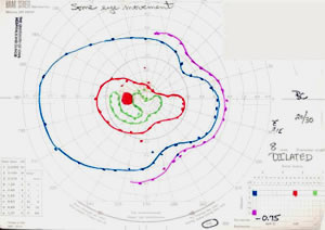
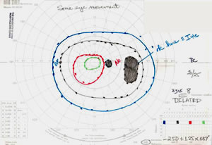
Figure 1: GVF
GVF OS with supero-nasal and infero-nasal constriction of all isopters. GVF OD with temporal scotoma.
GVF OS GVF OD

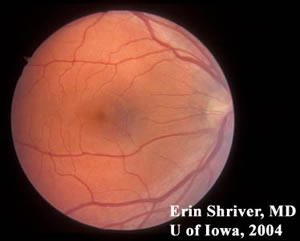
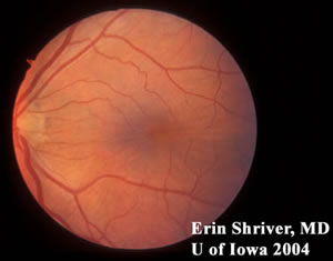
Figure 2: Fundus Photo
Fundus Photo OD Fundus Photo OS
Fundus OD Fundus OS
Right eye had peripheral atrophic holes corresponding to the temporal scotoma on GVF. Macular striae seen OS from intraconal mass.

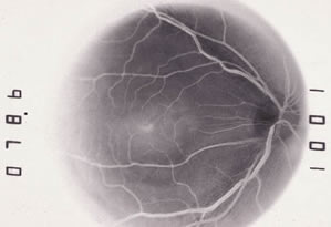
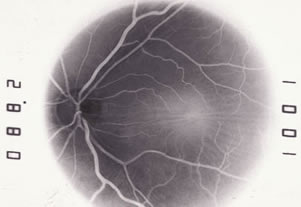
Figure 3: FFA
OD (Normal) OS
FFA OD FFA OS
Note: negative film FFA Macular striae OS seen on FFA.

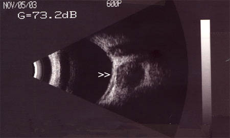
Figure 4: ECHO notable for round, encapsulated mass behind the globe OS (white arrow heads). A-Scan of lesion was highly reflective and suggestive of numerous vascular channels.
ECHO

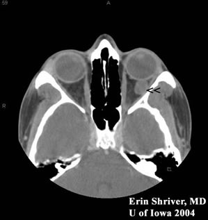
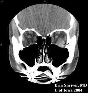
Figure 5. CT scan of orbits notable for a well-circumscribed, intraconal, round mass that was not eroding into surrounding tissues or bone. Mass was isodense to muscle (arrow heads).
CT CT

Figure 6: An orbitotomy was performed by the oculoplastics service, and an encapsulated mass was removed and submitted to ocular pathology.
Path Slide #1 Path Slide #2
Path Path

Path Slide #3
Path

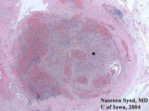
Path Slide #1: A large thrombus with inflammation (*) obscurred much of the large, dilated vascular spaces filled with blood. Immunohistochemistry confirmed that the thrombus contained mostly endothelial cells and fibroblasts, which ruled-out a neoplastic process. Thrombus formation is common because venous blood perfuses sluggishly through the cavernous hemangioma.

Path Slide #2: Large, dilated vascular spaces filled with blood. Cavernous hemangiomas have pseudoencapsulated vascular channels separated by fibrous septa.

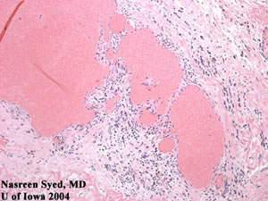
Path Slide #3: Some inflammatory cells were found in this lesion. However, significant stromal inflammation and lymphoid follicles may support the diagnosis of a lymphangioma.

Diagnosis: Cavernous Hemangioma

EPIDEMIOLOGY

  • Most common benign orbital tumor in adults.
  • Vascular hamartoma.
  • Average age of diagnosis: 40
  • Cause is unknown.

SIGNS

  • Unilateral, painless proptosis.
  • ECHO shows intraconal mass.
  • CT scan shows well-circumscribed, intraconal, round mass without invasion into adjacent soft tissue and bone.

SYMPTOMS

  • Vision not usually affected unless mass is pushing on the eye.
  • May cause optic nerve compression leading to visual loss, gaze-evoked amaurosis, or optic nerve swelling.
  • Low flow vascular lesion.

TREATMENT

  • May be observed with regular follow-up to confirm mass is not growing and there is no visual loss.
  • May be removed surgically via orbitotomy.

Differential Diagnosis:

  • lymphoid lesions of the orbit (benign reactive lymphoid hyperplasia, lymphoma, atypical lymphoid hyperplasia)
  • optic nerve meningioma
  • orbital metastasis
  • neurofibroma
  • neurilemoma (a.k.a. benign schwannoma)
  • fibrous histiocytoma
  • hemangiopericytoma
  • lymphangioma
Suggested citation format:

Shriver EM. Cavernous Hemangioma: 31-year-old caucasian female with a painless, dark spot in her left visual field. EyeRounds.org. Posted Feb. 21, 2005. Available from http://www.EyeRounds.org/cases/1-CavernousHemangioma.htm.


last updated: 02-21-2005

Image Permissions:
Creative Commons

Ophthalmic Atlas Images by EyeRounds.org, The University of Iowa are licensed under a Creative Commons Attribution-NonCommercial-NoDerivs 3.0 Unported License.